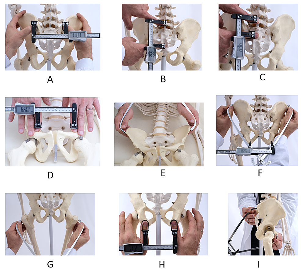
Did you know that the size and shape of a woman’s pelvis can greatly impact childbirth?
From the bispinous diameter to the various pelvic planes and axes, understanding these anatomical details is vital for both medical professionals and expectant mothers.
Join us as we delve into the fascinating world of gynaecoid, anthropoid, android, and platypelloid pelves.
Get ready to uncover the secrets behind a woman’s unique pelvic structure and its role in childbirth.
bispinous diameter
The bispinous diameter refers to the distance between the tips of the ischial spines in the pelvis, which is measured to be 10.5 cm.
Key Points:
- Bispinous diameter is a measurement of the distance between the tips of the ischial spines in the pelvis.
- The bispinous diameter is 10.5 cm.
- It provides information about the width of the pelvic outlet.
- The ischial spines are bony projections located in the pelvis.
- The bispinous diameter is an important parameter in obstetrics and gynecology.
- It is used to assess the size and shape of the pelvis for childbirth.
bispinous diameter – Watch Video
💡
Pro Tips:
1. The bispinous diameter is a term used in obstetrics to measure the widest diameter of the pelvic inlet, which is the smallest part of a woman’s pelvis through which a baby must pass during childbirth.
2. The average bispinous diameter in women is approximately 11 cm, but it can vary greatly based on factors such as body size, genetics, and previous pelvic injuries.
3. In most cases, a bispinous diameter of less than 9 cm is considered inadequate for a safe vaginal delivery. This condition, known as contracted pelvis, may require alternative birthing methods or even a cesarean section.
4. The bispinous diameter can be measured using specific instruments, such as a pelvimeter, which carefully measures the distance between the two bony prominences called the ischial spines in the pelvis.
5. Interestingly, the bispinous diameter tends to increase slightly during pregnancy due to hormonal changes and the softening of pelvic ligaments, which helps facilitate the passage of the baby during delivery.
Bispinous Diameter
The bispinous diameter is the distance between the tips of the ischial spines. It is an important measurement in evaluating the structure and dimensions of the female pelvis. The average bispinous diameter is approximately 10.5 cm.
The ischial spines are prominent bony structures located within the pelvic cavity. They are used as reference points for various obstetric measurements and play a crucial role during childbirth.
The bispinous diameter measurement helps determine the size and shape of the pelvis, which can impact the progress of labor and delivery outcomes.
Understanding the bispinous diameter is significant as it allows healthcare professionals to assess the adequacy of the pelvic inlet, mid cavity, and obstetric outlet. It provides valuable information about the passage for the fetus during childbirth, aiding in the assessment of potential obstacles or complications that may arise during delivery.
- The bispinous diameter is the distance between the tips of the ischial spines.
- The ischial spines are prominent bony structures located within the pelvic cavity.
- The measurement helps determine the size and shape of the pelvis.
- Understanding it allows healthcare professionals to assess the adequacy of the pelvic inlet, mid cavity, and obstetric outlet.
- It provides valuable information about the passage for the fetus during childbirth.
Pelvic Planes
Pelvic planes are critical anatomical landmarks used to assess and understand the structure and dimensions of the female pelvis. There are three main planes of the pelvis:
-
Plane of the pelvic inlet: This plane forms an angle of approximately 55 degrees with the horizon. It passes through the boundaries of the pelvic brim, which is the upper edge of the pelvic cavity. The plane of the pelvic inlet provides essential measurements of the size and shape of the upper part of the pelvis, offering insights into the space available for the fetal head to pass through during labor.
-
Plane of the mid-cavity: Another significant pelvic plane, it passes between the posterior surface of the symphysis pubis (the joint at the front of the pelvis) and the junction between the 2nd and 3rd sacral vertebrae (located in the lower back). It has a diameter of approximately 12.5 cm and helps evaluate the dimensions of the pelvic cavity.
-
Plane of the obstetric outlet: The final pelvic plane, passes from the lower border of the symphysis pubis anteriorly to the ischial spines laterally and to the tip of the sacrum posteriorly. The plane of the obstetric outlet is crucial in assessing the space available for the baby to pass through during childbirth.
These pelvic planes provide valuable information about the structure and dimensions of the female pelvis, contributing to a better understanding of childbirth and allowing for informed medical decisions.
- Bullet points are used to emphasize the main planes of the pelvis.
- Blockquote to highlight key information: “Pelvic planes are critical anatomical landmarks used to assess and understand the structure and dimensions of the female pelvis.”
Plane Of Pelvic Inlet
The plane of the pelvic inlet is a crucial factor in assessing the dimensions and structure of the pelvis. It is characterized by a unique angle of approximately 55 degrees with the horizon and defines the boundaries of the pelvic brim. This plane provides valuable insights into the available space for the descent and engagement of the fetal head during labor.
Determining the size and shape of the pelvic inlet allows healthcare professionals to anticipate any potential challenges or complications that may arise during delivery. A wider and more transverse oval inlet, such as the gynaecoid pelvis, typically facilitates an easier passage for the fetus. However, a more android or platypelloid-shaped inlet may pose difficulties and require additional medical interventions.
The plane of the pelvic inlet plays a vital role in obstetrics, guiding healthcare professionals in making informed decisions regarding the optimal birthing positions and necessary interventions during labor. Understanding the measurements of the pelvic inlet enables healthcare providers to tailor their approach for a safe and successful delivery.
Plane Of Mid Cavity
The plane of the mid-cavity is a significant pelvic plane used to assess the dimensions and structure of the pelvic cavity during childbirth. It extends between the posterior surface of the symphysis pubis (the joint at the front of the pelvis) and the junction between the 2nd and 3rd sacral vertebrae in the lower back.
The measurement of the mid-cavity plane helps healthcare professionals evaluate the amount of space available within the pelvic cavity for the movement and passage of the fetal head during labor. A mid-cavity diameter of approximately 12.5 cm is considered the average measurement.
Understanding the dimensions of the mid-cavity is crucial for determining the adequacy of the pelvic cavity and anticipating any potential challenges or complications during delivery. It aids in guiding healthcare providers in choosing the most suitable birthing positions and interventions to ensure a safe and successful birth.
The mid-cavity plane, along with other pelvic measurements, forms an integral part of obstetric assessments and is essential for identifying any abnormalities or deviations from the expected pelvic structure. This information enables healthcare professionals to provide appropriate care and support throughout the childbirth process.
Plane Of Obstetric Outlet
The plane of the obstetric outlet is a crucial pelvic plane used to evaluate the dimensions and structure of the lower part of the pelvis during childbirth. It starts at the lower border of the symphysis pubis at the front of the pelvis, extends laterally to the ischial spines, and continues posteriorly to the tip of the sacrum.
The measurements of the obstetric outlet plane play a crucial role in determining the available space for the fetus to pass through during delivery. The size and shape of the baby’s head are important factors in the safe navigation through the obstetric outlet.
Assessing the obstetric outlet plane enables healthcare professionals to anticipate potential challenges or complications during childbirth. A wider outlet with sufficient diameters allows for an easier passage of the fetus, while a narrow outlet may require additional interventions or accommodations to ensure a safe and successful delivery.
Understanding the plane of the obstetric outlet assists healthcare providers in making informed decisions regarding birthing positions and the need for medical interventions during labor. This knowledge contributes to optimizing outcomes for both the mother and the baby.
- The plane of the obstetric outlet is a key pelvic plane for evaluating the dimensions and structure of the lower part of the pelvis during childbirth.
- It begins at the lower border of the symphysis pubis, extends laterally to the ischial spines, and continues posteriorly to the tip of the sacrum.
- The measurements of the obstetric outlet plane are crucial in determining the space available for the fetus during delivery.
- The baby’s head size and shape influence its ability to navigate through the obstetric outlet safely.
- A wider outlet with sufficient diameters allows for an easier passage of the fetus, while a narrow outlet may require additional interventions.
- Understanding the obstetric outlet plane helps guide healthcare providers in making informed decisions during labor.
💡
You may need to know these questions about bispinous diameter
What is the bispinous diameter of the pelvis?
The bispinous diameter of the pelvis is commonly accepted to be around 10.5 cm. This measurement refers to the distance between the two spines (ischial spines) located in the pelvis. It plays a significant role in childbirth as it determines the space available for the baby to pass through the birth canal. A narrow bispinous diameter can potentially make childbirth more challenging and may require medical interventions.
What is the normal diameter of the pelvic inlet?
The normal diameter of the pelvic inlet, also known as the anatomical conjugate or true, measures approximately 11.0 cm. This measurement is taken from the sacral promontory to the upper edge of the pubic symphysis. It serves as an important reference for assessing the adequacy of the pelvic inlet for childbirth.
What is the widest diameter of the female pelvis?
The widest diameter of the female pelvis is determined by the pelvic inlet, measuring approximately 13 cm wide. However, the pelvic outlet has a narrower width of about 11 cm. As the fetus moves through the birth canal, it must rotate so that its head aligns with the widest dimension of the pelvic cavity, which spans 12.5 cm from top to bottom. This ensures a smoother passage during childbirth, accommodating the anatomical limitations of the pelvis.
What is the diameter of the maternal pelvis?
The maternal pelvis has varying dimensions, but the diameter in question is typically measured at the pelvic outlet. Based on the provided information, the AP diameter of the pelvic outlet is 13.5 cm, indicating the front-to-back measurement of the opening, while the transverse diameter is 11 cm, representing the side-to-side measurement. These dimensions provide crucial insights for understanding the capacity and structure of the maternal pelvis during childbirth.
Reference source
http://fourthstage2017.byethost16.com/[Obs]Nadia/[4]Anatomy%20of%20female%20pelvis%20and%20fetal%20head.pdf
https://digitalcommons.unmc.edu/cgi/viewcontent.cgi?article=3643&context=mdtheses
https://www.ncbi.nlm.nih.gov/books/NBK519068/
https://www.open.edu/openlearncreate/mod/oucontent/view.php?id=36&printable=1


