Hidden within the depths of the female reproductive system lies a crucial structure known as the cortex of ovary.
While it may seem like just another component, this enigmatic cortex holds the key to unlocking the mystical world of oogenesis, infertility, and the intricate processes that govern the creation of life itself.
Join us as we venture into the depths of the ovary, where the magic of conception and the secrets of the female body unravel before our very eyes.
cortex of ovary
The cortex of the ovary is the outer part of the ovary that contains the ovarian follicles, which are responsible for the development and maturation of female sex cells.
It is composed of connective tissue and is one of the primary female reproductive organs located in the pelvic cavity.
The cortex of the ovary contains the germinal epithelium and is surrounded by the tunica albuginea.
It is separated from the inner medulla, which contains blood vessels and connective tissue.
Oogenesis, the process of producing ova or eggs, occurs within the ovarian cortex.
Oogonia, the primary oocytes, undergo prophase in preparation for meiosis and are stimulated by follicle-stimulating hormone to develop into secondary oocytes.
During this process, polar bodies are formed, and one of the secondary oocytes becomes the ovum, which is ready for fertilization.
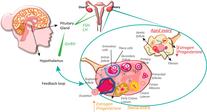
The development of ovarian follicles, which are composed of follicular cells and contain antrum and granulosa cells, is regulated by estrogen.
The dominant follicle, also known as the vesicular or Graafian follicle, eventually ruptures to release the secondary oocyte during ovulation.
After ovulation, the remaining follicle forms the corpus luteum, which secretes progesterone to support pregnancy.
If pregnancy does not occur, the corpus luteum regresses into the corpus albicans.
In summary, the cortex of the ovary is responsible for the formation and development of ovarian follicles and plays a crucial role in oogenesis and hormone secretion.
Key Points:
- The cortex of the ovary contains the ovarian follicles responsible for the development and maturation of female sex cells.
- It is composed of connective tissue and is located in the pelvic cavity.
- The cortex contains the germinal epithelium and is surrounded by the tunica albuginea.
- It is separated from the inner medulla which contains blood vessels and connective tissue.
- Oogenesis, the process of producing eggs, occurs within the ovarian cortex.
- The development of ovarian follicles is regulated by estrogen and eventually leads to ovulation.
cortex of ovary – Watch Video
💡
Pro Tips:
1. The cortex of the ovary is the outer layer of the organ and contains thousands of tiny structures called ovarian follicles.
2. The cortex of the ovary plays a crucial role in storing and releasing mature eggs during the process of ovulation.
3. Despite being the outer layer of the ovary, the cortex is actually responsible for producing hormones, such as estrogen and progesterone.
4. The cortex of the ovary is rich in connective tissue and blood vessels, which support the follicles and provide nutrients.
5. The cortex of the ovary is highly susceptible to damage caused by certain diseases, such as polycystic ovary syndrome (PCOS), which can disrupt the normal functioning of the ovary.
Introduction To The Cortex Of Ovary
The cortex of the ovary is a vital part of the female reproductive system. Located in the outer part of the ovary, it plays a crucial role in the development and maturation of ovarian follicles, which are sac-like structures that contain oocytes (immature eggs).
The cortex consists of connective tissue and is responsible for the production of hormones essential for reproduction.
In summary:
* The cortex of the ovary is located in the outer part of the ovary.
* It is responsible for the development and maturation of ovarian follicles.
* Ovarian follicles contain oocytes.
* The cortex of the ovary produces hormones essential for reproduction.
Note: The cortex of the ovary is an important component of the female reproductive system, as it contributes to the process of egg development and hormone production.
Structure And Function Of The Ovarian Cortex
The ovarian cortex is an important region of the ovary, alongside the inner medulla. It consists of various cell types, including ovarian follicles, connective tissue, and blood vessels. The ovarian follicles are crucial for the development and release of mature eggs during the menstrual cycle. These follicles undergo a series of stages, from primordial follicles to vesicular (graafian) follicles, before the rupture and release of the mature egg, also known as the ovum.
The connective tissue in the ovarian cortex offers support and nourishment for the follicles. It contains specialized cells called granulosa cells, which play a vital role in the maturation of the oocytes. Moreover, the ovarian cortex houses the tunica albuginea, a dense layer of fibrous tissue that surrounds and protects the ovarian follicles.
- Key Points:
- The ovarian cortex is one of the two main regions of the ovary, alongside the inner medulla.
- It consists of ovarian follicles, connective tissue, and blood vessels.
- Ovarian follicles are crucial for the development and release of mature eggs during the menstrual cycle.
- Follicles go through various stages before the rupture and release of the mature egg.
- The connective tissue supports and nourishes the follicles.
- Granulosa cells are specialized cells in the connective tissue that assist in the maturation of the oocytes.
- The ovarian cortex also contains the tunica albuginea, which protects the ovarian follicles.
Blockquote:
The ovarian follicles in the ovarian cortex are responsible for the development and release of mature eggs, ultimately leading to the release of the ovum. These follicles are supported by the connective tissue and protected by the surrounding tunica albuginea.
Ovarian Follicles In The Cortex
The ovarian follicles are the functional units of the cortex of the ovary. They play a crucial role in the production and development of oocytes. This process, known as oogenesis, takes place within the ovarian follicles and begins during embryonic development, continuing throughout a woman’s reproductive life.
The maturation of ovarian follicles occurs in several stages. Primordial follicles are present in the ovaries at birth and consist of primary oocytes surrounded by a single layer of follicular cells. As the follicles mature, they increase in size and develop multiple layers of granulosa cells. Additionally, fluid-filled spaces called antrum form within the follicles.
The growth and development of follicles are regulated by hormones, with follicle-stimulating hormone (FSH) being the most significant. FSH, produced by the pituitary gland, stimulates the growth and development of the follicles. Among the maturing follicles, one dominant follicle is ultimately selected and will release a secondary oocyte.
- Primordial follicles with primary oocytes are present at birth.
- Maturing follicles develop granulosa cells and antrum.
- Follicle-stimulating hormone (FSH) regulates follicle growth.
- One dominant follicle releases a secondary oocyte.
Note: The ovarian follicles are responsible for oocyte production and development. Oogenesis occurs within these follicles, with FSH playing a crucial role in their growth and maturation.
Connective Tissue In The Ovarian Cortex
Connective tissue is an important component of the ovarian cortex. It provides structural support to the follicles and facilitates the exchange of nutrients and waste products. The connective tissue contains blood vessels that supply oxygen and nutrients to the developing follicles.
The granulosa cells within the connective tissue play a critical role in the maturation of the oocytes. They secrete estrogen, a hormone that is responsible for the development of secondary sexual characteristics and the regulation of the menstrual cycle. Estrogen levels fluctuate throughout the menstrual cycle, with the highest levels occurring just before ovulation.
The connective tissue also serves as a site for the storage of hormones produced by the ovary. After ovulation, the ruptured follicle forms a structure called the corpus luteum, which secretes progesterone. Progesterone prepares the uterus for pregnancy and plays a critical role in maintaining a healthy pregnancy if fertilization occurs.
Ovarian Cortex Tissue Transplant For Infertility
Infertility is a common reproductive issue faced by many couples. Ovarian cortex tissue transplantation has emerged as a promising treatment option for couples with damaged or dysfunctional ovaries.
This procedure involves removing a small piece of ovarian cortex from either a donor or the patient herself and transplanting it into the affected ovary.
The transplanted ovarian cortex tissue has the ability to restore hormonal function and support the development of new follicles. This can potentially lead to the resumption of normal ovarian function and increase the possibility of natural conception.
Overall, ovarian cortex tissue transplantation has shown promising results in restoring fertility in women with premature ovarian failure or those who have undergone cancer treatments that damage the ovaries.
The Significance Of The Ovarian Cortex In Reproduction
The cortex of the ovary plays a critical role in the reproductive process. This is where the ovarian follicles develop, mature, and release oocytes for fertilization. A healthy ovarian cortex is vital for natural conception and the continuation of the human species.
Moreover, the ovarian cortex is responsible for regulating the menstrual cycle and producing essential hormones for reproductive health. The delicate balance of hormone secretion from the ovarian cortex is crucial for the proper functioning and synchronization of the female reproductive system.
The Role Of Germinal Epithelium In The Cortex
The germinal epithelium is a layer of cells that covers the surface of the ovary. It serves a protective function by separating the ovarian cortex from the surrounding pelvic cavity.
Recent research suggests that the germinal epithelium does not actually give rise to the oocytes. Instead, the primary source of new oocytes comes from the germ cells located within the ovarian cortex.
Oogenesis: Development Of Female Sex Cells In The Ovarian Cortex
Oogenesis is the process by which female sex cells, known as oocytes, develop within the ovarian cortex. It begins before birth when the primordial oogonia proliferate and differentiate into primary oocytes. These primary oocytes remain in a state of meiotic arrest until puberty.
Once puberty is reached, a small number of primary oocytes are activated each month to resume meiosis and enter the first meiotic division. During prophase, the primary oocyte undergoes significant changes, including DNA replication and chromosomal division. These changes prepare the oocyte for fertilization.
Upon ovulation, the primary oocyte completes the first meiotic division, yielding a secondary oocyte and the expulsion of the first polar body. The secondary oocyte is then arrested at metaphase of the second meiotic division until fertilization occurs.
Hormonal Regulation And Ovarian Cortex Function
The ovarian cortex is regulated by hormonal signals, including Follicle-stimulating hormone (FSH) which is secreted by the pituitary gland. FSH is vital for the development and maturation of ovarian follicles. It stimulates the growth and proliferation of granulosa cells within the follicles.
In addition, the ovarian cortex produces estrogen, a hormone that has several functions. It is responsible for the development of secondary sexual characteristics and helps regulate the menstrual cycle. Estrogen levels increase during the follicular phase of the menstrual cycle, which supports the development of the uterine lining in preparation for fertilization.
Corpus Luteum And Hormone Secretion In The Ovarian Cortex
After ovulation, the ruptured follicle transforms into the corpus luteum. The corpus luteum secretes progesterone, a hormone essential for the maintenance of a healthy pregnancy. Progesterone prepares the endometrium to receive a fertilized egg and prevents the shedding of the uterine lining.
If fertilization does not occur, the corpus luteum gradually degenerates, forming a scar tissue called the corpus albicans. The decline in progesterone levels triggers the shedding of the uterine lining, resulting in menstruation.
Understanding the structure and function of the ovarian cortex helps unravel the mysteries of female fertility and the intricate processes involved in human reproduction.
💡
You may need to know these questions about cortex of ovary
What is cortex and medulla in ovary?
The cortex and medulla are two distinct regions within the ovary. The cortex, which forms the outer portion, predominantly houses the ovarian follicles. These follicles are essential structures involved in the production and release of eggs during the menstrual cycle. On the other hand, the medulla resides in the inner portion of the ovary and primarily comprises larger blood vessels that supply oxygen and nutrients to the surrounding tissues. While there may be a slight difference in stromal texture, the key disparity lies in their composition, with the cortex being rich in follicles and the medulla being abundant in blood vessels.
What structures form the cortex of the ovary?
The cortex of the ovary is composed of a connective tissue stroma along with multiple ovarian follicles. These follicles are made up of an oocyte, which is surrounded by a single layer of follicular cells. Together, these structures form the intricate network of the ovarian cortex, contributing to the development and maturation of the follicles within the ovary.
Is cortex the functional part of the ovary?
No, the cortex is not the functional part of the ovary. While the cortex does contain the follicles and oocytes, which are important for the development and release of eggs, the functional part of the ovary is actually the medulla. The medulla contains the blood vessels, nerves, and connective tissue that support the overall function of the ovary. It is responsible for supplying nutrients and oxygen to the follicles and oocytes in the cortex, enabling them to develop and mature. Therefore, both the cortex and medulla work together to ensure the proper functioning of the ovary.
What is the function of cortex medulla?
The cortex and medulla have distinct functions within the kidney. The cortex is primarily responsible for filtering blood, ensuring that waste products and excess substances are removed from the bloodstream. On the other hand, the medulla is responsible for regulating the concentration of urine, by reabsorbing water and important substances back into the bloodstream while allowing waste products to continue on their path towards excretion. Together, these two regions of the kidney play crucial roles in maintaining fluid balance and eliminating waste from the body.
Reference source
https://www.ncbi.nlm.nih.gov/pmc/articles/PMC7246700/
https://histology.siu.edu/erg/ovary.htm
https://teachmeanatomy.info/pelvis/female-reproductive-tract/ovaries/
https://emedicine.medscape.com/article/1949171-overview


