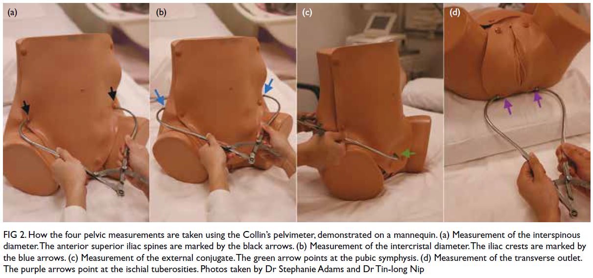
Step into the world of pregnancy and discover the fascinating method of external pelvimetry.
Imagine measuring the size and shape of the pelvis using calipers, providing valuable insights into the potential success of a vaginal delivery.
However, while this method has its limitations, it remains a key player in evaluating maternal pelvic dimensions and determining suitability for different delivery methods.
Join us as we delve deeper into the significance of external pelvimetry and its role in the world of obstetrics.
external pelvimetry
External pelvimetry is a method used to measure the size and shape of the pelvis during the third trimester of pregnancy.
It involves the use of calipers to measure various dimensions of the pelvis, including the inlet, mid-pelvis, and outlet.
This technique is used to predict the success of vaginal delivery and identify cases of cephalopelvic disproportion (CPD).
However, external pelvimetry has limitations, including low sensitivity and specificity, as well as a false positive rate.
To overcome these limitations, more reliable methods such as ultrasound pelvimetry and clinical assessment are often used.
Although external pelvimetry is a non-invasive method for evaluating maternal pelvic dimensions, it should be considered as an adjunct to traditional methods, such as internal pelvimetry, which provides more accurate information for determining the suitability of vaginal delivery versus cesarean section.
Factors that can affect the accuracy of external pelvimetry include obesity, fetal position, and pelvic soft tissue.
Despite its limitations, external pelvimetry can still provide valuable information for obstetric care.
Key Points:
- External pelvimetry measures size and shape of the pelvis during the third trimester
- Calipers are used to measure various dimensions of the pelvis
- Used to predict success of vaginal delivery and identify cases of cephalopelvic disproportion
- Limitations include low sensitivity, specificity, and false positive rate
- More reliable methods such as ultrasound pelvimetry and clinical assessment are often used
- Considered as an adjunct to traditional methods, such as internal pelvimetry
external pelvimetry – Watch Video
💡
Pro Tips:
1. External pelvimetry is a technique used to assess the dimensions of a woman’s pelvis before childbirth, helping doctors determine if a natural delivery is possible or if other measures need to be taken.
2. The practice of external pelvimetry dates back thousands of years, with evidence of its use found in ancient Egyptian papyrus scrolls dating back to 1550 BCE.
3. In the mid-20th century, external pelvimetry was often the only method available to evaluate pelvic dimensions before childbirth. However, advancements in technology have since allowed for more accurate and reliable methods, such as ultrasound and magnetic resonance imaging (MRI).
4. The external pelvimetry technique measures various pelvic dimensions, including the diagonal conjugate (the distance from the lower border of the pubic symphysis to the sacral promontory) and the interspinous diameter (the distance between the ischial spines). These measurements are crucial in assessing the pelvic capacity for childbirth.
5. Although external pelvimetry has been largely replaced by more advanced imaging techniques, it still holds some value in certain cases. In resource-limited settings where access to ultrasound or MRI is limited, external pelvimetry can still be an effective and affordable tool for evaluating pelvic dimensions.
Measuring Pelvis Size And Shape
Measuring the size and shape of the pelvis is an important aspect of obstetric care, especially in the third trimester of pregnancy. External pelvimetry is a method used to evaluate pelvic dimensions, with the primary goal of predicting the success of vaginal delivery and determining the need for interventions such as cesarean sections.
Key points:
- External pelvimetry is a technique that uses calipers to measure various dimensions of the pelvis from the outside.
- The significance of measuring the size and shape of the pelvis lies in its ability to assess the suitability of the pelvic anatomy for vaginal delivery.
- By determining the pelvic dimensions, healthcare providers can better anticipate any potential complications during labor and delivery.
- Successful vaginal delivery is associated with a pelvis that allows for the passage of the baby’s head without obstructing the birth canal.
- If external pelvimetry indicates any abnormalities or concerns regarding pelvic dimensions, healthcare providers may consider interventions such as cesarean sections to ensure the safe delivery of the baby.
Note: External pelvimetry plays a crucial role in obstetric care by providing valuable information on the pelvis’ size and shape. This aids in making informed decisions regarding the mode of delivery and potential interventions.
Predicting Success Of Vaginal Delivery
The main purpose of external pelvimetry is to predict the success of vaginal delivery. By assessing the dimensions and shape of the pelvis, healthcare providers can identify any potential obstructions or challenges that may arise during childbirth. If the pelvis is too narrow or misshapen, it may indicate a higher risk of complications and the need for alternative birthing methods like cesarean section. However, it is important to note that external pelvimetry is just one tool in a comprehensive assessment and should not be the sole determinant of delivery method.
Calipers For External Pelvimetry
External pelvimetry is a method that involves the use of calipers, which are medical instruments used for precise measurement of different points on the body. In the case of external pelvimetry, calipers are employed to measure distinct landmarks on the pelvic bones. These landmarks include the distance between the anterior superior iliac spines or the intertuberous diameter. By obtaining these measurements, healthcare professionals can gather essential information about the size and shape of the pelvis. Consequently, this data plays a crucial role in the decision-making process for childbirth.
- Calipers are medical instruments used for precise measurement.
- External pelvimetry measures specific landmarks on the pelvic bones.
- Landmarks include the distance between anterior superior iliac spines and the intertuberous diameter.
- Measurements provide valuable information about the size and shape of the pelvis.
Dimensions Of The Pelvis For Assessment
When using external pelvimetry, healthcare providers focus on assessing three main dimensions of the pelvis:
- The inlet: refers to the upper part of the pelvis, where the baby’s head enters during birth.
- The mid-pelvis: refers to the middle portion of the pelvis, where the baby descends and moves through the birth canal.
- The outlet: is the lower part of the pelvis, from which the baby is eventually delivered.
By measuring these dimensions, healthcare providers can determine if the pelvis is adequately sized and shaped for a vaginal delivery.
External pelvimetry is an important tool used by healthcare providers to assess the dimensions of the pelvis. By focusing on the inlet, mid-pelvis, and outlet, they can determine if the pelvis is suitable for a vaginal delivery.
- Inlet: upper part of the pelvis where the baby’s head enters
- Mid-pelvis: middle portion of the pelvis where the baby descends and moves through the birth canal
- Outlet: lower part of the pelvis from which the baby is delivered.
Inlet, Mid-Pelvis, Outlet Measurements
To determine the dimensions of the pelvis, healthcare providers utilize calipers or tape measures for accurate measurements. The landmarks involved in these measurements are the anterior superior iliac spines, ischial spines, and the sacrococcygeal joint. With these measurements, healthcare providers can assess whether the pelvis has enough room for the baby to pass through the birth canal without complications. This information plays a critical role in evaluating the feasibility of a vaginal delivery.
- Healthcare providers use calipers or tape measures to measure the inlet, mid-pelvis, and outlet dimensions of the pelvis.
- Landmarks such as the anterior superior iliac spines, ischial spines, and sacrococcygeal joint are used for these measurements.
- Assessing the available space in the pelvis is important in determining the possibility of a successful vaginal delivery.
Limitations Of External Pelvimetry
While external pelvimetry can provide valuable information about the size and shape of the pelvis, it is important to acknowledge its limitations. One such limitation is the low sensitivity and specificity of this method. External pelvimetry may not accurately identify all cases of cephalopelvic disproportion (CPD), which refers to a mismatch between the size of the baby’s head and the mother’s pelvis. Additionally, other factors such as fetal position and pelvic soft tissue can influence the success of vaginal delivery and are not assessed by external pelvimetry alone.
- It is important to acknowledge the limitations of external pelvimetry.
- Low sensitivity and specificity of this method.
- may not accurately identify all cases of cephalopelvic disproportion (CPD).
- Fetal position and pelvic soft tissue can influence the success of vaginal delivery.
Low Sensitivity And Specificity
Due to the limitations mentioned above, external pelvimetry has been associated with relatively low sensitivity and specificity in predicting the success of vaginal delivery. This means that it may result in both false positive and false negative results, leading to potential misinterpretation of the pelvis’s suitability for delivery. It is crucial for healthcare providers to consider other clinical assessments and more reliable methods to complement external pelvimetry and ensure accurate decisions regarding delivery.
Identifying Cephalopelvic Disproportion (CPD)
Cephalopelvic disproportion (CPD) is a condition where the baby’s head size is larger than the mother’s pelvis capacity, making it difficult or impossible for the baby to go through the birth canal. The screening tool commonly used for identifying potential CPD cases is external pelvimetry. However, it is crucial to remember that confirming the diagnosis and determining the best delivery plan may require additional procedures such as ultrasound or internal pelvimetry.
False Positive Rate For External Pelvimetry
External pelvimetry is a method used to assess cephalopelvic disproportion during childbirth. However, it is important to note that this procedure can produce false positive results, meaning it may incorrectly indicate the presence of disproportion when it is not actually present. This can lead to unnecessary interventions, such as cesarean sections or other alternate delivery methods. To minimize the risk of false positive results, healthcare providers should consider multiple factors and corroborate the findings of external pelvimetry with other assessments.
Ultrasound Pelvimetry For More Reliable Results
In cases where a more reliable assessment is required, ultrasound pelvimetry can be used. This non-invasive method provides a more detailed evaluation of maternal pelvic dimensions by measuring specific landmarks and utilizing imaging technology.
Ultrasound pelvimetry offers a more precise and accurate assessment compared to external pelvimetry. It is often used as an adjunct to traditional methods to further evaluate the suitability of the pelvis for vaginal delivery.
However, it should be noted that ultrasound pelvimetry also has its own limitations and should be interpreted in conjunction with other clinical findings.
- Ultrasound pelvimetry provides a detailed evaluation of maternal pelvic dimensions
- It uses imaging technology and measures specific landmarks
- More precise and accurate than external pelvimetry
- Used as an adjunct to traditional methods for evaluating pelvis suitability for vaginal delivery
- Should be interpreted alongside other clinical findings
“In cases where a more reliable assessment is required, ultrasound pelvimetry can be used.”
💡
You may need to know these questions about external pelvimetry
What is external pelvimetry?
External pelvimetry is a technique used to assess the dimensions of the pelvis from the outside of the body. It involves measuring the external diameter of the pelvis to determine its capacity for childbirth. By comparing these measurements with internal diameters, healthcare professionals can evaluate the risk of a contracted pelvis and potential complications during labor. External pelvimetry provides valuable insights into the adequacy of the birth canal without invasive procedures, helping healthcare providers plan for safe and successful deliveries.
What are the diameters of external pelvimetry?
The diameters of external pelvimetry play a crucial role in understanding the dimensions of the pelvic birth canal. These diameters are categorized into transverse, anteroposterior, and craniocaudal diameters. Interestingly, some of these external diameters correspond to the internal diameters of the pelvic birth canal, further highlighting their importance in assessing childbirth feasibility and potential complications [9,18]. The accurate measurement of these diameters is essential in obstetrics to ensure safe and successful delivery.
How is pelvimetry test done?
of the pelvis), or by imaging techniques such as magnetic resonance imaging (MRI) or ultrasound. The pelvimetry test involves the use of various methods to determine the dimensions and shape of the pelvis, which is crucial in predicting the likelihood of a successful vaginal delivery or the need for a cesarean section.
One common method of pelvimetry is through digital examination of the bony pelvis during a physical examination. The healthcare provider will use their fingers to palpate and measure the size of the pelvic bones and identify any bony prominences or abnormalities that may affect labor. Another method is through conventional X-rays, where the woman is positioned in different angles, and X-rays are taken to measure the dimensions of the pelvis. Lastly, imaging techniques such as MRI or ultrasound can provide more detailed images of the pelvis, allowing for a more accurate assessment of its size and shape. These tests enable healthcare professionals to make informed decisions regarding the mode of delivery and any potential risks or complications.
Is my pelvis too small for natural birth?
Having a pelvis that is considered too small for natural birth is actually a highly uncommon occurrence, and diagnosing it accurately can be a challenging task. Unfortunately, this misconception often leads to women being discouraged and denied the opportunity for a vaginal birth, resulting in subsequent c-sections for future pregnancies. It is crucial to remember that the human body is designed to accommodate childbirth, and medical professionals should always strive to provide women with the chance to experience a vaginal birth, even if they have been previously deemed to have a small pelvis.
Reference source
https://www.cureus.com/articles/52585-dynamic-external-pelvimetry-test-in-third-trimester-pregnant-women-shifting-positions-affect-pelvic-biomechanics-and-create-more-room-in-obstetric-diameters
https://www.ncbi.nlm.nih.gov/pmc/articles/PMC6901367/
https://www.ncbi.nlm.nih.gov/pmc/articles/PMC6464150/
https://www.sciencedirect.com/topics/medicine-and-dentistry/pelvimetry


