The Hidden Trauma of Genital Tract Injuries: A Comprehensive Guide

– Genital trauma refers to trauma to the genitalia, including the genital tract.
– Limited scientific data and evidence exist on genital injuries resulting from sexual assault.
– Studies on genital injuries have primarily focused on collecting evidence for legal purposes rather than medical purposes.
– Methods of studying and documenting genital injuries have improved through the use of tissue staining dyes and colposcopy.
– Vaginal trauma can occur during consensual and non-consensual intercourse, making it difficult to determine the circumstances based solely on a physical examination.
– Women are three times more likely to have vaginal injuries and intercourse-related injuries from a forced assault compared to consensual sexual experiences.
– Vaginal lacerations during intercourse may require surgery, while victims of forced assault may need additional services such as police intervention and trauma counseling.
– There is limited research on minor injuries in women of different age groups that do not require surgery or treatment.
– Factors that can predispose women to vaginal injury during consensual sex include first sexual experience, pregnancy, vigorous penetration, vaginal atrophy and spasm, previous operation or radiation therapy, disproportionate genitalia, penile ornamentation, and congenital anomalies.
– The missionary position with legs tilted all the way back during vaginal intercourse can lead to deep penetration and rotation of the uterus, potentially causing vaginal rupture.
– Vaginal tearing can occur in rape victims due to lack of vaginal lengthening and lubrication.
– Vulvar trauma is more common in prepubertal children and can occur from normal activities or sexual assault.
– Vaginal trauma can occur from the insertion of sharp objects, causing penetrating trauma.
– Severe vaginal injuries may require immediate medical attention if bleeding does not stop.
– Episiotomies can cause vaginal trauma.
– Penile trauma can occur in various forms, such as abrasions from zipper injuries or fractures from sexual activity.
– Penile amputation is a rare but emergency urological condition, often resulting from self-mutilation, accidents, or other causes.
– Micro-surgical repair is the most effective treatment method for penile trauma.
– Testicular trauma can occur from blunt, penetrating, or degloving injuries, particularly during contact sports.
– Wearing athletic cups can provide protection against testicular trauma.
– Testicular trauma can cause severe pain, bruising, swelling, and potential infertility.
Continue Reading

Unlocking the mysteries of microinvasive carcinoma: A comprehensive guide

– Microinvasive carcinoma of the breast is a rare tumor that occurs in a background of in situ carcinomas.
– The invasive component of the tumor is less than 1 mm.
– Signs and symptoms include a lump in the breast, swelling or skin thickening around the lump, and changes in breast profile.
– Complications can include the spread of cancer to other locations and treatment side effects such as nausea, vomiting, and hair loss.
– Treatment may involve surgery, chemotherapy, radiation therapy, and targeted therapy, depending on the stage of the tumor.
– The prognosis is generally excellent, as long as complete excision and removal of the tumor are achieved.
– Microinvasive carcinoma of the breast represents less than 1% of all breast cancer types.
– It is associated with ductal carcinoma in situ, and it is believed that 5-10% of DCIS cases may have microinvasion.
– The risk factors for microinvasive carcinoma of the breast include the presence of DCIS, being a woman, age over 40 years, a history of breast cancer, and a family history of breast cancer.
– Inherited gene mutations (BRCA1 or BRCA2) can increase the risk of microinvasive carcinoma.
– Radiation therapy to the chest or breast area can also increase the risk.
– Being overweight or obese increases the risk after menopause.
– Alcohol consumption, early onset of menstruation, and late menopause increase the risk.
– Postmenopausal hormone therapy with estrogen and progesterone increases the risk.
– Having the first child after the age of 35 or never having a child increases the risk.
– Lack of physical exercise and not breastfeeding the child increase the risk.
– The exact cause of microinvasive carcinoma is unknown, but hormonal influence and certain gene mutations may play a role.
– Symptoms may include a lump in the breast or underarm area, thickening or swelling of the breast, nipple inversion, bloody discharge from the nipple, changes to the skin covering the breast or nipple area, and breast pain.
– Diagnosis is typically done through a physical examination, evaluation of medical and family history, and potentially genetic testing for gene mutations.
– Microinvasive carcinoma can be diagnosed through various medical tests and procedures such as mammography, galactography, breast ultrasound scan, computerized tomography or magnetic resonance imaging, and positron emission tomography.
– The gold standard for diagnosing microinvasive carcinoma is a biopsy, which can be done through fine needle aspiration biopsy, core needle biopsy, or open tissue biopsy.
– Additional tests may be performed to rule out other conditions and obtain an accurate diagnosis.
– Complications of microinvasive carcinoma include emotional distress, rare metastasis to local lymph nodes, rare recurrence following surgery, and side effects of chemotherapy and radiation therapy.
– Treatment options depend on the type and staging of the cancer and the hormone sensitivity of the cancer cells.
– Surgical options include lumpectomy, mastectomy, sentinel node biopsy, and axillary node dissection.
– Chemotherapy may be used to kill remaining cancer cells or shrink the tumor.
– Radiation therapy uses high-energy beams to kill cancer cells.
– Hormone therapy may be recommended for hormone receptor-positive tumors.
– Targeted therapy drugs can be used to attack cancer cells in a specific manner.
– Lifestyle changes such as maintaining a healthy weight, exercising regularly, and following a well-balanced diet can help reduce the risk of microinvasive carcinoma.
– Regular cancer screenings, including breast self-exams, can help detect breast cancer at its earliest stages.
– Prophylactic measures such as taking estrogen-blocking drugs or undergoing prophylactic mastectomy may be suggested for women at high risk.
– Prognosis for microinvasive carcinoma is generally very good, especially with complete excision and removal of the tumor.
– Factors that can influence prognosis include grade, size, stage, hormone-receptor status, and response to treatment.

Continue Reading

Cervical Intraepithelial Neoplasm: Understanding Diagnosis, Treatment, and Prevention

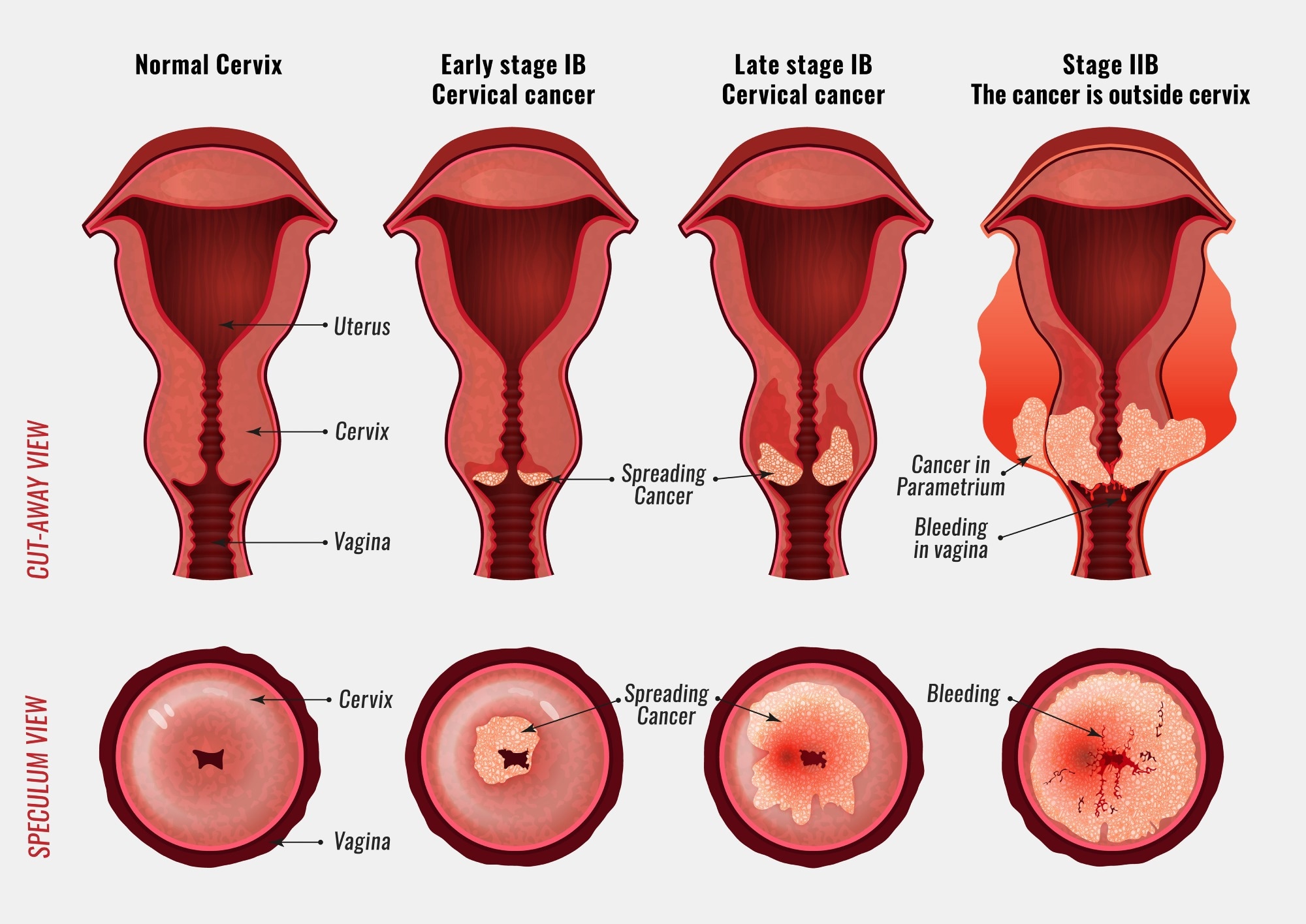
– Cervical dysplasia is a precancerous condition where abnormal cells grow on the surface of the cervix.
– Another name for cervical dysplasia is cervical intraepithelial neoplasia (CIN).
– Most people with cervical dysplasia do not develop cancer.
– Cervical dysplasia is classified on a scale from one to three, with CIN 1 affecting about one-third of the thickness of the epithelium, CIN 2 affecting about one-third to two-thirds of the epithelium, and CIN 3 affecting more than two-thirds.
– Cervical dysplasia primarily affects sexually active individuals assigned female at birth (AFAB) who have a cervix.
– It is most common among women of childbearing age, particularly aged 25 to 35.
– Approximately 250,000 to 1 million cisgender women in the U.S. are diagnosed with cervical dysplasia each year.
– Cervical intraepithelial neoplasia (CIN) is the abnormal growth of cells on the surface of the cervix that could potentially lead to cervical cancer. CIN is graded on a 1-3 scale, with 3 being the most abnormal.
– Human papillomavirus (HPV) infection is necessary for the development of CIN. Many women with HPV infection never develop CIN or cervical cancer. Typically, HPV resolves on its own. However, those with an HPV infection that lasts more than one or two years have a higher risk of developing a higher grade of CIN.
– Most cases of CIN either remain stable or are eliminated by the person’s immune system without the need for intervention. However, a small percentage of cases progress to cervical cancer if left untreated.
– There are no specific symptoms of CIN alone, but signs and symptoms of cervical cancer may include abnormal bleeding, abnormal discharge, changes in bladder or bowel function, pelvic pain, or abnormal appearance or palpation of the cervix.
– The cause of CIN is chronic infection of the cervix with HPV, especially infection with high-risk HPV types 16 or 18.
– Risk factors for developing CIN include infection with high-risk types of HPV, immunodeficiency, poor diet, multiple sex partners, lack of condom use, and cigarette smoking.
– Cervical intraepithelial neoplasia (CIN) is commonly associated with infection by human papillomavirus (HPV).
– Most women with HPV infection do not develop high-grade intraepithelial lesions or cancer.
– There are over 100 different types of HPV, with approximately 40 known to affect the anogenital area.
– The Digene HPV test is a highly accurate test for HPV, serving as both a direct diagnosis and adjuvant to the Pap smear.
– A colposcopy with directed biopsy is the standard for detecting CIN.
– Diagnosis of CIN or cervical carcinoma requires a biopsy for analysis.
– The Bethesda System for Reporting Cervical/Vaginal Cytologic Diagnoses provides a uniform way to describe abnormal epithelial cells.
– CIN is classified into grades: CIN 1 (mild dysplasia), CIN 2 (moderate dysplasia), and CIN 3 (severe dysplasia)
– CIN 3 can also be referred to as cervical carcinoma in situ.
– Locations of CIN findings can be described in terms of quadrants or clock face positions.
– Cervical intraepithelial neoplasm (CIN) is classified as LSIL or HSIL based on its severity.
– Screening for CIN can be done through Pap smear or testing for HPV.
– The accuracy of Pap smear results can vary.
– Abnormal Pap smear results may lead to colposcopy, which involves examining the cervix under magnification and taking a biopsy.
– HPV testing can identify high-risk HPV types responsible for CIN.
– HPV vaccination is the primary prevention method for CIN and cervical cancer, but it does not protect against all types of HPV known to cause cancer.
– Appropriate management and treatment are used as secondary prevention for cervical cancer cases.
– Treatment for CIN 1 is not recommended if it lasts fewer than two years, as it may clear on its own. Instead, close monitoring is advised.
– Treatment for higher-grade CIN involves removal or destruction of the abnormal cells.
– Retinoids may be effective in causing regression of CIN2.
– Therapeutic vaccines are being tested in clinical trials.
– The lifetime recurrence rate of CIN is about 20%.
– Surgical treatment of CIN may increase the risk of infertility or subfertility.
– Women receiving treatment for CIN during pregnancy may have an increased risk of premature birth.
– People with HIV and CIN 2+ should be managed according to general recommendations.
– Most cases of CIN spontaneously regress. Left untreated, about 70% of CIN 1 will regress within one year and 90% within two years. About 50% of CIN 2 cases will regress within two years. Progression to cervical carcinoma in situ (CIS) occurs in approximately 11% of CIN 1 and 22% of CIN 2 cases. Progression to invasive cancer occurs in approximately 1% of CIN 1, 5% of CIN 2, and at least 12% of CIN 3 cases.
– Treatment does not affect the chances of getting pregnant but is associated with an increased risk of miscarriage in the second trimester.
– Between 250,000 and 1 million American women are diagnosed with CIN annually.
– The estimated annual incidence of CIN in the United States is 4% for CIN 1 and 5% for CIN 2 and CIN 3.

Continue Reading

Exploring the Absence of Vagina: Causes, Treatments, and Resources

List of Pertinent Information for the Keyword ‘absence of vagina’:
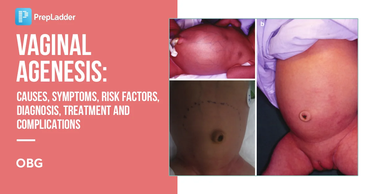
1. Vaginal agenesis is a condition in which a person is born without a vagina.
2. Diagnosis involves external genital exam, modified internal exam, ultrasound, and MRI.
3. Testing for MRKH syndrome involves a karyotype to examine chromosomes and pinpoint genetic causes.
4. Treatment options for vaginal agenesis include creating a vagina if sexual intercourse is desired.
5. Vaginal dilators are recommended as the first choice of treatment for MRKH.
6. Dilators are used to stretch the vaginal canal to a normal length.
7. Success rate of dilator treatment depends on consistent use and applied pressure.
8. Water-based lubricant may be necessary during intercourse.
9. Boston Children’s Hospital provides care for vaginal agenesis.
10. Anomalies of the Reproductive Tract specializes in treating females up to age 22 with reproductive organ anomalies, including vaginal agenesis.
11. The center has a multidisciplinary team consisting of gynecologists, radiologists, nurse specialists, and social workers.
12. Services provided include testing, treatment, counseling, and follow-up.
13. The Center for Young Women’s Health offers programs, resources, and services for young women’s health care.
14. Provides a team approach to accurate diagnoses and exceptional care and treatment options.
15. Offers information on gynecology, sexuality and health, development, fitness and nutrition, and emotional health.

Continue Reading

Understanding Abdominal Cellotomy: Exploring the Minimally Invasive Procedure

– Abdominal surgery
– Laparotomies
– Laparoscopic surgeries
– Ruptured appendix
– Cesarean sections
– Inguinal hernia surgery
– Exploratory laparotomy
– Cholecystectomy
– Appendectomy
– Exploratory celiotomy
– Diagnostic test
– Biopsy
– Ventral midline incision
– Linea alba
– Skin clipping
– Mammary chain
– Drain
– Feeding tube
– Lower urinary tract

Continue Reading

Key Considerations for Abdominal SalpingoOophorectomy: Benefits, Risks, Recovery, and Alternatives

– Abdominal salpingo-oophorectomy is a surgical procedure to remove the uterus, cervix, ovaries, and fallopian tubes.
– It is performed through an incision in the abdomen.
– Reasons for this surgery may include heavy periods, endometriosis, uterine fibroids, and cancer.
– The incision can be either horizontal or vertical.
– Risks of the procedure include bleeding, infection, damage to surrounding organs, and the possibility of further surgery.
– Preparation for the surgery includes fasting before the procedure and arranging for transportation.
– Recovery usually requires a hospital stay of about 2 nights and a full recovery time of 6 weeks.
– Vaginal bleeding and discharge are normal after surgery and should gradually decrease.
– Strenuous exercise, heavy lifting, and sexual activity should be avoided for 6 weeks after surgery.
– Medical attention should be sought if there is fever, severe nausea/vomiting, or abdominal pain, heavy bleeding, or redness/swelling/discharge from incisions.

Continue Reading

Abdominal Part: Uncovering the Core Muscles’ Essential Functions

The following list includes organs located in the abdominal part:

1. Stomach
2. Small intestine (jejunum and ileum)
3. Large intestine (colon)
4. Liver
5. Spleen
6. Gallbladder
7. Pancreas
8. Uterus
9. Fallopian tubes
10. Ovaries
11. Kidneys
12. Ureters
13. Bladder
14. Blood vessels (arteries and veins)

Please note that this information is not intended for emergencies, medical diagnosis, or treatment. It is essential to consult a licensed physician for any related concerns. The article also clarifies that links to other sites provided are for informational purposes only and should not be considered endorsements. Copyright restrictions apply, and unauthorized duplication or distribution is strictly prohibited.

Continue Reading

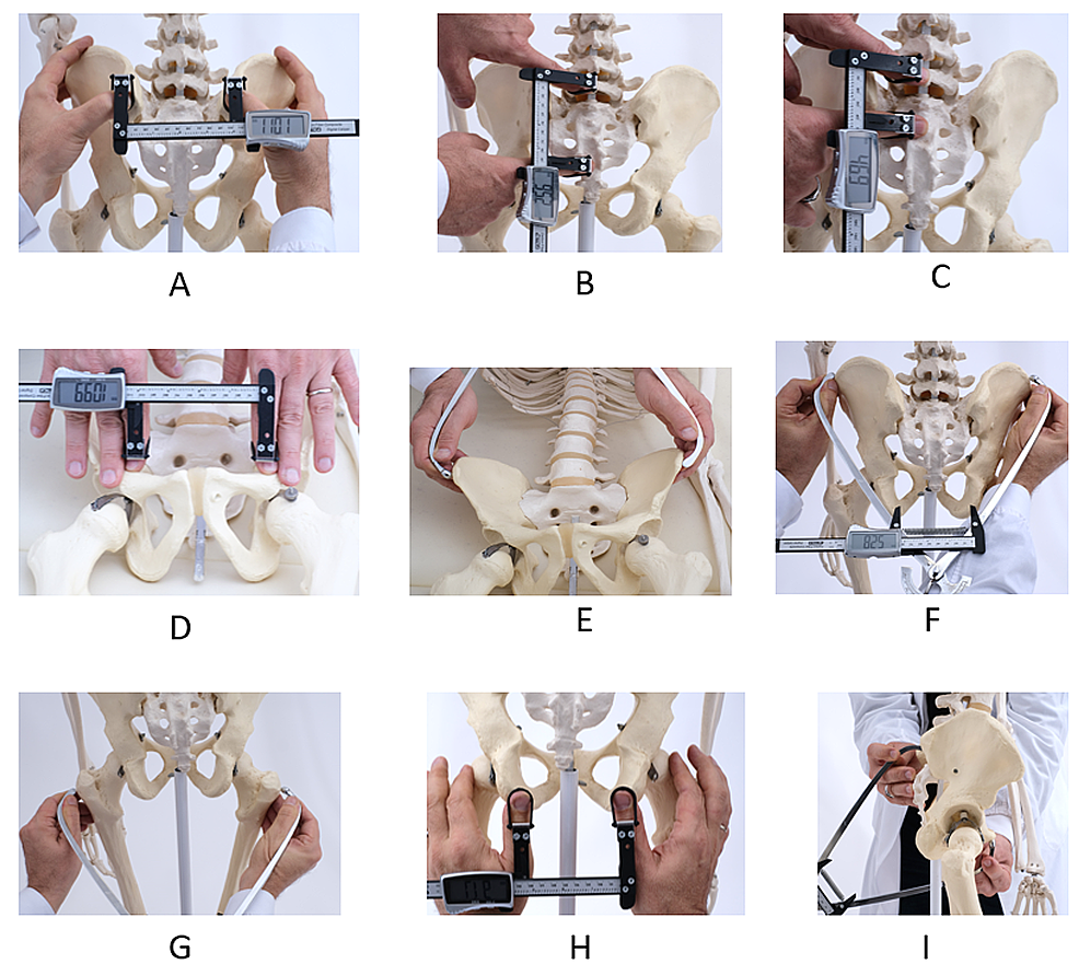
Conjugata Vera Obstetrica: Understanding Pelvic Measurements in Childbirth

List of relevant terms to the keyword “conjugata vera obstetrica”:

1. diameters and angles related to the pelvis
2. transverse diameters
3. dorsal transverse diameter
4. intermediary transverse diameter
5. ventral transverse diameter
6. cranial transverse diameter
7. caudal transverse diameter
8. medial transverse diameter
9. oblique diameters
10. right oblique diameter
11. left oblique diameter
12. right sacrocotyloid diameter
13. left sacrocotyloid diameter
14. conjugate diameters
15. conjugata vera
16. conjugata diagonalis
17. vertical diameter
18. pelvic inclination
19. angle between arcus ischiadicus
20. sacral promontory
21. symphysis pelvina/pubis

Continue Reading