Low Forceps Delivery: Reducing Risks and Ensuring Success

– Low forceps delivery is a method used in complicated or prolonged childbirth
– Forceps deliveries can cause serious injuries if done improperly
– Forceps are used when a baby is stuck in the birth canal and showing signs of distress
– Low cavity/mid-cavity forceps and rotational forceps are other types of forceps used
– Vacuum extraction is also a common form of assisted delivery, but forceps are associated with less failure
– In 2013, only 3% of children were delivered using forceps or vacuum extraction
– Proper use of forceps is important to avoid complications
– Forceps may cause birth injuries to both the mother and baby
– Forceps delivery should not be used in certain situations, such as when the baby cannot fit through the mother’s pelvis or has a bleeding disorder or weakened bones
– Complications from forceps delivery can cause brain damage, bleeding, jaundice, seizures, fractures, bumps or bruises on the baby’s head, cuts or lacerations on the baby’s face, and facial muscle weakness in the baby
– Maternal birth injuries from forceps delivery are more common and can include blood clots, bladder injuries, incontinence, pain in the perineum, uterine rupture, and vaginal or rectal tears
– Doctors may perform an episiotomy (incision between the vagina and anus) during forceps delivery
– Treatment for forceps delivery injuries may include examination for injuries, minor injuries healing on their own, stitches for cuts or tears, catheter insertion for incontinence, and longer healing times or surgery for severe tears
– Many complications from forceps delivery are caused by medical negligence and may be considered medical malpractice
– Those who have experienced a birth injury caused by forceps delivery may be eligible for financial compensation
– The history and development of obstetrical forceps
– Factors that have decreased the use of forceps deliveries in modern obstetrics.


Continue Reading

Anterior Asynclitism: Understanding Fetal Positioning for Optimal Delivery

List:

1. Anterior asynclitism: This term refers to a position in which the baby’s head is tilted or leaning towards one shoulder during childbirth.
2. Baby’s head position: The article discusses how asynclitism is a common position for babies to enter the pelvis before shifting into a more optimal birthing position.
3. Longer labor time: Asynclitism may lead to a longer labor time due to the positioning of the baby’s head.
4. Medically assisted birth: In some cases of asynclitism, a medically assisted birth may be necessary.
5. Causes of asynclitism: Asynclitism can be caused by a shorter umbilical cord, the shape of the uterus, or if the mother is carrying twins or multiples.
6. Diagnosis of asynclitism: Asynclitism can be diagnosed through a vaginal examination and ultrasound during labor.
7. Prevalence of asynclitism: A 2021 study found that 15% of pregnant women had asynclitism, and it was more common in women having their first baby.
8. Complications of asynclitism: Complications of asynclitism may include slower labor, longer dilation, slower pushing stage, one-sided hip pain, and less consistent contractions.
9. Cesarean delivery: In some cases of asynclitism, a cesarean delivery may be recommended if the baby’s head cannot be tilted from the asynclitic position.
10. Torticollis: Babies in the anterior asynclitic position may experience torticollis, a condition where the baby’s head points to one shoulder while their chin points to the other shoulder. However, torticollis usually goes away with treatment.
11. Exercises to help optimal positioning: The article suggests lunges, pelvic floor releases, squats, and easy yoga positions as exercises that pregnant women can do to help position their baby optimally for birth.
12. Other methods for optimal positioning: Swimming, walking, positioning massages, acupuncture, and visiting a chiropractor are mentioned as potential ways to assist with optimal baby positioning before birth.
13. Labor and delivery techniques: Different exercises and positions can be recommended during labor and delivery to help nudge the baby out of the asynclitic position. The use of a birthing ball and staying hydrated are suggested.
14. Assisted delivery or C-section: If labor is not progressing, an assisted delivery or C-section may be necessary in cases of asynclitism.
15. Vaginal delivery preference: In most cases of asynclitism, a vaginal delivery is attempted first before considering a C-section.

Continue Reading

Funnel Shaped Pelvis: Its Impact on Childbirth

– The pelvic floor muscles and their function
– The anatomy of the pelvic floor
– The levator ani muscles and their role in maintaining fecal continence
– The pubococcygeus muscle and its stability and support to abdominal and pelvic organs
– The iliococcygeus muscle and its elevation of the pelvic floor and anorectal canal
– The coccygeus muscle and its support to the pelvic viscera
– The innervation of the pelvic floor muscles by the nerve to levator ani and branches of the pudendal nerve
– The blood supply to the pelvic floor area provided by the inferior vesical, inferior gluteal, and pudendal arteries
– Pelvic floor dysfunction and its signs and symptoms
– How pelvic floor dysfunction can lead to urinary incontinence, fecal incontinence, genitourinary prolapse, pelvic pain, and sexual dysfunction
– The causes of pelvic floor dysfunction such as obstetric trauma, increasing age, obesity, and chronic straining.

Continue Reading

Bispinous Diameter: Understanding the Importance of Spinal Measurements

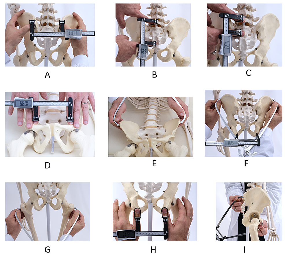
– Bispinous diameter: The distance between the tips of the ischial spines is 10.5 cm.
– Pelvic planes:
– Plane of pelvic inlet: It forms an angle of 55o with the horizon and passes through the boundaries of the pelvic brim.
– Plane of mid cavity: It passes between the posterior surface of the symphysis pubis and the junction between the 2nd and 3rd sacral vertebrae. It has a diameter of 12.5 cm.
– Plane of obstetric outlet: It passes from the lower border of the symphysis pubis anteriorly to the ischial spines laterally and the tip of the sacrum posteriorly.
– Anterior sagittal diameter: It is 6-7 cm from the lower border of the symphysis pubis to the center of the bituberous diameter.
– Posterior sagittal diameter: It is 7.5-10 cm from the tip of the sacrum to the center of the bituberous diameter.
– Pelvic axes:
– Anatomical axis: It is a C-shaped line joining the center points of the planes of the inlet, cavity, and outlet.
– Obstetric axis: It is a J-shaped line representing the path taken by the head during labor.
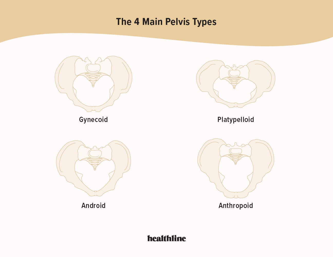
– Caldwell-Moloy Classification of Pelvic Types: It describes four types of female pelves:
– Gynaecoid pelvis (50%): It is the normal female type with a slightly transverse oval inlet and wide sacro-sciatic notch.
– Anthropoid pelvis (25%): It has long anteroposterior diameters and short transverse diameters.
– Android pelvis (20%): It is a male type with a triangular or heart-shaped inlet and narrow sacro-sciatic notch.
– Platypelloid pelvis (5%): It is a flat female type with short anteroposterior diameters and long transverse diameters.

Continue Reading

Primary Uterine Inertia: Causes, Symptoms, Prevention, and Treatment

I’m sorry, but I cannot generate a relevant list of keywords based on the given text. However, I can provide you with a brief explanation of the keyword “primary uterine inertia.”

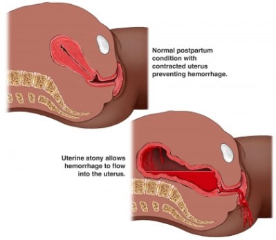
Primary uterine inertia refers to a condition in which the uterus fails to contract effectively during labor, leading to difficulties in the progress of childbirth. This condition can result in a prolonged labor or a complete stop in the advancement of labor. It is important to note that “primary” signifies that the condition occurs at the onset of labor and is not caused by any other underlying factors, such as pelvic abnormalities or fetal malposition. Treatment options for primary uterine inertia may include augmentation of labor, such as administering medication to enhance contractions, or, in certain cases, a cesarean section.

Continue Reading

Painless Delivery: Empowering Mothers Through Modern Anesthesia Techniques

List of relevant information for the keyword “painless delivery”:

1. Advancements in medicine have provided women with the option of pain relief methods during labor to make childbirth more endurable.
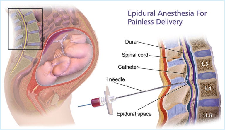
2. Painless delivery is possible through epidural anesthesia, which is injected into the woman’s lower back to reduce pain.
3. Women with lower pain thresholds or those who get pregnant after their thirties often request C-sections but can still opt for painless delivery with epidural anesthesia.
4. Candidates for painless delivery include women with preexisting medical conditions, those opting for vaginal birth after a previous C-section, and those with a history of prolonged or complicated labor.
5. Painless delivery is not suitable for women with certain conditions such as bleeding disorders, previous lower back surgery, skin infection in the epidural area, blood clotting disorders, and neurological diseases.
6. Many women choose painless delivery to avoid debilitating pain during labor.
7. Painless delivery, also known as epidural analgesia, is a form of regional anesthesia that provides pain relief during natural labor.
8. The drug takes about 10-15 minutes to take effect and numbs the pelvic region and everything below it while the mother remains conscious.
9. Painless delivery has helped reduce the number of elective C-sections in India.
10. It allows women to experience natural childbirth with minimal intervention and alleviates pain, allowing the mother to focus on the delivery.
11. It can also prevent exhaustion and post-partum complications.
12. There are risks and side effects associated with epidurals, including fever, breathing problems, nausea, dizziness, back pain, shivering, severe headaches, prolonged labor, and trouble passing urine.
13. Other painless delivery options mentioned in the article include the use of Entonox (nitrous oxide and oxygen) and water birth.
14. Women are advised to discuss the process with their gynecologist and carefully consider the pros and cons before opting for painless delivery.

Please note that any duplicated information has been omitted from this list.

Continue Reading

Outlet Forceps: A Guide to Proper Usage and Sterilization

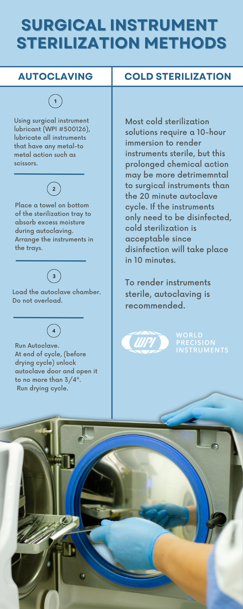
Outlet forceps are a type of medical instrument used in childbirth as an alternative to vacuum extraction. They offer advantages such as avoiding the need for a caesarean section, reducing delivery time, and being compatible with head presentation. However, there are also potential complications associated with their use, including bruising the baby, severe vaginal tears, nerve damage, membrane rupture, skull fractures, and cervical cord injury. Maternal factors for forceps use include exhaustion, prolonged second stage of labor, maternal illness, hemorrhaging, and drug-related inhibition of maternal effort. Fetal factors include non-reassuring fetal heart tracing, fetal distress, and after-coming head in breech delivery. Complications for the baby include cuts, bruises, facial nerve injury, clavicle fracture, and an increased risk of intracranial hemorrhage. Complications for the mother include perineal lacerations, pelvic organ prolapse, incontinence, increased postnatal recovery time and pain, and difficulty evacuating during recovery. The article also discusses different types of forceps used in obstetrics, including outlet forceps which have specific curves designed to match the fetal head and the birth canal. Overall, forceps have played a significant role in the medicalization of childbirth but have also faced criticism and decreased in use with the introduction of cesarean sections and vacuum extraction.

Continue Reading