Exploring Borderline Ovarian Tumors: Causes, Symptoms, Treatment

– Borderline ovarian tumors, also known as “low malignant potential” tumors, are not completely benign but also not invasive.
– Diagnosis of a borderline ovarian tumor can be suspected through imaging such as ultrasound or MRI, but the diagnosis is confirmed through microscopic assessment after surgery.
– Surgery is the primary treatment for borderline tumors, even if they have spread to other areas. Endocrine therapy may be considered in some cases.
– Fertility-sparing surgery or preservation of an unaffected ovary is often possible in young patients to avoid surgical menopause.
– Minimally invasive (laparoscopic) surgery is preferred for faster recovery and reduced risk of complications.
– Long-term follow-up care is necessary for patients with borderline ovarian tumors, with periodic imaging recommended for those with one ovary remaining.
– Approximately 10% of borderline tumors may recur, and surgical treatment is often used due to the limited response to systemic treatments like chemotherapy.
– Minimally invasive techniques are used for recurrent tumors whenever possible, and complex debulking surgeries may be performed to remove as much tumor as possible.

Continue Reading

Asphyxia Livida: Unveiling the Ominous Reality of Oxygen Deprivation

– The term “asphyxia livida” refers to a form of asphyxia neonatorum. In this condition, the skin appears cyanotic, indicating a lack of oxygen, but the heart functions normally and reflexes are intact.
– Birth asphyxia is a condition where a baby does not receive enough oxygen before, during, or directly after birth.
– It can cause serious complications and even be life-threatening.
– Birth asphyxia is also known as perinatal asphyxia and neonatal asphyxia.
– It can occur just before, during, or after birth.
– Insufficient oxygen supply can cause low levels of oxygen or excess acid in the baby’s blood.
– In mild or moderate cases, babies may fully recover, but in severe cases, it can cause permanent brain and organ damage or be fatal.
– Birth asphyxia rates are lower in developed countries at 2 in 1,000 births.
– In developing countries with limited access to neonatal care, the rate increases up to 10 times.
– Various factors can cause birth asphyxia, including umbilical cord prolapse, compression of the umbilical cord, meconium aspiration syndrome, premature birth, amniotic fluid embolism, uterine rupture, placental separation, infection during labor, prolonged or difficult labor, high or low blood pressure during pregnancy, and anemia in the baby.
– Risk factors for birth asphyxia include the pregnant person’s age between 20 and 25, multiple births, lack of prenatal care, low birth weight, abnormal fetal position, preeclampsia or eclampsia, and a history of birth asphyxia.
– Signs and symptoms of birth asphyxia can occur before, during, or after birth.
– Signs in the baby at birth may include unusual skin tone, silence, low heart rate, weak muscle tone and reflexes, lack of breathing, amniotic fluid stained with meconium, seizures, poor circulation, limpness or lethargy, low blood pressure, lack of urination, and abnormal blood clotting.
– A low Apgar score (between 0 and 3) that lasts for more than 5 minutes can indicate birth asphyxia.
– Immediate treatment can reduce the risk of long-term complications.
– Short-term effects can include acidosis, respiratory distress, high blood pressure, blood clotting problems, and kidney problems.
– Long-term effects of mild-to-moderate asphyxia can include cognitive and behavioral changes, hyperactivity, autism spectrum disorder, attention deficits, low intelligence quotient score, schizophrenia, and psychotic disorders.
– Severe asphyxia can cause intellectual disability, cerebral palsy, epilepsy, sight or hearing impairment.
– Treatment depends on the severity and cause of the asphyxia and may include providing extra oxygen, emergency or cesarean delivery, suctioning fluid from airways, putting the newborn on a respirator, placing the baby in a hyperbaric oxygen tank, and induced hypothermia.
– The article discusses the various treatments and prevention methods for asphyxia livida, a condition that can cause brain damage.
– Treatment options include medication to regulate blood pressure, dialysis to support the kidneys, medication to control seizures, IV nutrition, and the use of a breathing tube with nitric oxide.
– Life support with a heart and lung pump may also be necessary.
– Preventing asphyxia can be challenging, but proper care and monitoring before and after birth are crucial.
– Steps to prevent asphyxia include effective resuscitation, controlling body temperature, having the correct equipment available, ensuring trained healthcare providers are present for every birth, and pre-treatment with certain medications.
– Additionally, treatments like body cooling may be used to prevent complications from asphyxia.

Continue Reading

Episiotomy and Episiotitis: Understanding, Preventing, and Healing Tips

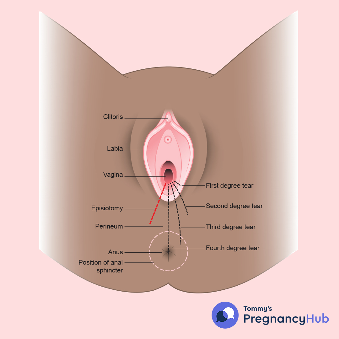
– An episiotomy is a cut made between the vaginal opening and the anus during childbirth.
– It was once thought to prevent larger vaginal tears and preserve the muscles and connective tissue in the pelvic floor, but research suggests it doesn’t prevent problems.
– Routine episiotomies are no longer recommended.
– An episiotomy may be needed if a baby needs to be quickly delivered due to a stuck shoulder, unusual heart rate pattern, or the need for forceps or vacuum extraction.
– There are two types of episiotomy incisions: midline and mediolateral.
– Midline incisions are easier to repair but have a higher risk of extending into the anal area.
– Mediolateral incisions are less likely to cause an extended tear into the anal area but are often more painful and difficult to repair.
– Episiotomy recovery is uncomfortable, and infection is possible.
– Some women may experience pain during sex after an episiotomy.
– A midline episiotomy puts women at risk of fourth-degree vaginal tearing.

Continue Reading

Understanding Metastatic Carcinoma of Ovary: Causes, Symptoms, Treatment

– Metastatic ovarian cancer is cancer that has spread from the ovaries or fallopian tubes to other parts of the body.
– It is classified as Stage IV cancer.
– Ovarian cancer typically spreads from the pelvis to the abdomen, peritoneal cavity, lymph nodes, and liver.
– Patients diagnosed with Stage I and Stage II ovarian cancer have survival rates of 90% and 70%, respectively.
– Patients diagnosed with Stage III ovarian cancer have a 5-year survival rate of 39%.
– Ovarian cancer can continue to spread to the lungs, fluid in the lungs, or tissue inside the liver if not successfully treated.
– At Stage IV, the disease becomes more difficult to treat, and the 5-year survival rate drops to approximately 17%.
– Ovarian cancer tumors that begin in the fallopian tubes take an average of 6.5 years to spread to the ovaries.
– Tumors can spread more quickly to nearby areas and eventually reach the spleen, intestines, brain, skin, and lymph nodes.
– Metastatic carcinoma of the ovary, particularly low-grade cases, is often resistant to therapies and challenging to treat.
– Achieving remission from metastatic ovarian cancer is difficult but not impossible.
– The course of treatment for metastatic ovarian cancer depends on various factors such as the type of ovarian cancer, patient’s age and overall health, presence of genetic mutations, and past response to treatment.
– Support for metastatic cancer patients is available through the OCRA (Ovarian Cancer Research Alliance) which offers resources such as a patient support team, online support groups, and peer-to-peer support programs.
– Early symptoms of ovarian cancer may include bloating, abdominal discomfort, trouble eating, and urinary urgency.
– Symptoms are more likely to occur when the cancer reaches an advanced stage and spreads to other areas of the body.
– Additional symptoms at this stage may include fatigue, constipation, vomiting and nausea, upset stomach, back pain, and abdominal swelling with weight loss.
– The five-year survival rate for metastatic ovarian cancer is approximately 18%.
– Treatment for metastatic ovarian cancer typically involves a combination of surgery and chemotherapy.
– Each patient’s case is unique and should be reviewed individually to determine the best treatment option.

Continue Reading

Conjugata Vera: Unraveling the Complexity of Verb Conjugation

List relevant terms extracted from the text for the keyword “conjugata vera”:

1. Study
2. Transverse diameters
3. Oblique diameters
4. Dorsal diameter
5. Intermediary diameter
6. Ventral diameter
7. Cranial diameter
8. Caudal diameter
9. Medial transverse diameter
10. Right oblique diameter
11. Left oblique diameter
12. Right sacrocotyloid diameter
13. Left sacrocotyloid diameter
14. Conjugata vera
15. Conjugata diagonalis
16. Vertical diameter
17. Pelvic inclination
18. Angle between arcus ischiadicus
19. Line
20. Diameter
21. Sacral promontory
22. Symphysis pelvina/pubis

Continue Reading

The Benefits and Risks of Abdominal Salpingectomy: A Comprehensive Guide

– Abdominal salpingectomy is the elective removal of both fallopian tubes during another abdominal surgery, such as gallbladder surgery, hernia operation, cesarean birth, or hysterectomy.
– The procedure is done to prevent cancer of the fallopian tube, ovary, or peritoneum.
– Salpingectomy may be recommended for the management of ectopic pregnancy, where a fertilized egg grows outside of the uterus and can cause life-threatening bleeding.
– Salpingectomy is also a method of permanent birth control, along with tubal ligation, but salpingectomy is more effective for contraception and has greater benefits in terms of cancer prevention.
– Some forms of ovarian cancer may start in the fallopian tubes, and removing the fallopian tubes can lower the risk of ovarian cancer, especially for individuals who do not desire future pregnancy.
– People with a hereditary risk of cancer, including those with BRCA1 and BRCA2 gene mutations or those associated with Lynch syndrome, have a higher lifetime risk of ovarian cancer. Abdominal salpingectomy is the recommended standard of care for people between the ages of 35 to 50 to lower the risk of ovarian cancer. It can also be beneficial for individuals without a known risk factor for ovarian cancer.
– Salpingectomy can be done at the same time as another planned abdominal surgery, such as gallbladder removal or hernia surgery. This is called opportunistic salpingectomy and is recommended for people who do not have a hereditary risk of ovarian cancer.
– Salpingectomy is a surgical procedure that involves the removal of the fallopian tubes.
– It can be done as either an endoscopic abdominal procedure (laparoscopic or robotic) or an open abdominal procedure.
– Laparoscopic salpingectomy requires three small incisions.
– The surgeon uses surgical instruments such as forceps to detach the fallopian tubes from the ovary and uterus.
– Recovery time typically takes one to three weeks, but it may be longer after a cesarean section or childbirth.
– Salpingectomy is effective for contraception right away and does not require additional contraceptive methods.
– The procedure does not protect against sexually transmitted infections, so condom use is still necessary.
– Laparoscopic salpingectomy is generally low risk, but rare complications may occur.

Continue Reading

Exploring Carcinoma Vulvae: Causes, Symptoms, Diagnoses, and Treatment

– Vulvar cancer is a cancer that occurs in any part of the external female genitals.
– It most commonly develops in the labia minora, labia majora, and perineum.
– There are several types of vulvar cancer: squamous cell carcinoma (90% of cases in Australia), vulvar melanoma (2-4% of cases), sarcoma (rare), adenocarcinoma (rare), and basal cell carcinoma (very rare).
– Vulvar cancer is not common.
– It mainly affects women who have gone through menopause, but can also occur in younger women.
– The average age at diagnosis is 69 years old.
– It is estimated that more than 400 people were diagnosed with vulvar cancer in 2023.

Continue Reading

Adenomyosis Interna: Understanding Symptoms, Causes, and Treatment Options

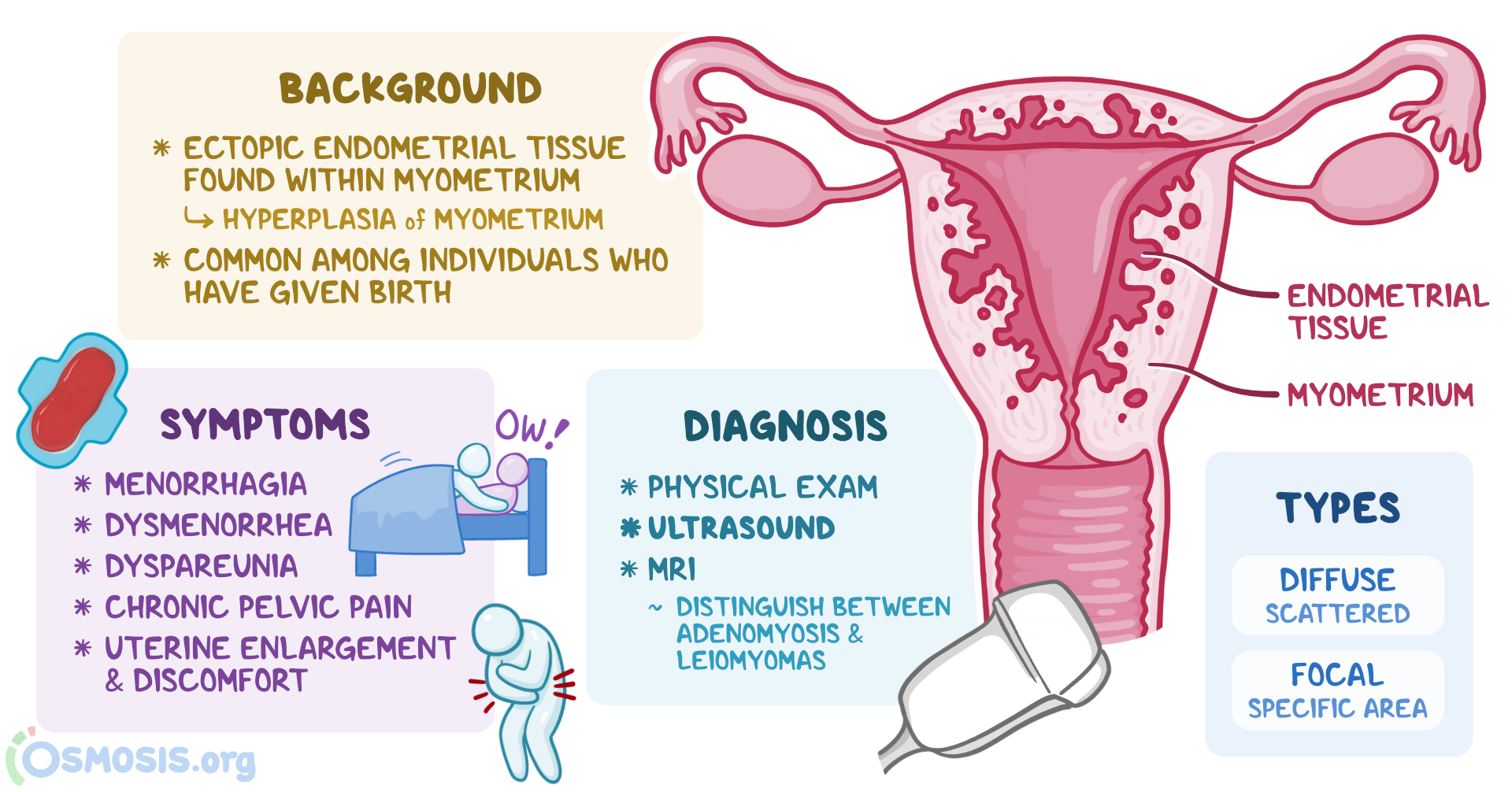
– Adenomyosis is a condition where the tissue that lines the uterus grows into the muscular wall of the uterus
– It causes the uterus to thicken and enlarge, sometimes up to double or triple its normal size
– Symptoms of adenomyosis include painful periods, heavy or prolonged menstrual bleeding with clotting, and abdominal/pelvic pain
– Many women and people assigned female at birth (AFAB) may not be aware they have adenomyosis because it doesn’t always cause symptoms
– The exact prevalence of adenomyosis is unknown
– Adenomyosis is more common in people who have had a procedure on their uterus and those who are older than 40
– Approximately 2% to 5% of adolescents with severely painful cycles have adenomyosis

Continue Reading

Understanding Atresia of Cervix: Causes, Symptoms, and Treatment

– Cervical agenesis
– Congenital disorder
– Absence of cervix
– Deformation of cervix
– Connecting structure between uterus and vagina
– Early adolescence
– Amenorrhea
– Cyclic pelvic pain
– Hematocolpos
– Endometriosis
– Pelvic adhesions
– Fetal development
– Paramesonephric duct
– Magnetic resonance imaging (MRI)
– Ultrasound
– Hormonal therapy
– Surgery
– Poor surgery outcomes
– Vaginal deformities
– Obstruction of menstrual flow
– Hematosalpinx
– Endometrioma
– Oral contraceptives
– Hysterectomy
– Neovaginoplasty
– Recanalization of cervix
– Low success rate
– 1 in 80,000 females

Continue Reading