The Importance of Arteria Ovarica in Female Reproduction:

1. The ovarian artery is an artery that supplies oxygenated blood to the ovary in females.
2. It arises from the abdominal aorta below the renal artery.
3. It can be found within the suspensory ligament of the ovary, anterior to the ovarian vein and ureter.
4. The ovarian arteries are paired structures that arise from the abdominal aorta, usually at the level of L2.
5. After emerging from the aorta, the artery travels within the suspensory ligament of the ovary and enters the mesovarium.
6. The ovarian arteries may anastomose with the uterine artery in the broad ligament.
7. Small branches are given to the ureter and the uterine tube.
8. One branch passes on to the side of the uterus and unites with the uterine artery.
9. Other offsets are continued on the round ligament of the uterus, through the inguinal canal, to the integument of the labium majus and groin.
10. In 20% of cases, the ovarian arteries arise from the renal arteries.
11. They may also arise from adrenal, lumbar, or internal iliac arteries.
12. The ovarian artery supplies blood to the ovary and uterus.
13. The ovarian arteries swell during pregnancy, in order to increase the uterine blood supply.

Continue Reading

Fetal Posture: Unlocking the Secrets of Optimal Development

– Fetal presentation before birth refers to the position of the baby in the uterus right before delivery.
– The most common position is cephalic occiput anterior, where the baby’s head is down and face down.
– Another position is cephalic occiput posterior, where the baby’s head is down but face up. This can make labor longer and may require manual rotation or assisted delivery.
– Breech presentation occurs when the baby’s feet or buttocks are in place to come out first during birth. This happens in about 3% to 4% of babies.
– The most common type of breech presentation is frank breech, where the baby’s knees aren’t bent and the feet are close to the baby’s head.
– A procedure called external cephalic version can be performed to try to move the baby into a head-down position if they are in a frank breech position.
– If the procedure is not successful or the baby moves back into a breech position, the delivery options should be discussed with the healthcare team.
– A complete breech presentation is when the baby has both knees bent and both legs pulled close to the body.
– An incomplete breech presentation is when one or both of the legs are not pulled close to the body, and one or both of the feet or knees are below the baby’s buttocks.
– If a baby is in a complete or incomplete breech presentation after 36 weeks of pregnancy, the health care professional may try to move the baby into a head-down position using external cephalic version.
– If the procedure is not successful or if the baby moves back into a breech position, alternative delivery options should be discussed with the health care team.
– A transverse lie is when the baby is lying horizontally across the uterus.
– If the baby is in a transverse lie at week 37 of pregnancy, the health care professional may try to move the baby into a head-down position using external cephalic version.
– If the procedure is not successful or if the baby moves back into a transverse lie, alternative delivery options should be discussed.
– If pregnant with twins and only one twin is head down, the health care provider may deliver the first twin vaginally and then suggest delivering the second twin in the breech position or try to move the second twin into a head-down position using external cephalic version.
– Delivery by C-section may be suggested for the second twin.

Continue Reading

Unraveling the Secrets of the Malignant Hydatidiform Mole

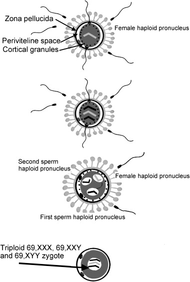
– Molar pregnancy, also known as HM or malignant hydatidiform mole, occurs due to abnormal fertilization of the egg.
– It results in an abnormal fetus and normal growth of the placenta with little or no fetal tissue growth.
– The placental tissue forms a mass in the uterus with a grape-like appearance on ultrasound, containing small cysts.
– Older women have a higher chance of developing a molar pregnancy.
– A history of molar pregnancy in earlier years is also a risk factor.
– There are two types of molar pregnancy: partial molar pregnancy, where there is an abnormal placenta and some fetal development, and complete molar pregnancy, where there is an abnormal placenta and no fetus.
– There is no known prevention for the formation of these masses.
– Mortality rate from hydatidiform mole is essentially zero due to early diagnosis and appropriate treatment.
– Approximately 20% of women with a complete mole develop a trophoblastic malignancy, which is almost 100% curable.
– Risk factors for malignant disease include advanced maternal age, high levels of hCG (>100,000 mIU/mL), eclampsia, hyperthyroidism, and bilateral theca lutein cysts.
– Predicting who will develop gestational trophoblastic neoplasia is difficult.
– Study suggests that outcomes of subsequent pregnancies in women who have had molar pregnancies are similar to those in the general population.
– Incidence of another molar pregnancy in women with a molar pregnancy is about 1.7%.
– Incidence of stillbirth in subsequent pregnancies in women with gestational trophoblastic neoplasia is 1.3%.
– Women with gestational trophoblastic neoplasia who conceive after chemotherapy have similar obstetric outcomes to those of the general population.
– Following a molar pregnancy, the risk of preterm birth is increased.
– Likelihood of large-for-gestational-age birth and stillbirth is greater if at least one birth occurs between the molar pregnancy and the index birth.
– Risk of adverse maternal outcomes is not increased following molar pregnancy.
– Malignancy is diagnosed in 15-20% of patients with a complete hydatidiform mole and 2-3% of partial moles.
– Lung metastases are found in 4-5% of patients.
– Perforation of the uterus during suction curettage is a potential complication, which may require laparoscopic guidance to complete the procedure.
– Hemorrhage is a common complication during the evacuation of a molar pregnancy, and intravenous oxytocin should be started at the beginning of suctioning.
– Other medications such as Methergine and Hemabate should also be available, and blood for possible transfusion should be readily available.
– Malignant trophoblastic disease develops in 20% of molar pregnancies, so quantitative hCG should be monitored regularly.
– Factors released by the molar tissue could trigger the coagulation cascade, leading to disseminated intravascular coagulopathy (DIC).
– Acute respiratory insufficiency can also occur due to trophoblastic embolism.
– The greatest risk factor for this complication is a larger uterus size compared to the expected gestational age.

Continue Reading

The Fascinating World of Syndactylus: Unraveling its Secrets

– Sumatran siamangs (Symphalangus syndactylus) are found in the Barisan Mountains of Sumatra and the mountains of the Malay Peninsula.
– They inhabit lowland, hill, and upper dipterocarp forests.
– Siamangs are the largest gibbons, weighing between 10 and 12 kg.
– Siamangs have a black fur coat, long arms, and a short-muzzled face.
– Siamangs have opposable thumbs and fused 2nd and 3rd toes.
– Siamangs are monogamous and highly territorial.
– Females give birth every 2 to 3 years, usually to one offspring, but twins can occur.
– Infants are weaned at 18 to 24 months and reach maturity at 6 to 7 years.
– Siamangs communicate through vocalizations, including bell-like tones, high yells, and high-pitched laughter.
– Territory size varies depending on food supply, averaging 28 to 95 acres.
– Siamangs primarily survive on leaves and fruit but also eat insects, bird eggs, and small vertebrates.
– Siamangs play a crucial role as seed dispersers in ecosystems.
– They have economic importance to humans as pets, subjects of primate studies, and for entertainment purposes in zoos.
– Siamang gibbons communicate through calls and songs to establish territory and reinforce family or mating bonds.
– Siamangs are the only gibbons with both opposable thumbs and opposable toes.
– They play a crucial role in their forest habitat by eating fruits from one tree and spreading seeds through their feces, leading to the growth of more trees elsewhere and a healthier tree population.

Continue Reading

Unraveling the Mystery: Foetus Papyraceus, an Extraordinary Phenomenon

– Fetus papyraceus
– Rare condition
– Compressed in-utero
– Resorption or paper thin
– Increased incidence with assisted reproductive techniques (ART)
– High morbidity and mortality for mother and fetus
– Incidence: 1 in 12,000 pregnancies
– Incidence in twin pregnancies: 1 in 190
– Can occur in monozygotic or dizygotic twins
– Associated with ovulation induction or in vitro fertilization
– Associated complications: pre-eclampsia, disseminated intravascular coagulation, thrombotic thrombocytopenic purpura
– Intrauterine complications: premature birth, low birth weight, hypoxic ischemic encephalopathy
– Case presentation of a 43-year-old primigravida with severe oligohydramnios and anemia
– Conceived through ART
– Three fetus papyraceus identified
– Emergency caesarean section performed
– Two live births
– Diagnosis through sonographic examinations
– Monitoring the effect on surviving fetus and mother

Continue Reading

Myoma of Uterus: Understanding Symptoms, Treatment, and Prevention

– A myoma, also known as a uterine fibroid or leiomyoma, is a common noncancerous tumor that grows in or around the uterus.
– Myomas can vary in size and may cause symptoms such as abdominal pain and heavy menstrual bleeding.
– Risk factors for myomas include a family history of myoma, obesity, and age.
– More than half of all people with a uterus will experience a myoma by age 50.
– Myomas can be categorized by location, size, and symptoms they may cause.
– Types of myomas include intramural myomas (located within the wall of the uterus), subserosal myomas (grow on the outside of the uterine wall), pedunculated myomas (develop a stalk or stem attaching them to the uterus), and submucosal myomas (found just under the lining of the uterus).
– A large myoma is considered to be 10 centimeters or more in diameter.
– Emergency room visits for myoma symptoms have increased from 2006 to 2017, including pelvic pain and heavy bleeding.
– Myomas are noncancerous growths that can cause various symptoms depending on their size and location.
– Symptoms of myomas include heavy and painful periods, bleeding between periods, pelvic pain, abdominal pressure, a feeling of fullness in the lower abdomen, constipation, diarrhea, frequent urination, pain during sex, lower back pain, trouble getting pregnant, fatigue, and weakness.
– Myomas are not life-threatening but can cause complications such as heavy blood loss or organ obstruction.
– Myomas are a top cause of hysterectomy surgeries.
– If a myoma bursts, immediate medical care is necessary.
– The exact cause of myomas is unknown but is likely associated with hormone activity, particularly high levels of estrogen and progesterone.
– Risk factors for myomas include a family history of the condition, obesity, high blood pressure, age, and certain dietary factors.
– Myomas are more common among Black people with a uterus.
– Diagnosis of myomas involves a series of steps, including medical history, physical examination, imaging tests such as ultrasound or MRI, and sometimes a biopsy.
– Treatment options for myomas include medication, noninvasive procedures, surgery, or a combination of therapies.
– Medications that may be used include over-the-counter pain medications, iron supplements, and birth control methods.
– Surgical options include laparoscopic myomectomy and uterine fibroid embolization (UFE).
– Lifestyle changes including dietary changes, exercise, stress management, and weight loss may improve symptoms and overall health.
– Complications of untreated myomas include fertility issues, pregnancy complications, and the need for cesarean delivery.
– It is important to discuss myomas with a healthcare provider before pregnancy for potential complications.
– Uterine fibroids, also known as myomas, are non-cancerous tumors that grow in the uterus.
– The most common symptom of myomas is vaginal bleeding.
– Other symptoms of myomas include heavy bleeding, anemia, fatigue, painful intercourse, pain, bleeding, or discharge from the vagina if myomas become infected, a feeling of pressure or lump in the abdomen, difficulties urinating, dribble at the end of urination, or urine retention if a myoma blocks the flow of urine.
– Myomas affect 20 percent of women in their childbearing years.
– The signs and symptoms of uterine fibroids include abnormal bleeding, pelvic masses, pelvic pain, infertility, and pregnancy complications.
– There are five types of uterine fibroids: intramural fibroids, subserosal fibroids, submucosal fibroids, pedunculated fibroids, and intracavitary fibroids.
– Between 70 and 80 percent of women develop a fibroid tumor by the time they reach age 50.
– Estrogen seems to activate the growth of uterine fibroids, and they shrink after menopause, but hormone therapy after menopause may cause their symptoms to continue.
– Factors associated with the development of uterine fibroids include race, age, early menstruation, caffeine and alcohol intake, genetics, obesity, high blood pressure, and diet.
– Nearly one-third of women with uterine fibroids seek treatment because of the severity of their symptoms.
– Treatment options for uterine fibroids include hormonal contraception, intrauterine devices, antifibrinolytic nonsteroidal agents, endometrial ablation, medications called gonadotropin-releasing hormone agonists, myomectomy, MRI-guided ultrasound surgery, and uterine fibroid embolization.
– Uterine fibroid embolization is a minimally invasive option that blocks the blood supply to fibroids, causing them to shrink and die.
– Myoma, leiomyoma, and uterine fibroids all refer to the same thing, a non-cancerous tumor of the uterus.
– The management of uterine fibroids, also known as myomas, requires further research to improve treatment outcomes.

Continue Reading

Endometriosis Interna: Breaking the Silence, Empowering Women

I apologize for the confusion. Since I don’t have the article you provided, I will generate a list of information related to the keyword “endometriosis interna” based on my training data. Here is a pertinent list of information related to endometriosis interna:

1. Definition of endometriosis interna
2. Symptoms of endometriosis interna
3. Causes and risk factors of endometriosis interna
4. Diagnosis of endometriosis interna
5. Treatment options for endometriosis interna
6. Complications and long-term effects of endometriosis interna
7. Lifestyle tips for managing endometriosis interna
8. Support groups and resources for individuals with endometriosis interna
9. Research and advancements in the field of endometriosis interna
10. Frequently asked questions about endometriosis interna

Please note, as an AI language model, I do not have access to real-time information or external sources, so this is a general list based on knowledge about endometriosis interna. It is always best to consult with a medical professional for accurate and specific information.

Continue Reading

The Art of Conservative Surgery: A Comprehensive Guide

– Conservative surgery, also known as breast-conserving surgery (BCS), removes the cancer while preserving as much normal breast tissue as possible.
– BCS is a good option for many women with early-stage breast cancer.
– Most women who undergo BCS will also need radiation therapy.
– BCS may be a good option for women who are concerned about losing a breast, are willing to have radiation therapy, have not had radiation therapy or BCS on the breast before, have a tumor smaller than 5 cm, are not pregnant or do not need immediate radiation therapy if pregnant, do not have certain gene mutations or connective tissue diseases, and do not have inflammatory breast cancer or positive margins.
– BCS is typically done in an outpatient surgery center.
– Post-surgery care instructions may include caring for the surgery site and dressing, caring for drains if applicable, recognizing signs of infection, bathing and showering tips, when to contact the doctor or nurse, and how to start using the affected arm again and perform arm exercises.
– After BCS, patients may experience pain, tenderness, or swelling in the breast.
– Formation of scar tissue and dimples at the surgical site is possible.
– Swelling of the breast due to fluid collection (seroma) might occur, requiring drainage.
– The shape of the breast may change after surgery.
– Nerve pain in the chest wall, armpit, and/or arm can persist after surgery.
– Patients who have axillary lymph nodes removed may be at risk of developing lymphedema.
– Pathologists examine the removed tissue to determine if all cancer cells were removed.
– Negative or clear margins indicate no cancer cells at the edges of the removed tissue.
– Close margins indicate cancer cells near the edges, while positive margins indicate cancer cells at the edge.
– If positive margins are found, further surgery may be needed, including a re-excision or mastectomy.
– Breast reconstruction surgery may be an option if significant changes in breast shape occur after BCS.
– Small metallic-like clips may be inserted during surgery to mark the area where the cancer was removed for radiation therapy planning.
– Most women will require radiation therapy and hormone therapy after BCS to reduce the risk of cancer recurrence.
– Some women may also require chemotherapy, in which case radiation therapy and hormone therapy are typically delayed until chemotherapy is completed.
– Risks of BCS include short-term breast swelling, changes in breast size and shape, scar tissue formation, wound infection or bleeding, and swelling of the arm.
– Reconstructive surgery may be an option to restore the appearance of the breast.
– Patients should discuss their expectations and options with their doctor before surgery.
– The recovery process after BCS depends on the type of procedure and anesthesia used.
– Patients may go home the same day or within 1 to 2 days after BCS.
– Pain levels vary and pain relievers should be taken as advised by the doctor.
– Wearing a supportive bra may be recommended.
– Normal activities can be resumed in 2 weeks, but strenuous activities should be avoided.
– Patients should follow the doctor’s instructions for driving, returning to work, and radiation therapy.
– Patients should notify the doctor if they experience certain symptoms or complications.
– Lymph node removal during BCS can affect lymphatic fluid drainage and increase the risk of infection and blood clots.
– Patients must follow safety steps for the rest of their lives after lymph node removal, including avoiding needle sticks and IVs in the affected arm, following arm exercise instructions, avoiding injuries, elevating the arm to drain fluid, wearing gloves during certain activities, avoiding sunburns, using a clean razor to shave, and avoiding tight clothing.
– Additional instructions may be provided by the doctor depending on the individual situation.

Continue Reading

Elderly Primipara: Overcoming Challenges and Navigating Motherhood Joyfully

List of pertinent information about ‘elderly primipara’:

– Incidence of elderly primigravida (women conceiving for the first time at the age of 35 or above) was 2.51%
– Most of these women (84.76%) married early but conceived late
– Majority of them belonged to high socioeconomic groups (62.86%)
– Common complications observed were anaemia (28.57%) and fibroids (5.71%)
– Other complications included pre-eclampsia (18.09%), eclampsia (3.81%), intrauterine growth restriction (12.38%), and twin pregnancies (5.72%)
– Complications during labor included fetal distress (33.33%), postpartum hemorrhage (3.81%), and retained placenta (2.86%)
– Gestational diabetes mellitus was observed in 0.95% of the cases
– Cesarean section rate was 29.52% while normal delivery accounted for 51.42%
– Incidence of congenital anomalies was 8.15%
– Elderly primigravida are at high risk for complications, but most pregnancies can result in healthy mothers and babies with proper supervision.

Continue Reading