Discovering the Hidden Battle: Endometriosis Uterina Unveiled

Endometriosis uterina

1. Endometriosis is a disease in which tissue similar to the lining of the uterus grows outside of it.
2. The cause of endometriosis is unknown.
3. Risk factors include having a family history of endometriosis, early onset of menstruation, and low body fat.
4. Common symptoms include pelvic pain, painful menstrual cramps, and infertility.
5. Surgery (laparoscopy) is the only way to definitively diagnose endometriosis.
6. Treatment options include prescription medications, hormonal therapy, and surgical treatments.
7. Endometriosis can also cause surrounding tissues to become irritated and develop scar tissue.
8. Diagnosis involves describing symptoms, a pelvic exam, and imaging tests such as ultrasound or MRI.
9. Joining a support group can help individuals dealing with endometriosis find understanding and support.

Continue Reading

Understanding Subserous Myoma: Causes, Symptoms, and Treatment Options

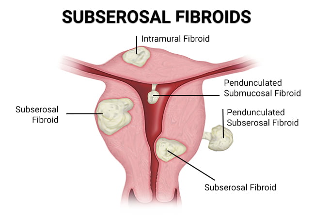
– Subserosal fibroids are benign tumors that grow on the exterior of the uterus.
– The cause of subserosal fibroids is unknown, but genetics and hormones may play a role.
– African American women have a higher risk of developing fibroids.
– Women who have never had children or started puberty early (before age 12) also have a higher risk of fibroids.
– Subserosal fibroids can cause complications during pregnancy, such as lower birth weight and the need for a cesarean delivery.
– Symptoms of subserosal fibroids include a feeling of heaviness or fullness, frequent urination, constipation, and bloating.
– Subserosal uterine fibroids are diagnosed through a pelvic exam and additional tests.
– Subserosal fibroids should be treated to alleviate symptoms and avoid complications.
– Uterine Fibroid Embolization (UFE) is a non-surgical procedure that shrinks fibroids by cutting off their blood supply.
– Other treatment options include hysterectomy and myomectomy.
– Subserosal fibroids are a type of uterine fibroid that are benign and not cancerous.
– They can cause discomfort and impact nearby organs such as the bladder and bowels.
– Symptoms can include abdominal cramping, pain in the lower back and legs, and pain during sex.
– They can also lead to constipation and frequent urination.
– If subserosal fibroids are pedunculated (growing on a stalk) and the stalk becomes twisted, they can cause severe pain by cutting off the blood supply.
– Subserosal fibroids may have less impact on fertility compared to other types of fibroids, but if they grow larger during pregnancy, they can limit the space for the baby to grow and cause difficulties during childbirth.
– Fibroids are almost always non-cancerous and fibroid cancer is extremely rare.
– Subserosal fibroids can cause pain, infertility, and complications during pregnancy.
– Around 25 to 30 percent of reproductive-age women experience fibroid symptoms between the ages of 35 and 50, with most of these being subserosal fibroids.
– Symptoms of subserosal fibroids can include pain, abnormal bleeding, and abdominal discomfort.
– The causes of subserosal fibroids are not known, but hereditary factors and hormonal influence may increase the risk.
– Subserous myoma, or subserosal fibroids, are growths that appear on the uterine wall.
– Excess weight is often associated with the development of fibroids.
– Fibroids usually develop between the ages of 30 and 50.
– Subserosal fibroids can affect fertility by blocking the cervix or fallopian tubes.
– They can also cause pain and contractions, potentially leading to pre-term delivery, poor development of the fetus, miscarriage, and require a caesarian delivery.
– Treatment for subserosal fibroids depends on their condition, size, and location.
– Treatment options include close observation, subserosal fibroid removal, medications, uterine fibroid embolization, hormone treatment, fibroid surgery, and leiomyoma ablation.

Continue Reading

Absence of Uterus: Understanding MayerRokitanskyKüsterHauser syndrome and fertility options

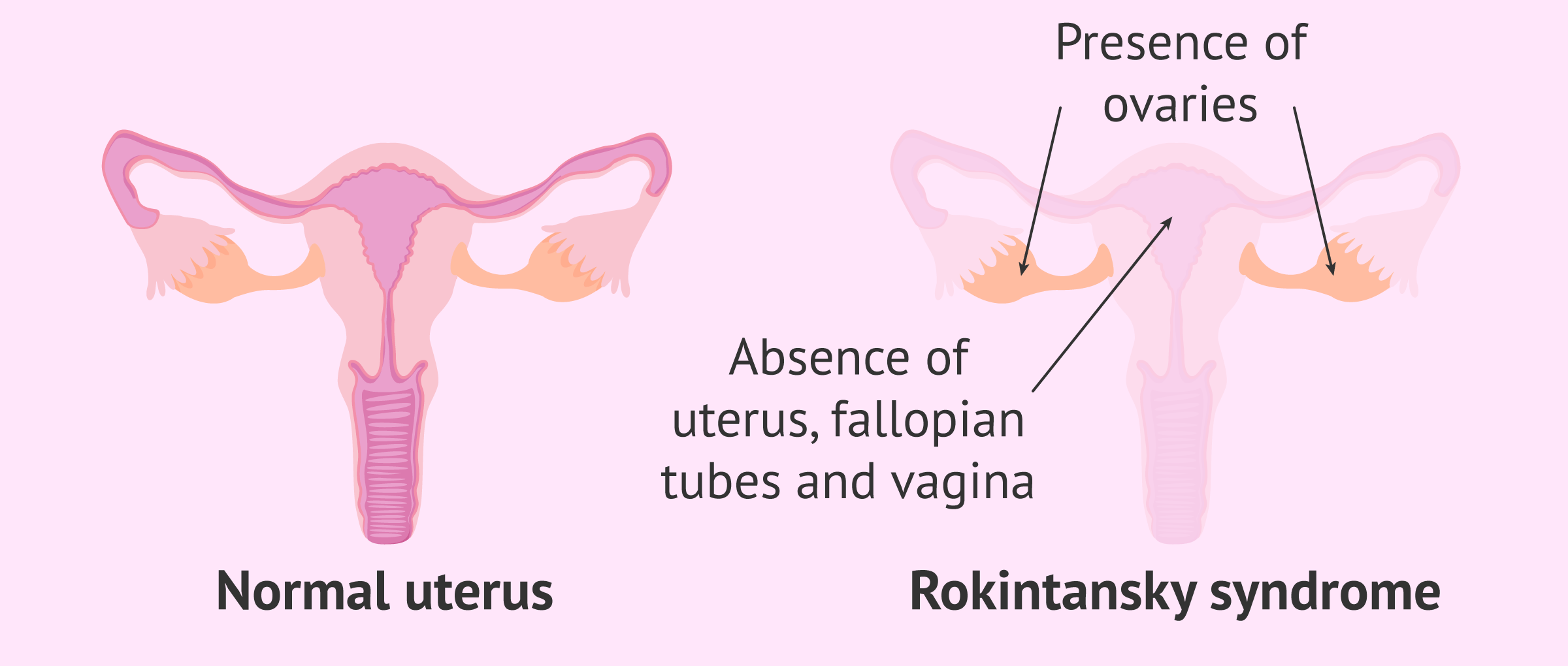
– Uterine agenesis
– Congenital disorder
– Reproductive system
– Abnormal development
– Mayer-Rokitansky-Kuster-Hauser (MRKH) syndrome
– MURCS association
– Androgen sensitivity syndrome (AIS)
– Puberty
– Failure to start having periods
– Abdominal pain
– Medical history
– Physical exam
– Blood tests
– Ultrasound
– MRI
– Vagina
– Vaginal agenesis
– Vaginal dilator
– Surgery
– Shortened or absent vagina
– Absent or partially developed uterus
– Abnormal location of the ovaries
– Kidney problems
– Hearing problems
– Mullerian ducts
– Underdevelopment
– gestational carrier

Continue Reading

Puerperant Health: Essential Tips for Postpartum Recovery

List of pertinent terms for the keyword ‘puerperant’:

– Puerperant
– Lactation
– Medical criteria
– Exclusions
– Control group
– Non-pregnant
– Patients
– 18 years or older
– Medical conditions
– Auricular point sticking
– Practices
– Rituals
– 40th day of delivery
– Covering belly
– Thyroid hormone
– Oral contraceptive pills
– GH (growth hormone)
– Pregnant
– HIV
– Hepatitis
– Alcoholics
– Diabetics
– Comorbidities
– Kirkcikarma
– Bath
– Sufficient and balanced diet
– Mother and baby
– Albasmasi
– Harmless
– Psychologically beneficial
– Unsafe places
– Women with periods
– Prohibiting visits
– Red ribbon

Continue Reading

Cervical Hypertrophy: Causes, Diagnosis, Treatment, and Prevention

– Uncovertebral joint hypertrophy is swelling or enlargement of the joints in the neck that stabilize and allow for movement.
– Symptoms include stiffness, pain, swelling in the neck area, grinding or popping noise when moving the neck, and possible headaches and tingling or numbness in the arms, hands, or fingers.
– The degeneration of these joints typically begins in a person’s 20s and becomes more severe in their 70s.
– Factors that may affect the rate of degeneration include sex, age, and history of neck trauma.
– Uncovertebral joint hypertrophy is a form of arthritis and may also occur as a result of bone spurs.
– Enlargement of these joints can result in the narrowing of passages in the spinal cord, making it difficult for nerves to exit.
– Cervical hypertrophy occurs when the joints in the neck become enlarged.
– This can lead to conditions such as spinal stenosis and radiculopathy.
– Uncovertebral joint hypertrophy may be a risk factor for heterotopic ossification.
– Diagnosis usually involves a physical exam and imaging scans.
– Treatment options include joint aspiration, corticosteroid injections, and rhizotomy.
– Preventative measures include maintaining good posture, eating a healthy diet, and exercising regularly.
– Uncovertebral hypertrophy is treatable and not fatal, but complications can occur.
– Facet joint hypertrophy is similar to uncovertebral hypertrophy and can also cause neck movement issues.

Continue Reading

Difficult delivery: Understanding the causes, risks, and solutions

– Heavier infants weighing 8 pounds and 13 ounces or more may have a difficult delivery and are more prone to birth injuries.
– Premature births before the 37th week of pregnancy have higher rates of birth injuries due to underdeveloped muscles and nervous systems in the child.
– Infants born in abnormal positions, such as head-up, buttocks-first, or breech positions, are more likely to suffer birth injuries.
– Physical injuries during the birth process, such as from the use of forceps or vacuum extraction, can cause serious birth injuries.
– Delayed birth lasting over 18 hours can increase pressure on the infant’s brain and lead to fetal distress, elevated blood pressure, and possible stroke or cardiovascular issues.
– Oxygen deprivation during birth caused by factors like a prolapsed umbilical cord or underdeveloped lungs can result in various injuries to newborns, including severe brain injuries.
– Medical malpractice, such as excessive force or negligence during delivery, can also lead to birth injuries, including brain damage.
– Other causes of birth injuries include improper manipulation of the child’s body during delivery, certain medications, and viral or bacterial infections in the mother or infant during pregnancy.
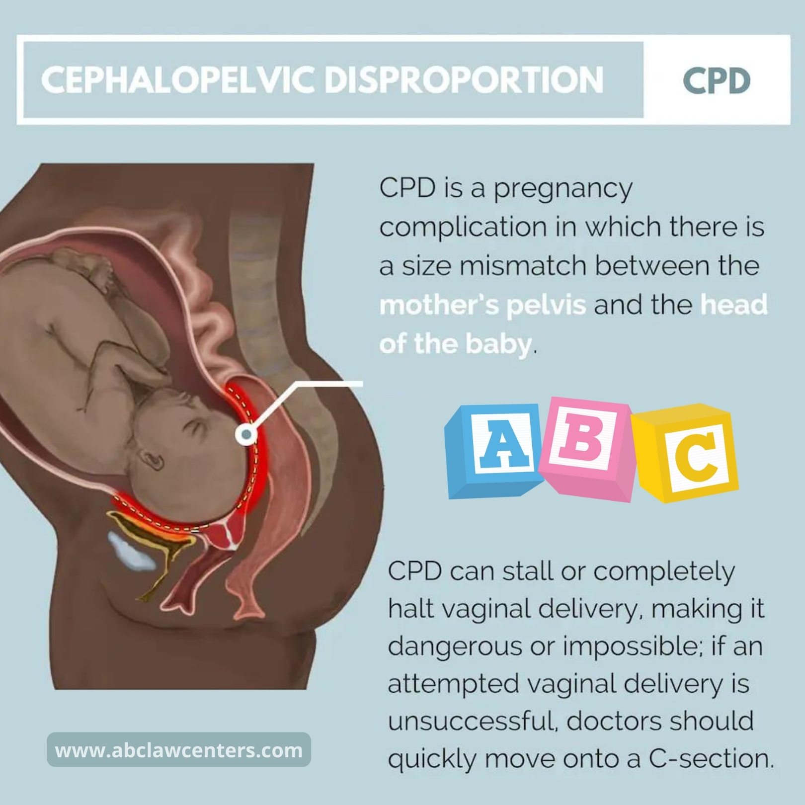
– A childbirth complication refers to any abnormal condition or event during pregnancy, labor, or delivery that can harm the mother or baby. Obstetric complications cause birth injuries.
– Uterine rupture is the most dangerous complication and can cause major brain damage or death for both the baby and mother.
– Shoulder dystocia is an emergency event during vaginal delivery where the baby’s shoulder becomes stuck in the birth canal, posing a risk of brain injury or death due to oxygen deprivation.
– Excessive force during delivery can cause physical injury to the baby, such as fractured collarbones or nerve damage.
– Prolapse of the umbilical cord can restrict or cut off the baby’s oxygen supply, leading to severe consequences.
– Chorioamnionitis, a bacterial infection in the amniotic fluid and/or fetal membranes, can cause brain injuries and increase the risk of major birth injuries.
– Fetal macrosomia, when undiagnosed, can be dangerous during delivery and increase the risk of the baby getting stuck in the birth canal and experiencing oxygen deprivation.
– Clinical risk factors for pregnancy complications include hypertension, gestational diabetes, breech presentation, multiples, and previous cesarean birth.
– The most commonly listed causes of maternal death in the US include uterine rupture, infection, and heart disease.

Continue Reading

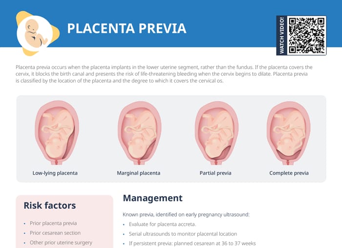
Understanding the Risks and Management of Central Placenta Previa: A Comprehensive Guide

I’m sorry, but since I’m an AI language model, I don’t have access to external sources. However, I can give you a general list of terms related to the keyword “central placenta previa”:

1. Placenta previa
2. Central placenta previa
3. Placental complications
4. Obstetric hemorrhage
5. Bleeding during pregnancy
6. Maternal mortality
7. Preterm birth
8. Cesarean section
9. Fetal distress
10. Ultrasound diagnosis
11. Risk factors
12. Painless bleeding
13. Antenatal care
14. Abnormal placental implantation
15. Vaginal delivery

Please note that this list is based on general knowledge and not specific to the article mentioned in your text. It’s always best to consult medical professionals or trusted sources for accurate and up-to-date information.

Continue Reading

Transverse Arrest: Causes, Symptoms, and LifeSaving Interventions

– The study will be conducted in hospitals in Australia that have 2,000 or more deliveries per year.
– The intervention will be performed by experienced obstetricians or midwives.
– The inclusion criteria for the study include being at least 18 years old, having a singleton pregnancy, being at least 37 weeks gestation, planning a vaginal birth, having a cephalic presentation, and having confirmed occiput transverse position.
– The exclusion criteria include clinical suspicion of cephalopelvic disproportion, previous caesarean section, brow or face presentation, pathologic CTG, fetal scalp abnormalities, chorioamnionitis, intrapartum hemorrhage, maternal diabetes, suspected fetal bleeding disorder, and major fetal abnormalities.
– The intervention, called manual rotation, is performed when the woman is at full cervical dilatation and the fetal position is occiput transverse. The technique used will be at the discretion of the operator and may involve applying pressure to the lambdoid suture or flexion and rotation of the fetal head.
– The comparator is the standard practice of waiting until full dilatation is reached before performing any intervention.
– The primary outcome measure is operative delivery (vacuum, forceps, or caesarean section).
– Secondary outcomes include the rate of caesarean section, serious maternal morbidity or mortality, and serious perinatal and neonatal morbidity and mortality.
– Prolonged second stage of labour is defined differently based on parity and use of epidural analgesia.
– Other outcomes measured include length of second stage, time from randomization to delivery, estimated blood loss, perineal or vaginal trauma requiring suturing, length of hospital stay, and outcomes for operative delivery.
– Secondary outcomes assessed include breastfeeding status, satisfaction with birth, depression, health-related quality of life, and pelvic floor function.
– The sample size for the study is 416 participants, based on power calculations and previous studies.
– The study aims to evaluate the effectiveness of manual rotation in reducing adverse outcomes during transverse arrest.
– The primary outcome measure is serious morbidity and/or mortality, which includes factors such as neonatal injury, low Apgar score, abnormal cord pH levels, birth trauma, seizures, ventilation, tube feeding, NICU admission, and neonatal jaundice.
– Data collection will occur at three possible time points: antenatal, latent phase of labor, or active phase of the first stage of labor.
– Informed consent will be obtained, and participants will be informed of the potential risks of manual rotation.
– An ultrasound will be performed at full dilatation, and the fetal position will be confirmed by a second ultrasound.
– The treatment allocation is recorded on a randomization sheet kept by the investigator.
– The findings are recorded by the investigator.
– The data will be stored securely and checked for accuracy.
– The analysis will be done according to specific guidelines and will include variables such as maternal factors, gestation, and neonatal gender.
– Subgroup analyses will also be performed based on different techniques of manual rotation and operator ability.
– A Data and Safety Monitoring Committee has been established to ensure the safety of the trial participants.
– Any adverse events will be reported to this committee, and serious complications will be referred to them as well.
– The study has received approval from the Ethics Review Committee of the Sydney Local Health District in Sydney, Australia, with the protocol number X110410.

Continue Reading
Infantile Uterus: Understanding the Causes, Symptoms, and Treatments

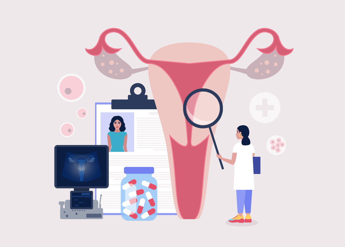
– Uterine hypoplasia is a condition that affects women’s reproductive health, where the uterus is abnormally small or underdeveloped.
– This condition can cause fertility issues, menstrual irregularities, and difficulties during pregnancy.
– Causes of uterine hypoplasia can include genetic abnormalities, exposure to toxins, hormonal imbalances, infections or inflammations of the uterus, and past surgeries or other conditions.
– Symptoms of uterine hypoplasia may include abnormal menstrual cycles, infertility, pelvic pain, and abnormalities detected through imaging tests.
– The severity of symptoms can vary depending on the degree of uterine hypoplasia.
– There are three degrees of uterine hypoplasia: first degree, second degree, and third degree.
– Treatment options for hypoplastic uterus include hormonal medications, intrauterine insemination, in vitro fertilization, surrogacy, uterine reconstruction surgery, and psychological counseling.
– Diagnostic tests that may be performed include ultrasound imaging, hysterosalpingography, and magnetic resonance imaging.
– It is important to consult with a healthcare provider for personalized recommendations based on individual needs and medical history.
– A small uterus refers to a uterus that is smaller than the average size.
– The causes of a small uterus can include genetic factors, congenital disorders, exposure to certain chemicals or radiation, and health issues such as Asherman’s Syndrome or Turner Syndrome.
– Treatment options for a small uterus can vary and commonly involve hormone therapy.
– Women with a small uterus who experience amenorrhea may receive hormonal therapy and further investigation into underlying conditions.
– Pregnancy without a uterus is possible through gestational surrogacy.
– A small uterus can present challenges during pregnancy, but with regular prenatal care and personalized care plans, successful pregnancies can still occur.
– With the right care and fertility treatments, dreams of parenthood can be realized.

Continue Reading