Bispinous Diameter: Understanding the Importance of Spinal Measurements

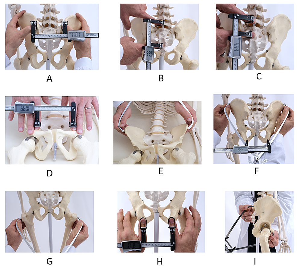
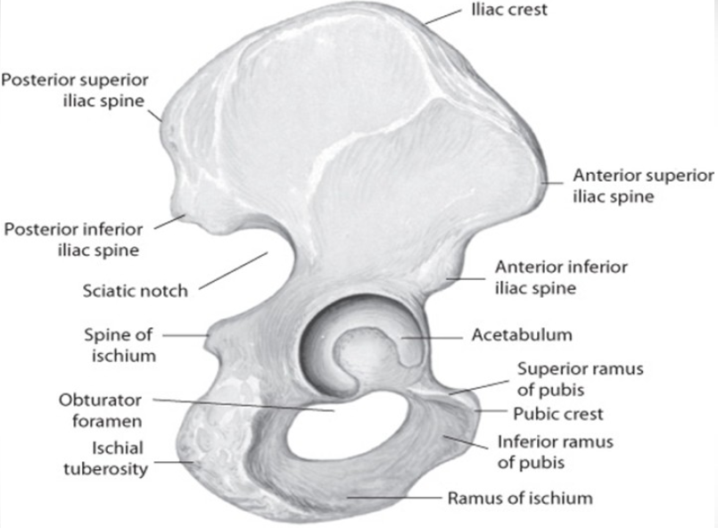
– Bispinous diameter: The distance between the tips of the ischial spines is 10.5 cm.
– Pelvic planes:
– Plane of pelvic inlet: It forms an angle of 55o with the horizon and passes through the boundaries of the pelvic brim.
– Plane of mid cavity: It passes between the posterior surface of the symphysis pubis and the junction between the 2nd and 3rd sacral vertebrae. It has a diameter of 12.5 cm.
– Plane of obstetric outlet: It passes from the lower border of the symphysis pubis anteriorly to the ischial spines laterally and the tip of the sacrum posteriorly.
– Anterior sagittal diameter: It is 6-7 cm from the lower border of the symphysis pubis to the center of the bituberous diameter.
– Posterior sagittal diameter: It is 7.5-10 cm from the tip of the sacrum to the center of the bituberous diameter.
– Pelvic axes:
– Anatomical axis: It is a C-shaped line joining the center points of the planes of the inlet, cavity, and outlet.
– Obstetric axis: It is a J-shaped line representing the path taken by the head during labor.
– Caldwell-Moloy Classification of Pelvic Types: It describes four types of female pelves:
– Gynaecoid pelvis (50%): It is the normal female type with a slightly transverse oval inlet and wide sacro-sciatic notch.
– Anthropoid pelvis (25%): It has long anteroposterior diameters and short transverse diameters.
– Android pelvis (20%): It is a male type with a triangular or heart-shaped inlet and narrow sacro-sciatic notch.
– Platypelloid pelvis (5%): It is a flat female type with short anteroposterior diameters and long transverse diameters.

Continue Reading

Unlocking the Mysteries of Uterus Subseptus: A Comprehensive Guide

– A subseptate uterus is a mild form of congenital uterine anomaly.
– It is often considered a normal variant.
– The condition involves the presence of a partial septum within the uterus.
– The septum does not extend to the cervix.
– The angle of the central point of the septum is acute (<90°). - The external uterine contour is uniformly convex or has an indentation <10 mm. - The prevalence of a septate uterus is approximately 55% among uterine anomalies. - It is classified as a class V Müllerian duct anomaly. - A septate uterus is associated with subfertility, preterm labor, and reproductive failure in approximately 67% of cases. - 15% of women with recurrent pregnancy loss have a septate uterus. - Concurrent renal anomalies may be associated with a septate uterus. - A septate uterus is considered a type of uterine duplication anomaly that results from the partial or complete failure of resorption of the uterovaginal septum after fusion of the paramesonephric ducts. - There are different subtypes of a septate uterus, including a partial septum (subseptate uterus) and a complete septum that extends to either the internal or external cervical os. - Hysterosalpingogram alone has an accuracy of only 55% in differentiating a septate uterus from a bicornuate uterus. - Ultrasound can show that the echogenic endometrial stripe is separated at the fundus by the septum, which is isoechoic to the myometrium. The external uterine contour should be convex, flat, or mildly concave. - MRI is considered the preferred imaging modality for diagnosing a septate uterus. On MR images, the septate uterus appears normal in size, and each endometrial cavity appears smaller than a normal cavity. The septum may be composed of fibrous tissue, myometrial tissue, or both. - The treatment for a septate uterus involves shaving off the septum during hysteroscopy (metroplasty) to form a single uterine cavity without perforating the uterus. Resection of the septum in the uterus has been shown to improve outcomes, with a reported decrease in the spontaneous abortion rate from 88% to 6% after hysteroscopic metroplasty. Differential diagnosis considerations for a septate uterus include a bicornuate uterus, and it is important to differentiate between the two due to different clinical and interventional approaches. Ultrasound or MRI may also be used for diagnosis.

Continue Reading

Exploring the Benefits of Extraperitoneal Cesarean Section: Insights

– extraperitoneal cesarean section
– asepsis protocol
– preoperative urinary catheterization
– surgical analgesia
– mobilization after surgery
– reduced doses of anesthetics
– EMLA cream
– fine 27 Gauge epidural catheter
– ropivacaine and sufentanyl
– midazolam and ketamine
– rapid absorbing braided Vicryl 2/0 stitches
– dermal adhesive for scar closure
– continuous suture for aponeurosis closure
– cruciform aponeurotic incision
– continuous or interrupted stitches for wound closure
– intramyometrial sutures with Vicryl 1 thread
– subserous layer closure
– uterus purse closure
– forceps or spatulas for extraction facilitation
– pressing on the base of the uterus
– anatomical triangle for lower segment approach
– emptying the bladder before surgery
– importance of appropriate bladder identification
– vertical paramedian opening of rectus abdominis’ aponeurosis anterior sheath
– surveillance period in recovery room
– acetaminophen for pain management
– prevention of reflex paralytic ileus and peritoneal adhesions
– early mobilization after surgery
– monitoring signs of hypotension during mobilization
– care of newborn immediately after leaving recovery room
– prevention of post-surgical thromboembolic events
– showering immediately after surgery
– less painful glued skin closure
– reduced scarring with glued skin closure
– gradual elimination of glue through desquamation

Continue Reading

Residual Ovary Syndrome: Unraveling its Causes, Treatment, and Prevention

– Oophorectomy is the surgical removal of one or both ovaries.
– It can be done to treat pelvic inflammatory disease, endometriosis, chronic pelvic pain, ectopic pregnancy, benign tumors, and large ovarian cysts.
– Oophorectomy can also be done to lower the risk of ovarian cancer in women with BRCA1 or BRCA2 gene mutations.
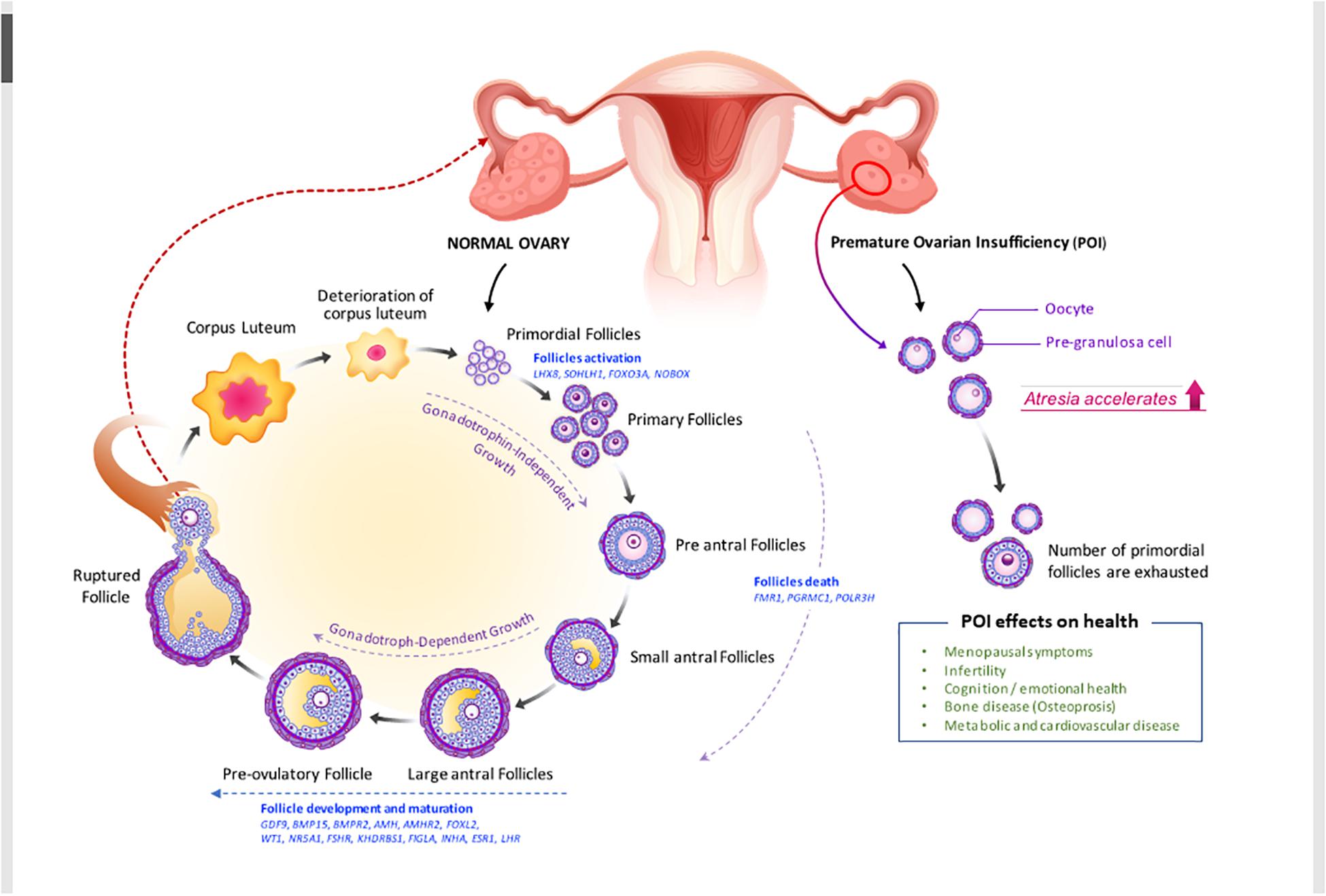
– Residual ovary syndrome occurs when pieces of ovarian tissue are left in the body after oophorectomy.
– The remnants can implant themselves in the abdominal cavity and cause pain or develop into cysts.
– The risk of residual ovary syndrome increases if the ovarian tissue is not completely removed during surgery.
– Causes of incomplete removal include pelvic adhesions, anatomical variations, and poor surgical procedures.
– Patients with a previous history of endometriosis or pelvic adhesions are at higher risk for residual ovary syndrome.
– Lack of menopause and continuous production of estrogen and progesterone are symptoms of residual ovary syndrome.
– Symptoms of residual ovary syndrome include irregular menstrual cycles, cyclic pelvic pain, formation of a pelvic mass, painful intercourse, difficulty in urination, and painful bowel movements.
– Diagnosis of residual ovary syndrome is done through a pelvic ultrasound or surgical exploration and biopsy of the remnant ovarian tissue.
– Treatment for residual ovary syndrome involves surgery to remove the residual ovarian tissue and hormonal therapy as an alternative.
– Preventive measures to avoid residual ovary syndrome include early surgical treatment of endometriosis and skilled surgery for ovary removal.
– Regular follow-ups with a doctor are recommended after oophorectomy to prevent the development of residual ovary syndrome.
– Risk factors for residual ovary syndrome include adhesions that make complete removal difficult and abnormal location of ovaries.
– Residual ovary syndrome is an uncommon condition, with an incidence of 2% to 3%.
– Symptoms of residual ovary syndrome include pelvic pain, pain during intercourse and bowel movements, and difficulty urinating.
– Ovarian remnant syndrome does not cause symptoms initially but can lead to the growth of cysts if left untreated.
– Ovaries may be removed during a hysterectomy depending on the underlying condition.
– If ovaries are not removed during a hysterectomy, they remain in place but hormone production slightly decreases.
– Ovaries reduce in size after menopause but do not disappear.
– Smaller ovaries may not be visualized during ultrasound imaging, but the risk of ovarian cancer is lower in these cases.
– Treatment options for ovarian remnant syndrome include surgical removal of leftover ovarian tissue, hormonal replacement therapy, and laparoscopy method.
– Residual ovary syndrome occurs after the removal of ovaries in surgery.
– The leftover tissue can grow as a cyst, causing pain.
– The leftover tissue may also get reimplanted on other organs such as the ureters, bowel, and bladder.
– Stem cells have been studied for ovary regeneration, but it has not been approved yet.

Continue Reading

Cancer of Cervix: Causes, Symptoms, Prevention, Treatment Options

– Cervical cancer is the growth of abnormal cells in the lining of the cervix.
– Squamous cell carcinoma is the most common type of cervical cancer, accounting for 70% of cases. Adenocarcinoma is less common, accounting for about 25% of cases.
– In 2023, over 900 people were diagnosed with cervical cancer, with an average age of 50 years old.
– The incidence of cervical cancer has decreased since the introduction of the National Cervical Screening Program in 1991 and a national HPV vaccine program in 2007.
– Signs of cervical cancer include vaginal bleeding between periods, longer or heavier menstrual bleeding, pain during intercourse, bleeding after intercourse, pelvic pain, changes in vaginal discharge, and vaginal bleeding after menopause.
– Persistent infection with high-risk types of HPV is the cause of almost all cases of cervical cancer.
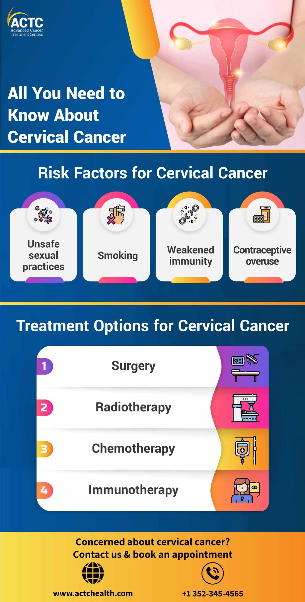
– Other risk factors include smoking and long-term use of the contraceptive pill.
– Diagnosis involves a colposcopy with biopsy, which allows doctors to locate and examine abnormal cells in the cervix.
– Treatment options depend on the stage of the disease, ranging from surgery to chemotherapy and radiation therapy.
– The Pap smear test has been replaced by the new Cervical Screening Test.
– The rate of cervical cancer in Australia has halved since the introduction of the National Cervical Screening Program.
– The HPV vaccine has been introduced as part of the National Cervical Screening Program and is offered to Australian children aged 12 to 13 for free.
– Australia aims to be the first country to eliminate cervical cancer as a public health issue.
– Gardasil 9 is the vaccine offered in Australia, protecting against nine types of HPV that cause around 90% of cervical cancers.
– Having the HPV vaccine does not replace the need for regular Cervical Cancer Screening Tests.
– Cervical cancer can be effectively treated if detected early, but treatment may impact fertility.

Continue Reading

Lyingin: The Importance, Challenges, and Benefits Explored

– The practice of lying-in
– Postpartum confinement
– Old-fashioned practice
– Essential practice
– No medical complications during childbirth
– Lying-in period
– Duration of lying-in
– Recommendations for not getting out of bed
– Care during lying-in
– Female relatives
– Monthly nurse
– Maternity hospitals
– Standard postpartum care
– Caudle – hot drink for new mothers
– Congratulatory visits during lying-in
– Desco da parto – painted tray for new mothers
– Representation of lying-in in art
– Depictions of the Birth of Jesus
– Virgin Mary reclining on a couch
– Ideal images of lying-in in well-off households
– Secular images on desci da parto
– Experiences and challenges faced by women during lying-in
– Preparing for lying-in
– Dilemma of preparing for lying-in
– After-pains during lying-in
– Mixtures for lying-in women after delivery

Continue Reading