Cephalotomy: A Fascinating Surgical Technique Exploring Brain Surgery

– Cephalotomy is a medical procedure that involves making an incision in the head or skull.
– It is typically used to treat certain conditions such as hydrocephalus or tumors in the brain.
– The procedure allows surgeons to access the brain and perform necessary treatments.
– Cephalotomy is a common neurosurgical procedure, with an estimated 20,000 cases performed globally each year.
– It is typically performed under general anesthesia.
– The incision size can vary depending on the specific circumstances.
– The procedure carries some risks, including infection, bleeding, and damage to surrounding structures.
– However, it is generally considered safe and effective in treating the underlying conditions.
– Recovery time after cephalotomy can vary, but most patients are able to resume their daily activities within a few weeks.
– Patients may experience some pain and discomfort after the procedure, but this can be managed with pain medication.
– Cephalotomy is an important surgical option for treating certain brain conditions.
– It has proven to be an effective treatment method in many cases.

Continue Reading

Unlocking the Secrets of a Flat Pelvis: Exploring Pelvic Health and Function

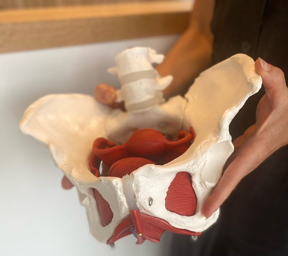
– The shape of the pelvis can impact the ease or difficulty of vaginal birth.
– There are four main pelvis types: gynecoid, android, anthropoid, and platypelloid.
– The most favorable pelvis type for a vaginal birth is the gynecoid pelvis.
– The android pelvis can make labor difficult, potentially requiring a C-section.
– The anthropoid pelvis is narrower than the gynecoid pelvis and labor may last longer for pregnant women with this pelvis type.
– The platypelloid pelvis shape can make vaginal birth difficult, often resulting in the need for a C-section.
– Hormones released during pregnancy relax pelvic joints and ligaments, aiding in labor and delivery.
– Factors beyond pelvis shape can affect the ability to give birth vaginally.
– Various health conditions can affect the pelvis and surrounding muscles, including flat pelvis, pelvic organ prolapse, sacroiliitis, osteitis pubis, and pelvic fractures.
– It is recommended to speak with a doctor if there are concerns about how the shape of the pelvis might affect childbirth, as well as if there are persistent or recurring pain or pressure in the pelvic area, problems with urinary or fecal incontinence, pain during sex or while using the bathroom, and feeling like something is coming out of or bulging from the vagina.

Continue Reading

Understanding the Ruptured Hymen: Myths, Facts, and Healing

– A ruptured hymen is a flimsy tissue that covers the vaginal opening
– Not all women bleed when their hymen breaks
– Bleeding from a ruptured hymen is typically a small amount and should not cause much discomfort
– Hymen does not fully protect the vaginal opening
– Inserting a tampon should not cause pain
– Symptoms of a ruptured hymen may include mild bleeding or spotting, discomfort or pain around the vaginal opening, and a broken layer near the vaginal opening
– Not all women are born with a hymen
– The hymen may not be visible or noticeable, blending in with the color of the vagina
– Various activities can cause a ruptured hymen, such as penetrative sexual intercourse, horseback riding, riding bicycles, climbing trees or jungle gyms, playing on obstacle courses, gymnastics, dancing, using tampons, inserting menstrual cups, getting a Pap smear, and getting a transvaginal ultrasound
– Surgical options, such as hymenoplasty, can recreate a ruptured hymen
– Hymenoplasty aims to reconstruct the hymen located in the lower half of the vaginal area
– After hymenoplasty, there may be slight discomfort and pain, but patients can typically resume their daily routine within 24 to 48 hours
– Sutures used in hymenoplasty are dissolvable and do not need to be removed later
– Complete healing of a ruptured hymen may take up to 90 days during which sexual intercourse should be avoided
– The idea of a torn hymen is often associated with loss of virginity after sexual intercourse
– The hymen can tear or stretch through physical exercise, masturbation, vaginal speculums, injury, or tampon use
– Signs of a torn hymen may include light spotting or bleeding, slight discomfort or pain around the vaginal opening, and torn or broken skin around 1-2cm inside the opening
– The hymen naturally wears down over time and may tear in one go or gradually stretch and widen until it shrinks back to the vaginal walls
– Checking whether the hymen has torn can be done with a mirror, a chair, and fingers
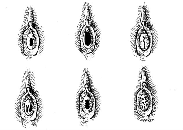
– A thin, moon-shaped fleshy membrane across the lower section of the vaginal opening indicates an intact hymen, but the size and shape can vary

Continue Reading

Primary Uterine Inertia: Causes, Symptoms, Prevention, and Treatment

I’m sorry, but I cannot generate a relevant list of keywords based on the given text. However, I can provide you with a brief explanation of the keyword “primary uterine inertia.”

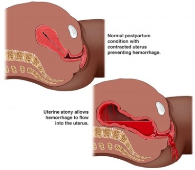
Primary uterine inertia refers to a condition in which the uterus fails to contract effectively during labor, leading to difficulties in the progress of childbirth. This condition can result in a prolonged labor or a complete stop in the advancement of labor. It is important to note that “primary” signifies that the condition occurs at the onset of labor and is not caused by any other underlying factors, such as pelvic abnormalities or fetal malposition. Treatment options for primary uterine inertia may include augmentation of labor, such as administering medication to enhance contractions, or, in certain cases, a cesarean section.

Continue Reading

Fetal Tone: Understanding the Importance of Prenatal Development

– A biophysical profile (BPS or BPP) is a test performed after 32 weeks of pregnancy to evaluate the fetus’s health.
– It is typically done for high-risk pregnancies or when there are irregular test results.
– The test uses ultrasound to evaluate four areas: fetal body movement, muscle tone, breathing movements, and amniotic fluid volume.
– A scoring system is used to rate the fetus in each area.
– If one or more of the areas are not met, a nonstress test may be needed to assess the fetus’s heart rate.
– A modified biophysical profile combines a nonstress test with an ultrasound assessment of amniotic fluid.
– The purpose of a biophysical profile is to assess the fetus’s well-being.
– It may be ordered for high-risk pregnancies or pregnancies that go beyond 40 weeks.
– The five parameters of a biophysical profile include a nonstress test and four ultrasound assessments.
– A biophysical profile is a test used in the third trimester of pregnancy to assess the unborn baby’s overall health.
– It combines a nonstress test, which checks the baby’s heart rate and contractions, with an ultrasound evaluation.
– Five areas are assessed during the profile: body movements, muscle tone, breathing movements, amniotic fluid, and heartbeat.
– Each area is given a score of either 0 (abnormal) or 2 (normal), and the total score ranges from 0 to 10.
– A score of 8 or 10 is considered normal, while 6 is borderline and below 6 indicates possible problems.
– The test can help determine if the baby needs to be born early.
– Reasons for needing a biophysical profile include concerns about the baby’s health, decreased fetal movement, fetal growth problems, or pregnancy going past 42 weeks.
– The procedure is safe and painless, involving sensors attached to belts for the nonstress test and an ultrasound wand with gel for the ultrasound.
– The test poses very little risk to the mother and baby, and concerns about ultrasounds over a long period of time have not been proven.
– Mothers should discuss any concerns with their healthcare provider.
– A biophysical profile is a test done during pregnancy to assess the health of the fetus.
– It is typically done after 32 to 34 weeks of pregnancy.
– The test involves a nonstress test and an ultrasound.
– The results of the test are scored based on the baby’s body movements, muscle tone, breathing movements, amniotic fluid, and heartbeat.
– Depending on the score, the healthcare provider may suggest inducing labor or delivering the baby by C-section if the score is low, while if the score is normal, the pregnancy can continue as usual.
– There are no special precautions to take after the test.
– The article advises individuals to discuss the test and any instructions with their healthcare provider before agreeing to it.

Continue Reading

Unintended Consequences: Understanding Accidental Abortion and Prevention

– A missed abortion is also known as a missed miscarriage or spontaneous abortion.
– It’s a miscarriage in which the fetus didn’t form or is no longer developing, but the placenta and embryonic tissues are still in the uterus.
– A missed abortion does not cause symptoms of bleeding and cramps like other types of miscarriages.
– Common symptoms of a missed abortion include brownish discharge, lessening or disappearing early pregnancy symptoms like nausea and breast soreness.
– Typical miscarriages can cause vaginal bleeding, abdominal cramps or pain, discharge of fluid or tissue, and lack of pregnancy symptoms.
– About 50% of miscarriages happen because the embryo has the wrong number of chromosomes.
– Uterine problems like scarring can also cause missed abortion. Endocrine or autoimmune disorders and heavy smoking can increase the risk.
– Physical trauma can cause missed miscarriage as well.
– Stress, exercise, sex, and travel do not cause miscarriage.
– It’s important to see a doctor if any miscarriage symptoms occur.
– A lack of pregnancy symptoms may be the only sign of a missed miscarriage.
– A missed miscarriage is usually diagnosed through ultrasound before 20 weeks of gestation.
– Doctors typically diagnose it when they can’t detect a heartbeat during a prenatal checkup.
– If the pregnancy hormone hCG doesn’t rise at a typical rate, it indicates that the pregnancy has ended.
– A follow-up ultrasound may be ordered a week later to check for the heartbeat.
– There are different treatment options for a missed miscarriage.
– Expectant management, where the patient waits for the tissue to pass naturally, is successful in more than 65% of cases.
– Medical management involves taking a medication called misoprostol to trigger the body to pass the tissue.
– Surgical management may be necessary if the tissue doesn’t pass on its own or with medication. Dilation and curettage (D&C) surgery is a common option.
– Physical recovery time after a miscarriage can range from a few weeks to a month or longer.
– Emotional recovery can take longer, and people may choose to perform religious or cultural traditions or seek counseling support.
– It is important to be understanding and supportive of someone who has experienced a miscarriage, giving them time and space to grieve in their own way.
– A miscarriage is the loss of a pregnancy before 20 weeks gestation.
– Most spontaneous miscarriages occur in the first 12 weeks of pregnancy.
– It is estimated that 1 in 4 pregnancies end in miscarriage.
– Miscarriages usually occur because the pregnancy is not developing properly.
– Miscarriages are more common in older women than younger women.
– Another cause of miscarriage may be improper embedding of the developing pregnancy in the uterus lining.
– Symptoms of a miscarriage can include pain and bleeding in early pregnancy, but not always.
– Treatment for a miscarriage is aimed at avoiding heavy bleeding and infection and providing emotional support.
– Once a miscarriage has begun, nothing can be done to stop it.
– If heavy bleeding, severe abdominal pain, fever, dizziness, or other concerning symptoms occur, medical attention should be sought.
– Types of miscarriage include missed miscarriage, threatened miscarriage, incomplete miscarriage, and complete miscarriage. There are different types of miscarriages, including blighted ovum and ectopic pregnancy. Blighted ovum occurs when a pregnancy sac is formed, but there is no developing baby within the sac. Ectopic pregnancy happens when the developing pregnancy implants in the fallopian tubes instead of the uterus. 1-2% of all pregnancies are ectopic.
– Reactions to miscarriage can include feelings of emptiness, anger, disbelief, disappointment, sadness, and isolation. Grief is common after a miscarriage, and partners may react differently. Hormonal changes may cause emotional distress. It is important not to blame yourself for a miscarriage as it is rarely caused by anything the mother did.
– After a miscarriage, it is necessary to remove any remaining pregnancy tissue to avoid complications such as prolonged bleeding or infection. This can be done with a curette under general anesthesia. Women may experience bleeding for 5-10 days after a curette and should contact a doctor if they experience prolonged or heavy bleeding, blood clots, abdominal pain, changes in vaginal discharge, or fever/flu-like symptoms.
– After a miscarriage, the first period should occur within 4 to 6 weeks.
– A check-up with a doctor is recommended 6 weeks after a miscarriage to ensure there are no problems and to check the size of the uterus.
– Most miscarriages happen by chance and are not likely to happen again in future pregnancies.
– Testing is not usually offered to women who have miscarried once or twice.
– Women who have had 3 consecutive miscarriages are at risk of miscarrying again and can seek further investigations and counseling.
– There is no right time to try for another pregnancy after a miscarriage, it varies for each individual.
– It is suggested to wait until after the next period before trying for another pregnancy.
– If a person has an Rh negative blood group, they will require an injection of anti-D immunoglobulin following a miscarriage to prevent problems with the Rh factor in future pregnancies.
– Preparing for another pregnancy after a miscarriage includes stopping smoking, exercising, having a balanced diet, reducing stress, and maintaining a healthy weight.
– Taking folic acid is recommended for all women planning a pregnancy as it helps promote normal development of a baby’s nervous system.

Continue Reading

Exploring the Rudimentary Horn of Uterus: Anatomy, Risks, and Treatment

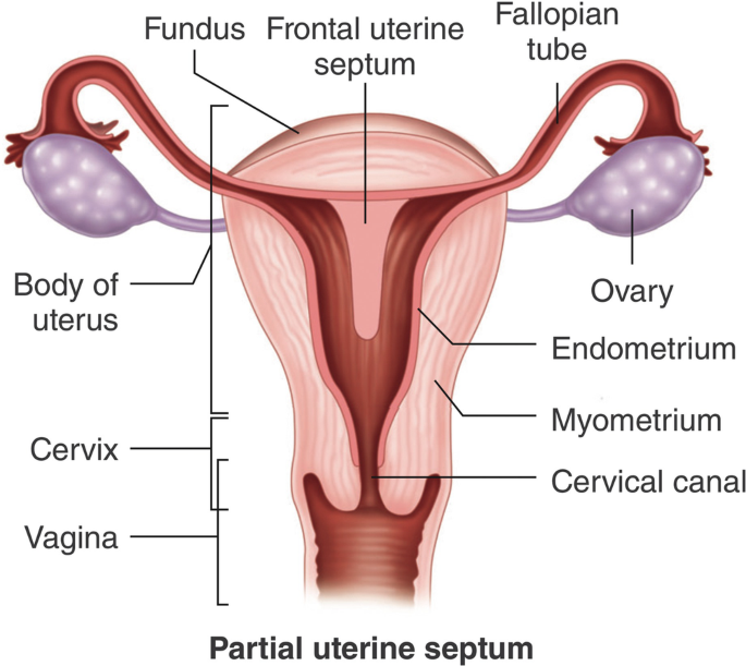
– Rudimentary horn pregnancy occurs when a fertilized egg grows in an underdeveloped part of the uterus called the rudimentary horn of a unicornuate uterus.
– Congenital uterine anomalies, including the unicornuate uterus, occur in less than 5% of all women.
– The unicornuate uterus comprises approximately 10-20% of all uterine malformations.
– Rudimentary horn pregnancy is an extremely rare type of ectopic pregnancy with an incidence of 1 in 75,000 – 150,000 pregnancies.
– Uterine anomalies result from abnormal development of embryonic structures called Mullerian ducts during fetal life.
– A unicornuate uterus results from incomplete development and failure of fusion with the opposite side of a Müllerian duct. Two-thirds of women with a unicornuate uterus may also have a rudimentary horn.
– 85% of rudimentary horn pregnancies occur in non-communicating rudimentary horns.
– Symptoms of a rudimentary horn pregnancy may include amenorrhea, vaginal bleeding (light or prolonged/intermittent), pain in the lower abdomen/pelvis/lower back, and gastrointestinal symptoms (nausea/vomiting).
– Diagnosis of a rudimentary horn pregnancy is difficult and may not be detected during regular pelvic exams.
– Transvaginal ultrasound scan (TVS) is the preferred tool for diagnosing ectopic pregnancies.
– In equivocal cases, three-dimensional ultrasound or MRI can help confirm the diagnosis.
– If left untreated, a rudimentary horn pregnancy can cause life-threatening bleeding.
– Treatment options include medical treatment with drugs, laparoscopic surgery, or abdominal surgery.
– The risk of recurrence of a pregnancy in the rudimentary horn is extremely rare with medical treatment.
– Excision of the rudimentary horn and fallopian tube is recommended to prevent future complications.
– Follow-up appointments should be scheduled, and the chances of a healthy future pregnancy can be discussed.
– The timing for attempting another pregnancy and any special precautions may be advised.
– A rudimentary horn pregnancy may not always cause symptoms and can be detected during a routine pregnancy scan.
– Diagnosis of a rudimentary horn pregnancy can be difficult and may require further medical examination.
– Symptoms of a rudimentary horn pregnancy include severe abdominal or pelvic pain, fainting, and shock.
– Prompt treatment is necessary to prevent life-threatening complications, and options include medical treatment, laparoscopic surgery, or abdominal surgery.
– Recurrence of a pregnancy in a rudimentary horn is extremely rare but possible, and routine excision of the rudimentary horn and fallopian tube may be recommended.
– Important questions to ask include the timing of follow-up appointments, chances of having a healthy future pregnancy, when to try for pregnancy again, and any special precautions to take if becoming pregnant again.

Continue Reading

Myoma of the uterus: Understanding causes, symptoms, treatment

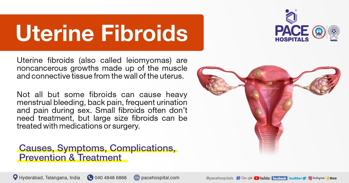
– Uterine fibroids are non-cancerous tumors that grow in the uterus, also known as myomas.
– Myomas are smooth, non-cancerous tumors made partly of muscle tissue that may develop in or around the uterus.
– Myomas in the larger, upper part of the uterus are called fibroids or leiomyomas.
– Most myomas can be seen or felt during a pelvic examination and those causing symptoms can be removed surgically or through less invasive procedures.
– The most common symptom of myomas is vaginal bleeding, which may be irregular or heavy.
– Other symptoms may include heavy bleeding, anemia, fatigue, weakness, painful intercourse, pain, bleeding, or discharge from the vagina if myomas become infected, pressure or lump in the abdomen, difficulties urinating, and urinary tract infections.
– Uterine fibroids, or myomas, affect 20 percent of women in their childbearing years.
– Uterine fibroids can cause abnormal bleeding, pelvic masses, pelvic pain, infertility, and pregnancy complications.
– There are five types of uterine fibroids: intramural, subserosal, submucosal, pedunculated, and intracavitary fibroids.
– Between 70 and 80 percent of women develop a fibroid tumor by the age of 50.
– Estrogen seems to activate the growth of uterine fibroids, and they usually shrink after menopause.
– Hormone therapy after menopause may cause fibroid symptoms to continue.
– African American women are more susceptible to developing fibroids.
– Fibroids tend to grow faster in white women younger than 35 years compared to those older than 45 years.
– Delaying pregnancy until age 30 or older increases the risk of developing fibroids.
– Early menstruation increases the risk of developing fibroids.
– Alcohol and caffeine intake may increase the risk of developing fibroids.
– Specific genetic alterations are linked to fibroid growth.
– Obesity and high blood pressure may play a role in fibroid development and growth.
– A diet rich in red meat may increase the chance of developing fibroids.
– Treatment options for uterine fibroids vary depending on the severity of symptoms and the patient’s desire to have children.
– Hormonal contraception, intrauterine devices, antifibrinolytic drugs, and nonsteroidal agents are options for managing heavy bleeding.
– Endometrial ablation can be performed if the patient does not want to have children.
– Gonadotropin-releasing hormone agonists can shrink fibroids, but they may grow back.
– Myomectomy is a procedure that removes fibroids while preserving the uterus, but fibroids may grow back.
– MRI-guided ultrasound surgery can shrink fibroids and reduce heavy bleeding.
– Uterine fibroid embolization is a minimally invasive procedure that blocks the blood supply to fibroids, causing them to shrink and die.
– Myomas can cause symptoms such as abdominal pain and heavy menstrual bleeding, but some people may remain symptom-free.
– The cause of myomas is unclear, but risk factors include a family history of myoma, obesity, and age.
– More than half of all people with a uterus will experience a myoma by age 50.
– Myomas can vary in size, from as small as a pea to as large as a melon.
– They can be located inside the uterus (intramural myomas), on the outside of the uterine wall (subserosal myomas), have a stalk or stem attaching them to the uterus (pedunculated myomas), or be found just under the lining of the uterus (submucosal myomas).
– Large myomas are considered to be 10 centimeters (cm) or more in diameter.
– Emergency room visits for myoma symptoms, such as pelvic pain and heavy bleeding, have increased from 2006 to 2017, according to a recent study.
– Myomas of the uterus are noncancerous tumors that can cause a variety of symptoms.
– Symptoms may include heavy or prolonged periods, bleeding between periods, pelvic pain, abdominal pressure, a feeling of fullness in the lower abdomen, constipation or diarrhea, frequent urination or trouble urinating, pain during sex, lower back pain, reproductive issues, fatigue, and weakness.
– Myomas are a top cause of hysterectomy surgeries, which come with their own risks and complications.
– The exact cause of myomas is unknown, but they are likely associated with hormone activity.
– High levels of estrogen and progesterone may stimulate their growth, and myomas tend to shrink when hormone levels decrease after menopause.
– Risk factors for developing myomas include a family history of myomas, obesity, high blood pressure, age, and dietary factors such as a diet high in red meat or vitamin D deficiency.
– Myomas are more common among Black individuals with a uterus, and factors such as low vitamin D levels, obesity, stress, genetics, and unequal access to healthcare have been proposed as potential risk factors.
– Medical tests used to diagnose myomas include pelvic examination, ultrasound or transvaginal ultrasound, and magnetic resonance imaging (MRI).
– Treatment for myomas depends on factors such as the severity of symptoms, the size and location of the myomas, the desire for future pregnancy, age, and proximity to menopause.
– Medications can be used to treat myomas, including over-the-counter pain medications, iron supplements for depleted iron levels, and birth control methods to control heavy menstrual bleeding.
– GnRH agonists (hormone-stimulating medications) can be used to temporarily shrink myomas, especially if surgery is planned.
– Surgical options for myomas include laparoscopic myomectomy and uterine fibroid embolization (UFE).
– No specific figures or statistics are mentioned in the article.
– One treatment option is a radiology procedure that uses injections to block blood flow to the myomas, causing them to shrink and sometimes die.
– Another option is MRI-guided ultrasound surgery, which uses ultrasound waves to shrink myomas.
– In more severe cases, other surgical options may be considered.
– Hysterectomy is a surgery to completely remove the uterus, eliminating the fibroids but also making pregnancy impossible in the future.
– Abdominal myomectomy is a surgical procedure that removes the fibroids without removing the uterus, with the possibility of future pregnancy but a risk of the fibroids returning.
– While there are no home remedies that directly treat fibroids, certain complementary therapies like acupuncture, yoga, massage, traditional Chinese medicine, and heating pads may help manage symptoms.
– Lifestyle changes such as dietary changes, exercise, stress management, and weight loss can also be beneficial.
– Myomas can cause complications related to fertility, pregnancy, and childbirth.
– These complications can include fertility issues, pregnancy complications such as miscarriage or early labor, and the need for a cesarean delivery.
– It’s important for those with myomas who want to become pregnant to discuss the condition with their healthcare provider to assess potential risks.
– It is recommended to communicate symptoms affecting one’s life to healthcare providers to determine the most suitable treatments.

Continue Reading