Unraveling the Mysteries: Cortex of Ovary Explored

List of keywords related to ‘cortex of ovary’:

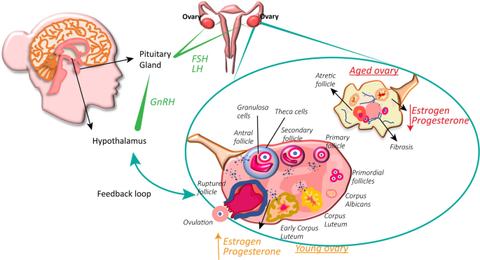
– Ovarian cortex
– Outer part of the ovary
– Ovarian follicles
– Connective tissue
– Ovarian cortex tissue transplant
– Infertility
– Primary female reproductive organs
– Pelvic cavity
– Germinal epithelium
– Tunica albuginea
– Inner medulla
– Oogenesis
– Female sex cells
– Oogonia
– Primary oocytes
– Prophase
– Follicle-stimulating hormone
– Secondary oocyte
– First polar body
– Second polar body
– Ovum
– Metaphase
– Fertilization
– Meiosis II
– Formation of an ovum
– Polar bodies
– Development of ovarian follicles
– Follicular cells
– Primordial follicles
– Antrum
– Granulosa cells
– Estrogen
– Vesicular (graafian) follicle
– Rupture of follicle
– Corpus luteum
– Progesterone
– Placenta
– Hormone secretion
– Corpus albicans.

Continue Reading

Unlocking the Wonders of Uteroscopy: A Glimpse inside

– Ureteroscopy is a procedure used to address kidney stones.
– It involves the use of a small telescope called a ureteroscope.
– The procedure is typically performed under general anesthesia.
– The duration of the procedure is usually between one to three hours.
– Small stones can be removed whole using a basket device.
– Large stones or narrow ureters may require fragmentation, often done with a laser.
– Once the stone is broken into small pieces, they are removed.
– The use of the ureteroscope may cause swelling in the ureter.
– To ensure proper drainage of urine, a small tube called a ureteral stent may be temporarily left inside the ureter.
– Ureteroscopy is usually an outpatient procedure but may require an overnight hospital stay if it is lengthy or difficult.
– Ureteroscopy is a minimally invasive method to treat kidney stones and stones in the ureter.
– It is performed in the operating room with general or spinal anesthesia.
– Pre-op lab tests and X-rays are done to ensure safety.
– An antibiotic is given before the procedure.
– Monitoring devices are attached to the patient.
– The inside of the bladder is examined with an optical cystoscope.
– X-ray images with contrast may be taken to locate the stone.
– The ureteroscope is passed through the natural urinary channel to reach the stone.
– A wire basket can be used to extract the stone, or it can be fragmented with laser or electrohydraulic energy.
– The procedure takes 1-2 hours.
– The patient is monitored in the post-operative recovery area for about 2 hours before being discharged.
– Some patients may have a temporary stent placed in the ureter to prevent blockage.
– The stent is usually removed 1 to 2 weeks after the procedure.
– Urine may be bloody for several days after the procedure.
– Pain relief medication is provided.
– Activity should be limited after the procedure.
– Most people can resume normal activities without pain several days after the procedure or once the stent is removed.
– A follow-up visit with a Urologist will be scheduled within 1-2 weeks to remove the stent if one was placed without a thread.
– If a stent was not required or was already removed at home, the follow-up visit will be scheduled within 4-6 weeks after surgery.
– During the follow-up visit, an X-ray will be performed to determine the success of the procedure and check for any complications.
– Depending on the individual’s risk of stone recurrence, further testing may be offered to prevent future stones.

Continue Reading

Alba Lochia: An Insightful Guide to Postpartum Discharge

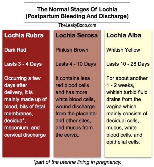
– alba lochia is the third stage of postpartum bleeding, lasting 10-28 days postpartum and characterized by whitish yellow bleeding
– stage 1: lochia rubra is the first stage of postpartum bleeding, lasting 3-4 days postpartum and characterized by bright or dark red bleeding
– stage 2: lochia serosa is the second stage of postpartum bleeding, lasting 4-10 days postpartum and characterized by pinkish brown bleeding
– heavy bleeding beyond the third day after birth may be a sign of postpartum hemorrhage
– blood clots bigger than a plum may be a sign of postpartum hemorrhage
– bleeding that soaks more than one sanitary pad an hour and doesn’t slow down or stop may be a sign of postpartum hemorrhage
– blurred vision, chills, clammy skin, rapid heartbeat, dizziness, weakness, nausea, and a faint feeling are signs of postpartum hemorrhage
– postpartum hemorrhage can cause death if left untreated
– postpartum hemorrhage can occur within the first 24 hours after delivery or anytime within the first 12 weeks after delivery
– if experiencing postpartum hemorrhage, it is important to contact a doctor or head to the emergency room immediately
– regular menstrual pads may not be sufficient for managing bleeding after birth
– postpartum underwear, like Always Discreet, may be more effective for managing heavy bleeding after giving birth than regular pads or tampons
– Always Discreet postpartum underwear comes in different sizes and absorbency levels
– Always Discreet postpartum underwear is thin and flexible, providing comfort to the wearer
– Always Discreet postpartum underwear use gel technology to keep the skin dry and retain moisture.

Continue Reading

Acyeterion: Exploring the Origins and Significance of Greek Tragedy

Emergency contraception, also known as morning-after pills or certain IUDs, refers to methods that can be used to prevent pregnancy after unprotected sex. It should be used within 5 days (120 hours) of unprotected sex for best results. There are two types of morning-after pills: ulipristal acetate (ella) and levonorgestrel (Plan B, Take Action, etc.). Ulipristal acetate requires a prescription, while levonorgestrel pills can be bought over the counter. Morning-after pills work best when taken as soon as possible and may not be as effective if the body has already started ovulating.

Emergency contraception should not be used as a regular form of birth control. It does not cause long-term side effects, harm existing pregnancies, or affect future fertility. It is a safe method to prevent pregnancy after unprotected sex. Different types of morning-after pills should not be used simultaneously or within 5 days of each other as they may counteract each other.

The effectiveness of emergency contraception can be affected by factors such as body weight. Levonorgestrel pills may be less effective for individuals weighing over 165 pounds, while ulipristal acetate is less effective for those weighing 195 pounds or more. The Paragard, Mirena, and Liletta IUDs are the most effective at preventing pregnancy and can be used within 120 hours of unprotected sex.

Emergency contraception can be purchased over the counter at drugstores and pharmacies without a prescription. It is recommended to use whichever method is available, even if not the most effective. The cost of emergency contraception varies, with Plan B One-Step typically costing around $40-$50 and levonorgestrel pills costing about $11-$45. ella, a different brand of morning-after pill, usually costs about $50 or more at the pharmacy but may be free with health insurance or Medicaid. The cost of getting an IUD can range from $0 to $1,300, but they may be free or low cost with health insurance plans, Medicaid, and some government programs.

Emergency contraception may cause temporary side effects such as nausea, vomiting, slight irregular vaginal bleeding, and fatigue. It does not have long-term side effects. It is not advisable to rely on home remedies for emergency contraception, as they are not effective. Morning-after pills do not require age or gender restrictions for purchase. Sometimes they may be locked up or kept behind the counter at stores, requiring assistance from a pharmacist or store clerk.

Insurance usually covers the cost of emergency contraception, but a prescription from a doctor or nurse may be required for coverage. Emergency contraception can also be obtained at family planning or health department clinics, as well as Planned Parenthood health centers. The first period after taking emergency contraception may come earlier or later than expected.

Continue Reading
Perinatal Cardiomyopathy: Causes, Symptoms, Treatment, and Prevention Guidelines

Perinatal cardiomyopathy is a rare condition where weakness in the heart muscle occurs sometime during the final month of pregnancy through about five months after delivery. It can have mild or severe symptoms. The seriousness of the condition can be measured by the ejection fraction, which is the percentage of blood the heart pumps out with each beat. A normal ejection fraction is about 60%. The degree of severity does not affect the rate of recovery. Some patients with a low ejection fraction can fully recover from perinatal cardiomyopathy. Recovery can occur over a period of six months or longer, but in some cases, the heart can return to full strength in as little as two weeks. Perinatal cardiomyopathy has a relatively high recovery rate compared to other forms of cardiomyopathy. Symptoms of perinatal cardiomyopathy, such as swelling in the feet and legs and shortness of breath, can be similar to the symptoms of the third trimester of a normal pregnancy. Mild cases may go undiagnosed and recover without medical attention. Severe cases can cause shortness of breath and swollen feet. Cardiomyopathy can be detected through an echocardiogram which shows the diminished functioning of the heart.

Continue Reading

Tubal Abortion: Understanding the Risks, Procedures, and Recovery

List of pertinent topics to the keyword ‘tubal abortion’:

1. Ectopic pregnancy and its treatment
2. Debate surrounding treatment for ectopic pregnancy
3. Misconceptions about ectopic pregnancy management
4. Distinction between ectopic pregnancy treatment and abortion
5. Timing for trying to conceive after treatment for ectopic pregnancy
6. Methotrexate injections and their effects on fertility
7. Importance of folic acid supplementation after ectopic pregnancy treatment
8. Lack of information on tubal abortion in the article

Continue Reading

Painless Delivery: Empowering Mothers Through Modern Anesthesia Techniques

List of relevant information for the keyword “painless delivery”:

1. Advancements in medicine have provided women with the option of pain relief methods during labor to make childbirth more endurable.
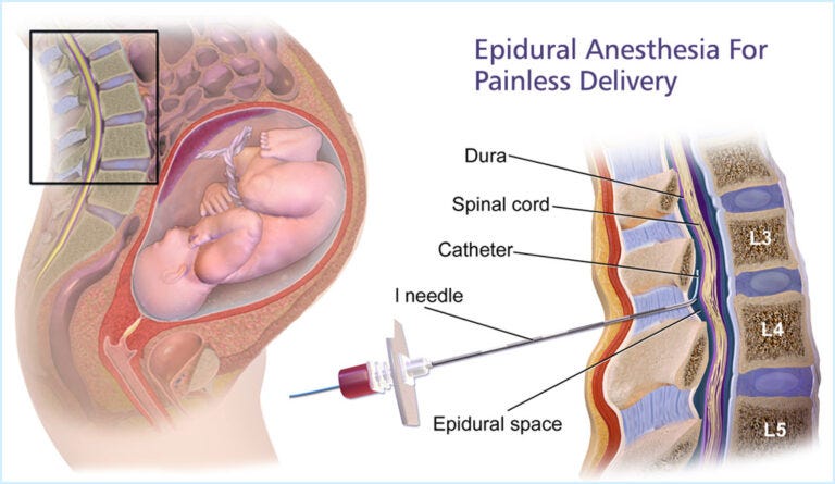
2. Painless delivery is possible through epidural anesthesia, which is injected into the woman’s lower back to reduce pain.
3. Women with lower pain thresholds or those who get pregnant after their thirties often request C-sections but can still opt for painless delivery with epidural anesthesia.
4. Candidates for painless delivery include women with preexisting medical conditions, those opting for vaginal birth after a previous C-section, and those with a history of prolonged or complicated labor.
5. Painless delivery is not suitable for women with certain conditions such as bleeding disorders, previous lower back surgery, skin infection in the epidural area, blood clotting disorders, and neurological diseases.
6. Many women choose painless delivery to avoid debilitating pain during labor.
7. Painless delivery, also known as epidural analgesia, is a form of regional anesthesia that provides pain relief during natural labor.
8. The drug takes about 10-15 minutes to take effect and numbs the pelvic region and everything below it while the mother remains conscious.
9. Painless delivery has helped reduce the number of elective C-sections in India.
10. It allows women to experience natural childbirth with minimal intervention and alleviates pain, allowing the mother to focus on the delivery.
11. It can also prevent exhaustion and post-partum complications.
12. There are risks and side effects associated with epidurals, including fever, breathing problems, nausea, dizziness, back pain, shivering, severe headaches, prolonged labor, and trouble passing urine.
13. Other painless delivery options mentioned in the article include the use of Entonox (nitrous oxide and oxygen) and water birth.
14. Women are advised to discuss the process with their gynecologist and carefully consider the pros and cons before opting for painless delivery.

Please note that any duplicated information has been omitted from this list.

Continue Reading

Decidua vera: Understanding the Biology and Significance Explained

List of pertinent information about ‘decidua vera’:

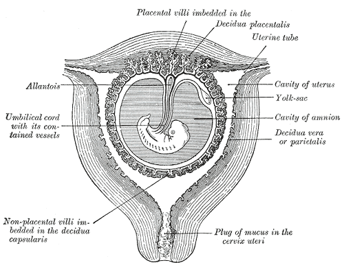
1. Decidua vera, also known as parietal decidua or true decidua, is the decidua that does not include the area occupied by the implanted ovum and chorion.
2. Decidua vera is the exclusive name for the decidua that is not occupied by the implanted ovum and chorion.
3. The decidua is the mucosal lining of the uterus that forms every month in preparation for pregnancy.
4. Decidua vera is shed after childbirth, except for the deepest layer.
5. The decidua forms the maternal part of the placenta during pregnancy and is shed together with the placenta after birth.
6. The decidua has distinct histological features, including large decidual cells.
7. The decidua plays a role in nutrient exchange, gas exchange, and waste removal during pregnancy.
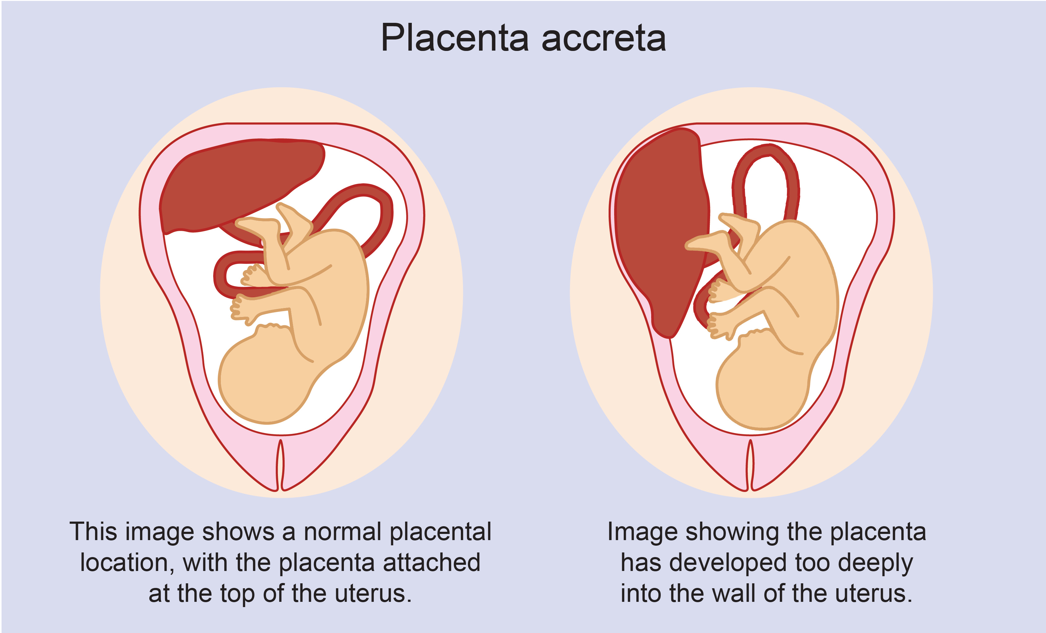
8. In invasive placental disorders like placenta accreta, the decidua is found to be deficient.
9. The decidua secretes hormones, growth factors, and cytokines.
10. The role and interplay of these hormones and factors is not well understood.
11. Chronic deciduitis, a long-lasting infection of the decidua, is associated with pre-term labor.

Continue Reading

Adherent Placenta: Understanding risks, symptoms, and treatment options

In the intricate world of childbirth, there is a fascinating condition that occasionally arises – the adherent placenta. This mysterious and intricate phenomenon captivates medical professionals and expectant parents alike. As we delve into the complexities of this condition, prepare to uncover the intriguing secrets hidden within the realm of childbirth complications. adherent placenta An […]

Continue Reading

Outlet Forceps: A Guide to Proper Usage and Sterilization

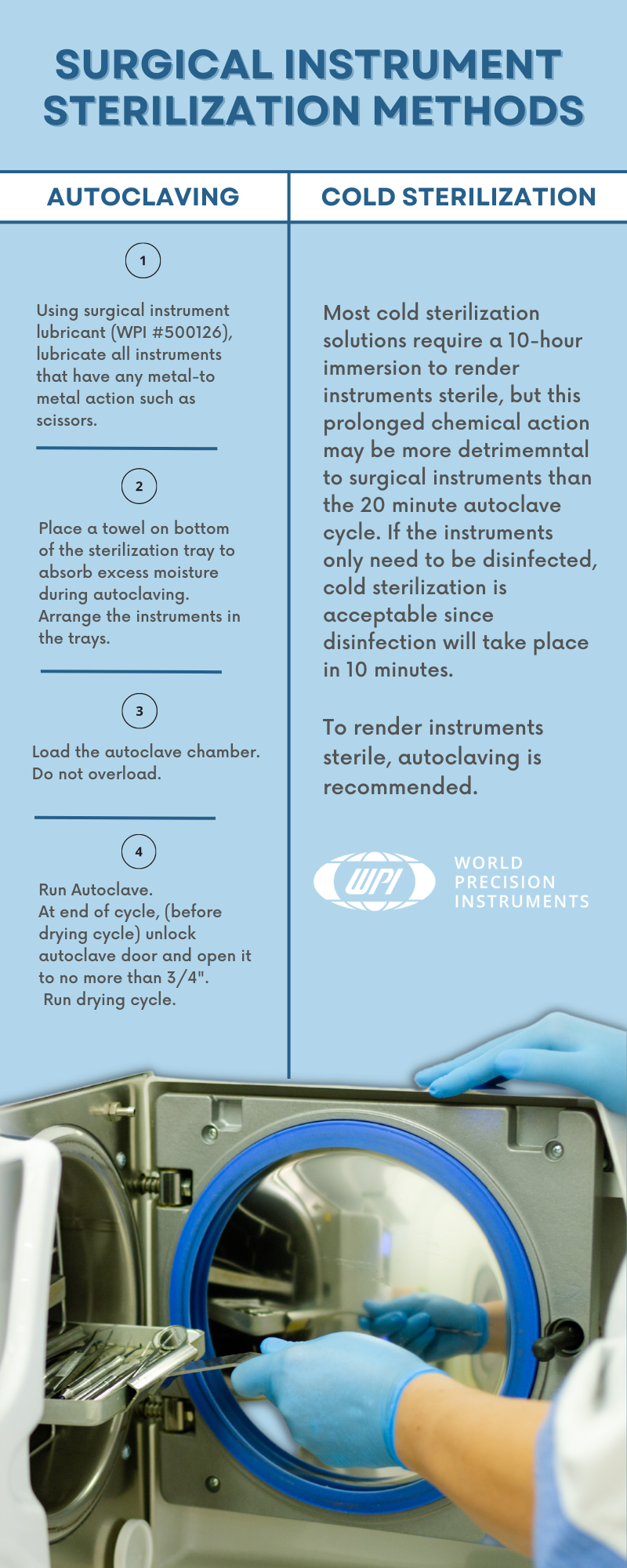
Outlet forceps are a type of medical instrument used in childbirth as an alternative to vacuum extraction. They offer advantages such as avoiding the need for a caesarean section, reducing delivery time, and being compatible with head presentation. However, there are also potential complications associated with their use, including bruising the baby, severe vaginal tears, nerve damage, membrane rupture, skull fractures, and cervical cord injury. Maternal factors for forceps use include exhaustion, prolonged second stage of labor, maternal illness, hemorrhaging, and drug-related inhibition of maternal effort. Fetal factors include non-reassuring fetal heart tracing, fetal distress, and after-coming head in breech delivery. Complications for the baby include cuts, bruises, facial nerve injury, clavicle fracture, and an increased risk of intracranial hemorrhage. Complications for the mother include perineal lacerations, pelvic organ prolapse, incontinence, increased postnatal recovery time and pain, and difficulty evacuating during recovery. The article also discusses different types of forceps used in obstetrics, including outlet forceps which have specific curves designed to match the fetal head and the birth canal. Overall, forceps have played a significant role in the medicalization of childbirth but have also faced criticism and decreased in use with the introduction of cesarean sections and vacuum extraction.

Continue Reading