Effacement: Understanding the Process of Cervical Labor Dilation

– Stages of dilation and effacement during labor and delivery
– Symptoms associated with dilation and effacement
– Effacement and dilatation as processes during childbirth
– Stretching and thinning of the cervix during effacement
– Opening of the cervix during dilatation
– Variation of effacement and dilatation speed between individuals
– Effacement and dilatation starting during labor for first-time mothers
– Checking the cervix for effacement and dilatation status
– Contractions opening the cervix during labor
– Movement of the baby into the birthing position
– Mucus plug coming loose during labor
– Tinged mucus plug potentially indicating effacement
– Measuring effacement in percentages (0-100)
– Measuring dilatation in centimeters (0-10)
– Full dilatation necessary for the pushing stage of labor.

Continue Reading

Discover the Hidden Secrets: Amniotic Fluid Unveiled

Amniotic fluid is a vital component of pregnancy, surrounding and protecting the baby in the womb. It provides cushioning, helps regulate temperature, promotes lung and digestive system development, aids in muscle and bone growth, and prevents compression of the umbilical cord.

The fluid is primarily water in the early weeks of pregnancy but is later composed mostly of the baby’s urine. It also contains nutrients, hormones, and antibodies.

The amount of amniotic fluid increases until around 36 weeks of pregnancy and then gradually decreases. Too little (oligohydramnios) or too much (polyhydramnios) amniotic fluid can be problematic for both the mother and the baby, although most babies are born healthy even with these conditions.

Normal amniotic fluid is clear or tinted yellow, but green or brown fluid indicates that the baby may have passed their first bowel movement (meconium) in the womb. Meconium in the amniotic fluid can lead to breathing difficulties, known as meconium aspiration syndrome. Some babies may require immediate treatment at birth to prevent complications, while others may be healthy and not require treatment.

Continue Reading

Unveiling the Mystery: Macrosomia and Its Associated Risks

– Fetal macrosomia is a term used to describe a newborn who is much larger than average.
– A baby with fetal macrosomia weighs more than 8 pounds, 13 ounces (4,000 grams), regardless of gestational age.
– Approximately 9% of babies worldwide are born weighing more than 8 pounds, 13 ounces.
– The risks associated with fetal macrosomia increase significantly when birth weight is more than 9 pounds, 15 ounces (4,500 grams).
– Vaginal delivery can be complicated by fetal macrosomia and the baby is at risk of birth injuries.
– Fetal macrosomia increases the baby’s risk of health problems after birth.

Continue Reading

Congenital Heart Disease: Understanding, Treatment, and Prevention Strategies

– Stenosis
– Complex defects
– Tetralogy of Fallot
– Transposition of the great arteries
– Hypoplastic left heart syndrome
– Most common birth defect
– 1% of all newborns affected
– Possible causes
– Risk factors
– Maternal age over 40
– Certain genetic conditions
– Family history of heart defects
– Symptoms
– Difficulty breathing
– Poor feeding
– Bluish tint to skin or lips
– Early diagnosis and treatment
– Treatment options
– Medications
– Catheter procedures
– Surgery
– Prognosis improvement
– Normal, healthy lives
– Long-term follow-up care
– Potential complications

Continue Reading

Uterine Prolapse: Causes, Symptoms, Treatment, and Prevention Strategies

– Uterine prolapse occurs when muscles and tissue in the pelvis weaken, allowing the uterus to drop into the vagina
– Nearly half of all women between ages 50 and 79 have uterine prolapse
– The main cause is weakened muscles and tissue in the pelvic floor that can’t support the weight of the uterus
– Risk factors include giving birth (highest risk), vaginal delivery, menopause, being Caucasian, being overweight, and smoking
– Many women with uterine prolapse have no symptoms, but possible symptoms include leakage of urine, inability to completely empty the bladder, feeling of heaviness or fullness in the pelvis, bulging in the vagina, lower-back pain, aching or pressure in the lower abdomen or pelvis, and constipation
– Diagnosing uterine prolapse involves a physical exam, possibly a cystoscopy to examine the bladder and urethra, and an MRI to get a better look at the kidneys and other pelvic organs.

Continue Reading

LH: Discover the Magnificent World of Lighthouses

– Luteinizing hormone (LH) is produced by both men and women and affects the sex organs
– LH plays a role in puberty, menstruation, and fertility
– LH is produced in the pituitary gland, located at the base of the brain
– In women, LH is involved in the menstrual cycle and works with follicle-stimulating hormone (FSH)
– LH levels can be measured through blood tests or urine samples
– LH tests can be used to track hormone levels for fertility purposes or diagnose issues with the pituitary gland or other parts of the body
– Reasons for requesting an LH blood test include difficulty getting pregnant, irregular menstrual periods, suspected menopause in women, and signs of low testosterone levels or pituitary disorders in men
– LH blood test can be used to determine if a person is entering puberty too late or too soon, as well as to find underlying causes for delayed or early puberty
– LH blood test can also be used to determine the amount of LH in the bloodstream for individuals with absent or irregular periods, as well as after menopause
– LH blood test can indicate a problem with the supply of eggs in a woman’s ovaries and a man’s sperm count, affecting fertility
– LH levels in urine can be used to determine when ovulation will likely occur, often used for enhancing chances of conceiving
– The test is administered by drawing a small amount of blood from the arm, either in a doctor’s office or at a lab
– There are minimal risks associated with the test, such as bruising or rare phlebitis. Individuals with bleeding disorders should inform their healthcare provider
– A luteinizing hormone (LH) blood test is mentioned in the article
– Patients should follow exact directions given by their doctor to prepare for the test
– Certain medications that can affect the results may need to be stopped prior to the test
– Women may need to stop taking birth control or other hormone pills for up to four weeks before the test
– The date of the patient’s last period is important information for the doctor
– Patients may need to avoid eating or drinking for up to eight hours prior to the test
– If the patient has had any tests or procedures involving radioactive substances within seven days before the LH blood test, the doctor should be informed as it can interfere with the results
– High LH levels in women may indicate problems with ovulation, polycystic ovary syndrome (PCOS), or Turner syndrome
– Low LH levels in women may suggest issues with the pituitary gland, eating disorders, or malnutrition
– High LH levels in men may be caused by damage to the testicles or Klinefelter’s syndrome
– Low LH levels in men may indicate a disorder of the pituitary gland or hypothalamus
– High LH levels in children, along with high levels of follicle-stimulating hormone, may suggest early puberty
– Low LH and follicle-stimulating hormone levels in children may indicate delayed puberty
– It is recommended to consult a healthcare provider for further understanding or questions about the results.

Continue Reading

Unveiling the Dangers and Causes of Placental Abruption

– Placental abruption is when the placenta detaches partially or completely from the wall of the uterus, causing bleeding in the mother.
– It can interfere with the baby’s supply of oxygen and nutrients from the mother’s bloodstream through the lining of the uterus.
– Prompt medical treatment is necessary to prevent dire consequences for both the mother and baby, including death.
– Worldwide, placental abruption occurs in about one pregnancy in every 100.
– About 50% of cases are mild and can be managed with ongoing monitoring. 25% are moderate, and 25% threaten the life of both the baby and the mother.
– Symptoms of moderate to severe placental abruption include bleeding, continuous abdominal and lower back pain, tender and hard uterus, very frequent uterine contractions, and fetal distress.
– In some cases, bleeding may be scanty or non-existent due to a retroplacental clot.
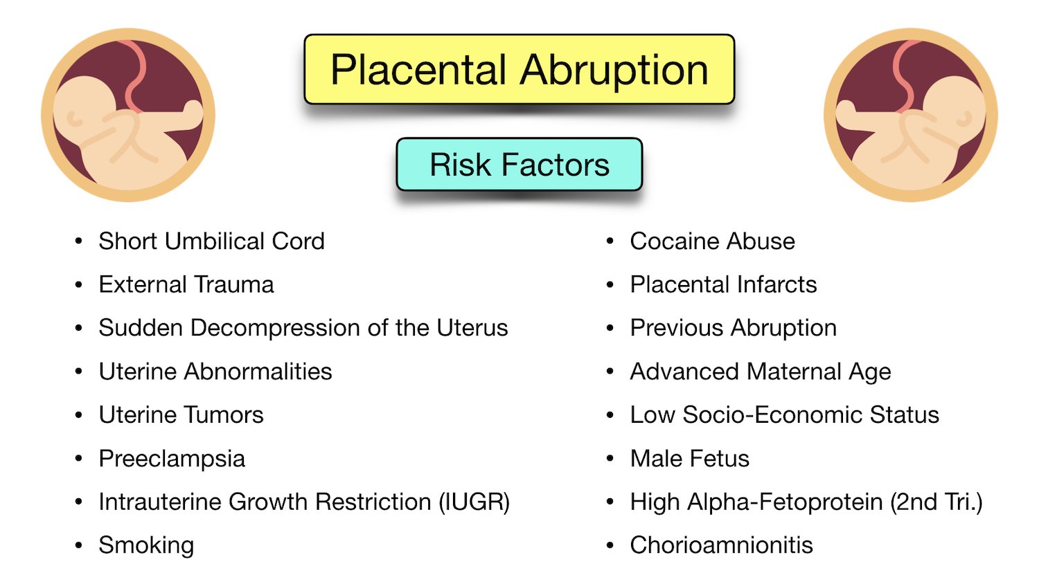
– The exact cause of placental abruption is unknown in most cases, but it is thought that abnormal blood supply in the uterus or placenta may play a role.
– Known causes of placental abruption include abdominal trauma and uterine decompression.
– Certain factors, such as advanced maternal age, prior pregnancies, and carrying multiple fetuses, increase the risk of placental abruption.
– High blood pressure increases the risk of abnormal bleeding between the placenta and the uterine wall.
– Excessive amniotic fluid increases the risk of bleeding between the placenta and the uterine wall.
– Substance use during pregnancy, such as smoking, alcohol use, and taking drugs like methamphetamine or cocaine, increase the risk of placental abruption.
– Any blood condition that affects the blood’s ability to clot can increase the risk of placental abruption.
– Procedures such as amniocentesis and amnioreduction involve a needle inserted through the mother’s abdomen into the uterus and can rarely cause bleeding.
– External cephalic version, a procedure to turn a breech baby to a head-down position, can occasionally dislodge the placenta.
– Complications in severe cases of placental abruption can include decreased oxygen to the baby, stillbirth, and maternal blood loss.
– Diagnosis of placental abruption can be done through medical history, physical examination, blood tests, ultrasound, and fetal heartbeat monitoring.
– Treatment depends on the severity of the condition and may include rest, induction of labor, vaginal birth or caesarean section, and immediate delivery.
– Severe cases may require supportive care, blood transfusion, or emergency hysterectomy.
– It is impossible to prevent placental abruption, but the risk can be reduced by avoiding substances such as cigarettes, alcohol, and street drugs, and controlling high blood pressure.

Continue Reading

Ovary Health: Understanding the Key to Fertility Success

– The ovaries are located on either side of the womb in the pelvis.
– Each woman has two ovaries.
– The ovaries are oval in shape and about four centimeters long.
– The ovaries produce eggs for fertilization.
– The ovaries produce reproductive hormones such as estrogen, progesterone, and androgens.
– The functions of the ovaries are controlled by hormones released from the hypothalamus and pituitary gland.
– Ovulation is the process of releasing an egg from the ovary.
– A female baby is born with around two million eggs, which decreases to about 400,000 by puberty.
– Menopause refers to the end of a woman’s reproductive years, usually around 51 years old.
– Menopause is caused by the loss of follicles in the ovary.
– The ovaries produce the hormones estrogen and progesterone, which regulate the menstrual cycle.
– Estrogen production dominates in the first half of the menstrual cycle, progesterone production dominates in the second half.
– Ovaries also produce small amounts of male hormones called androgens.
– Medical conditions that affect the ovaries can decrease fertility.
– Premature ovarian insufficiency is when the ovaries stop functioning before the age of 40.
– Hormone replacement therapy is a common treatment for restoring missing ovarian hormones.
– Conditions like Turner syndrome or damage from treatments like chemotherapy can affect ovarian function.
– Polycystic ovary syndrome affects 8-13% of women of childbearing age.
– PCOS can cause stunted follicles, cysts in the ovaries, excess male hormones, irregular or absent periods, and a higher risk of type 2 diabetes.
– Amenorrhea is the absence of menstrual periods during reproductive years and can be caused by various factors.
– Factors like low body weight, excessive exercise, and psychological stress can affect ovarian function.
– Disorders of the pituitary gland, such as hypopituitarism caused by pituitary tumors or excess prolactin, can also impact ovarian function.

Continue Reading

Abdominal Pain: Understanding Causes, Symptoms, and Treatment Options

– The abdomen is the largest cavity in the body, located between the chest and the pelvis.
– It contains organs such as the liver, stomach, and intestines.
– The groin is where the upper thighs meet the lowest part of the abdomen.
– The abdomen and the groin are separated by a wall of muscle and tissue.
– The wall has small tunnels called inguinal and femoral canals.
– These canals allow nerves, blood vessels, and other structures to pass between the abdomen and groin.
– Abdominal pain is a common and often non-serious issue that almost everyone experiences at some point.
– Causes of abdominal pain can range from minor issues like constipation and food allergies to more serious conditions like appendicitis, bowel blockage, and cancer.
– Abdominal pain can also be caused by problems outside of the belly, such as heart attacks, menstrual cramps, and pneumonia.
– For mild abdominal pain, helpful tips include sipping water or clear fluids, avoiding solid food for a few hours, and trying antacids if experiencing heartburn or indigestion.
– Seek immediate medical attention if abdominal pain is being treated alongside cancer, if unable to pass stool, if vomiting blood, or if experiencing chest, neck, or shoulder pain.
– Call a doctor if abdominal pain lasts more than 1 week, does not improve within 24 to 48 hours, bloating lasts for more than 2 days, or if diarrhea persists for more than 5 days.

Continue Reading

Cinematic Evolution: A Journey through Film History’s Milestones

– The main cause of CIN (cervical intraepithelial neoplasia) is an infection called the human papilloma virus (HPV).
– There are over 100 types of HPV, and some types can affect the cervix.
– The types of HPV that cause abnormal cell changes in the cervix are called high-risk HPV.
– The body’s immune system usually gets rid of the HPV infection naturally, often without causing any symptoms or damage.
– In some cases, the immune system does not clear the infection and the virus remains in the body for longer periods.
– If the cervix is affected by HPV for a long time, it can cause damage that may eventually develop into CIN.
– HPV is very common, and most people are infected with it at some point.
– HPV can affect all sexual orientations and anyone who has been sexually active, including individuals in long-term relationships.
– HPV can still affect individuals who haven’t been sexually active for some time.
– HPV can live on the skin around the entire genital area and can spread easily through any type of sexual contact, including skin-to-skin genital contact or sharing sex toys.
– Using a condom or other barrier contraception may reduce the risk of HPV infection, but it does not provide complete protection.
– Cervical dysplasia is a precancerous condition where abnormal cells grow on the surface of the cervix.
– It is also known as cervical intraepithelial neoplasia (CIN).
– Most people with cervical dysplasia do not develop cancer, and it takes years for cancer to form.
– The severity of cervical dysplasia is classified on a scale from one to three.
– CIN 1 refers to abnormal cells affecting about one-third of the thickness of the epithelium.
– CIN 2 refers to abnormal cells affecting about one-third to two-thirds of the epithelium.
– CIN 3 refers to abnormal cells affecting more than two-thirds of the epithelium.
– Cervical dysplasia affects sexually active individuals assigned female at birth, including cisgender women, transgender men, and nonbinary people with a cervix.
– It is most common among women aged 25 to 35, with 250,000 to 1 million cisgender women in the U.S. diagnosed each year.

Continue Reading