Craniotomy: A Fascinating Journey into Brain Surgery Techniques

– A craniotomy is a surgical procedure that involves removing part of the skull bone to access the brain.
– Specialized tools are used to remove the bone flap, which is temporarily replaced after the brain surgery.
– Some craniotomy procedures use computer imaging (MRI or CT scans) to guide the surgery to the specific location in the brain.
– This technique is called stereotactic craniotomy and provides a three-dimensional image of the brain, useful for distinguishing tumor tissue from healthy tissue.
– Other uses of stereotactic craniotomy include biopsy, aspiration, and radiosurgery.
– Endoscopic craniotomy involves inserting a lighted scope with a camera into the brain through a small incision in the skull.
– Aneurysm clipping is a surgical procedure that may require a craniotomy to isolate and prevent the rupture of a bulging weakened area in an artery in the brain.
– Craniectomy is a similar procedure where a portion of the skull is permanently removed or replaced later.
– Other related procedures for diagnosing brain disorders include cerebral arteriogram, CT scan, EEG, MRI, PET scan, and X-rays of the skull.
– Johns Hopkins neurosurgeons are skilled in all types of craniotomy and offer less invasive options for brain tumor surgery, aneurysm surgery, and skull deformity repair after brain surgery.
– The extended bifrontal craniotomy involves removing bone from the front of the brain to safely access and remove tumors.
– The supra-orbital ?€?eyebrow?€? craniotomy is a minimally invasive procedure that involves making a small incision within the eyebrow to access tumors in the front of the brain or around the pituitary gland.
– The retro-sigmoid ?€?keyhole?€? craniotomy is also minimally invasive and involves removing tumors through an incision behind the ear, providing access to the cerebellum and brainstem.
– The orbitozygomatic craniotomy is a traditional approach that involves removing bone from the orbit and cheek to reach difficult tumors and aneurysms.
– The translabyrinthine craniotomy involves making an incision behind the ear to access tumors.
– Before the procedure, patients may need to fast and inform healthcare providers of any allergies or sensitivities to medications, latex, tape, and anesthetic agents.
– Patients should also disclose all medications, including over-the-counter drugs and herbal supplements, as well as any history of bleeding disorders or the use of anticoagulant medications.
– Smoking should be stopped before the procedure to improve chances of successful recovery and overall health.
– Patients may be required to wash their hair with a special antiseptic shampoo the night before surgery.
– Sedatives may be administered to help patients relax before the procedure.
– The length of hospital stay for a craniotomy is usually 3 to 7 days, followed by possible rehabilitation.
– The specific procedures during a craniotomy may vary depending on the patient’s condition and the doctor’s practices.
– The patient will have to remove clothing, jewelry, and other objects that may interfere with the procedure and wear a gown.
– An intravenous (IV) line and urinary catheter will be inserted.
– Patients will be positioned on the operating table to provide the best access to the affected area of the brain.
– The anesthesiologist will monitor heart rate, blood pressure, breathing, and blood oxygen levels throughout the surgery.
– The scalp over the surgical site will be cleansed with an antiseptic solution.
– Different incision types may be used depending on the location of the affected brain area. Endoscopes may be used to make smaller incisions.
– A device may be used to hold the head in place, which will be removed at the end of the surgery.
– The scalp will be pulled up and clipped to control bleeding and provide access to the brain.
– A medical drill may be used to create burr holes in the skull, and a special saw may be used to carefully cut the bone.
– The bone flap will be removed and saved.
– The dura mater, the outer covering of the brain, will be separated from the bone and carefully cut.
– After the surgery, the layers of tissue are sewn together and the bone flap is reattached. If a tumor or infection is found, the bone flap may not be replaced.
– Recovery time after brain tumour surgery varies for each individual
– Hospital stay after surgery can be between 3 to 10 days
– Risks after surgery include infection, blood clots, breathing problems, bleeding, and wound problems
– Immediate side effects of brain surgery can include swelling in the brain (oedema)
– Swelling can result in symptoms such as headaches, weakness, dizzy spells, poor balance, personality or behaviour changes, confusion, speech problems, seizures, and blurred vision
– Steroids may be given to reduce swelling and pressure around the brain
– Medication to prevent seizures may also be prescribed
– After brain surgery, it may be difficult to return to work immediately, especially in jobs that require mental skills or involve operating heavy machinery.
– Alcohol consumption may have a greater effect after brain surgery, and certain medicines may require avoiding alcohol.
– There are no medical reasons to avoid sexual activities after brain surgery, but individuals may experience less interest in sex due to tiredness or changes in libido.
– The healthcare team is available to help with any concerns related to sexual problems after brain surgery.

Continue Reading

Octopuses: The Masters of Camouflage in the Ocean

– Optical Coherence Tomography (OCT) is a non-invasive diagnostic technique that provides a cross-sectional view of the retina.
– OCT was introduced in 1991 and has been used in ophthalmology and other fields.
– Time domain OCT acquires approximately 400 A-scans per second.
– Spectral domain technology scans 20,000-40,000 A-scans per second.
– Spectral domain systems have higher resolution and diminish the chance of missing lesions.
– Swept-source technology uses a wavelength-sweeping laser and can acquire 100,000-400,000 A-scans per second.
– OCT angiography is a recent development that uses motion contrast to detect blood flow.
– The term “band” refers to the three-dimensional structure of the retinal layers anatomically.
– The term “zone” describes regions on OCT where the anatomical correlation is not clearly delineated.
– The RPE (retinal pigment epithelium)/Bruch’s complex is one of the layers considered a zone because they are inseparable due to cellular interdigitation.
– Mirror artifacts in OCT occur when the area of interest crosses the zero delay line and result in an inverted image.
– Vignetting or cut edge artifact occurs when part of the OCT beam is blocked by structures like the iris, resulting in a loss of signal on one side of the image.
– Misalignment or off-center artifact occurs when the fovea is not properly aligned during a volumetric scan, leading to incorrect measurements.
– Out of Range Error or out of register artifact happens when the area of interest is not at the center of the image, leading to cutoff or improper positioning of structures.
– Blink artifacts result in partial loss of data due to momentary blockage of OCT image acquisition during blinking.
– Motion artifact occurs when there is movement of the eye during OCT scanning, leading to distortion or double scanning of the same area.
– Segmentation error can occur in horizontal scans when the software of the OCT machine automatically detects the border of the inner retina.
– Cystoid macular edema appears as multiple circular hyporeflective spaces in the retina.
– Senile retinoschisis involves a splitting of retinal layers at the outer plexiform layer.
– The “pearl necklace sign” is seen in exudative macular diseases and appears as hyperreflective dots arranged in a ring around the inner wall of cystoid spaces in the retina.
– Paracentral acute middle maculopathy (PAMM) is characterized by a hyperreflective band at the level of the inner nuclear layer in OCT scans.
– Disruption of the ISOS line (separating the inner and outer segments of photoreceptor cells) is correlated with retinal function loss in various retinal disorders.
– The ILM drape sign is seen in macular telangiectasia 2 and occurs when a thin membrane overhangs a cystoid lesion at the base of the fovea

Continue Reading

Hormone Replacement Therapy: Empowering Women Through Menopause

– Hormone replacement therapy (HRT) is medication that contains female hormones used to replace estrogen during menopause.
– It is primarily used to treat menopausal symptoms such as hot flashes, night sweats, and vaginal discomfort.
– HRT has been proven to prevent bone loss and reduce fractures in postmenopausal women.
– There are risks associated with HRT, including an increased risk of heart disease, stroke, blood clots, and breast cancer.
– There are two main types of HRT: estrogen-only therapy (ET) and a combination of estrogen and progestin therapy (EPT).
– ET is recommended for women who have had a hysterectomy, while EPT is recommended for women with an intact uterus.
– HRT can be administered in several forms, including pills, patches, creams, gels, and injections.
– The optimal dosage, duration, and route of administration of HRT should be individualized for each woman based on her symptoms, medical history, and risk factors.
– Regular follow-up care and screenings, such as mammograms and pelvic exams, are important for monitoring the benefits and risks of HRT.
– Making healthy lifestyle choices, including exercise, a healthy diet, maintaining a healthy weight, not smoking, limiting alcohol, managing stress, and managing chronic health conditions, is recommended alongside HRT.
– Nonhormonal approaches, such as keeping cool, limiting caffeine and alcohol, and using relaxation techniques, can help manage hot flashes.
– There are nonhormonal prescription medications, vaginal moisturizers/lubricants, and a prescription medication called ospemifene (Osphena) that may help with menopausal symptoms.
– Individual symptoms and health risks should be discussed with a doctor to determine if HRT is a suitable treatment option.
– Regular conversations with a doctor are important as recommendations for menopausal treatments may change over time.

Continue Reading

Proteinuria: Understanding Causes, Symptoms, and Treatment Options Explored

Keywords: proteinuria, albuminuria, condition, elevated level, protein, urine, disease, symptom, kidney, glomeruli, functioning, filters, nephritis, hypertension, heart disease, diabetes, kidney disease, severity, damage, risk, kidney failure, visible symptoms, dipstick urine test, detect, protein levels, underlying cause, kidney inflammation, high blood pressure, blood sugar levels, African Americans, American Indians, Hispanics/Latinos, Pacific Islander Americans, routine protein screening, foamy urine, swelling, eyes, face, hands, feet, pediatrician

Revised list:

– proteinuria
– albuminuria
– elevated level
– urine
– kidney
– glomeruli
– functioning
– filters
– nephritis
– hypertension
– heart disease
– diabetes
– kidney disease
– severity
– damage
– risk
– kidney failure
– visible symptoms
– dipstick urine test
– detect
– protein levels
– underlying cause
– kidney inflammation
– high blood pressure
– blood sugar levels
– African Americans
– American Indians
– Hispanics/Latinos
– Pacific Islander Americans
– routine protein screening
– foamy urine
– swelling
– eyes
– face
– hands
– feet
– pediatrician

Continue Reading

Flexion: The Key to Unlocking Your Body’s Potential

Pertinent List for Keyword ‘flexion’:

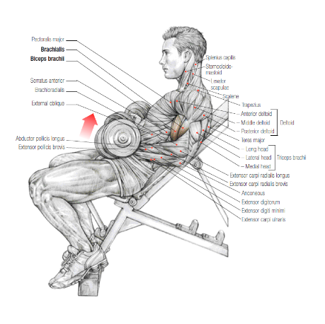
– Flexion is the bending of an arm or leg, decreasing the angle between bones at a joint.
– Hyperflexion is when a joint is bent beyond its normal range of motion, potentially causing injury.
– The opposite of flexion is extension, which is the straightening of a joint.
– Normal extension is typically limited to 180 degrees or less.
– Hyperextension is an extreme version of extension that can result in injury.
– Joints have a predetermined range of motion, measured in degrees.
– Flexion refers to a decrease in the angle between two body parts, such as the elbow and the knee.
– Extension refers to an increase in the angle between two body parts, such as the elbow and the knee.

Continue Reading

Infertility: Understanding the Science, Causes, and Treatment

– Infertility is defined as not being able to get pregnant after one year (or longer) of unprotected sex.
– Infertility can be frustrating and unexpected for couples hoping to become parents.
– Many couples who struggle with infertility end up having children, sometimes with medical help.
– Fertility in women declines with age, so providers may evaluate and treat women aged 35 years or older after 6 months of unprotected sex.
– Couples unable to conceive should consider making an appointment with a reproductive endocrinologist, a doctor specializing in managing infertility.
– Reproductive endocrinologists may also help women with recurrent pregnancy loss, defined as having two or more spontaneous miscarriages.

Continue Reading

Myomectomy: Understanding the Surgical Procedure for Uterine Fibroids

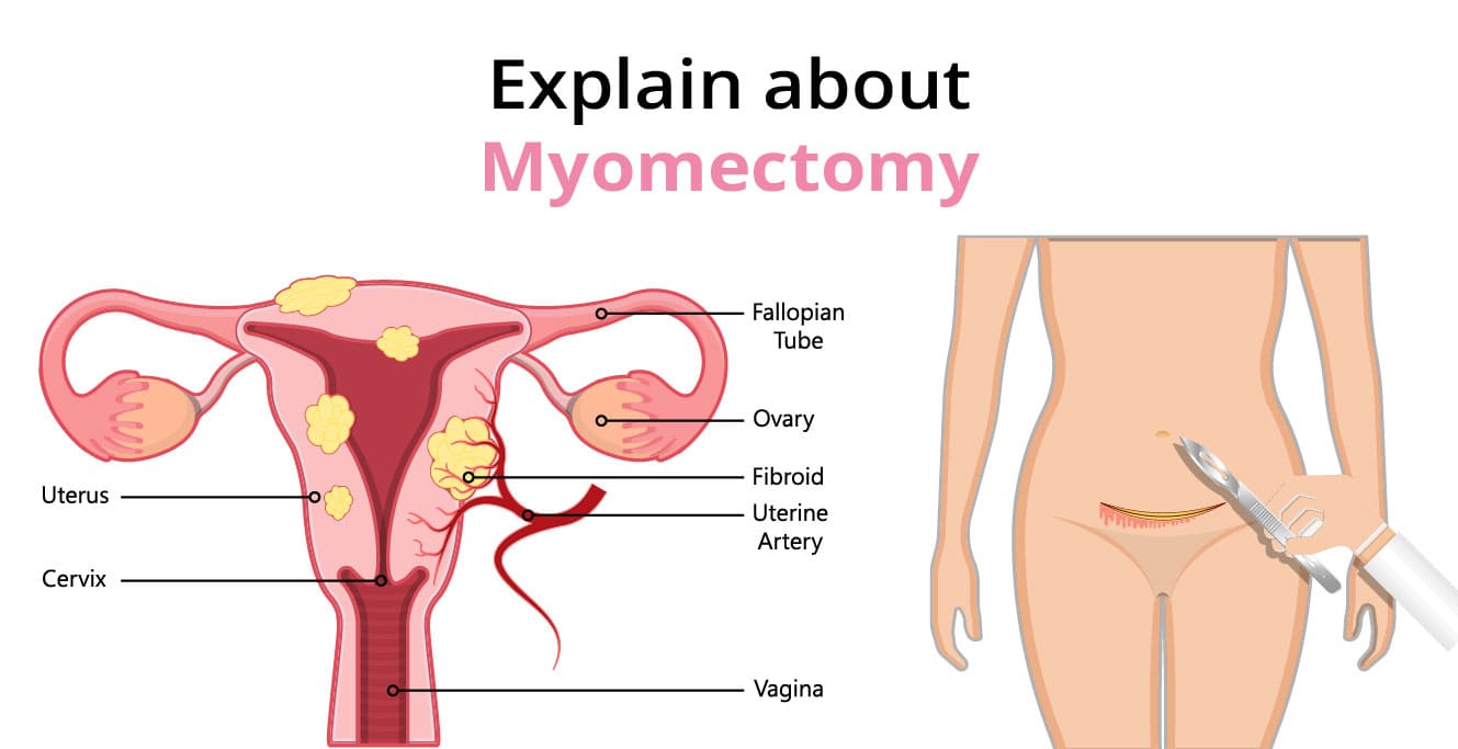
– Myomectomy: a procedure to remove fibroids from the uterus
– Fibroids: non-cancerous tumors in the uterus
– Uterus: female reproductive organ where the baby grows during pregnancy
– Recurring problems: likelihood of fibroids re-growing after myomectomy
– Abdominal myomectomy: major surgical procedure with incision in lower abdomen
– Laparoscopic myomectomy: removal of certain fibroids using small incisions and laparoscope
– Hysteroscopic myomectomy: not mentioned in the article
– Recovery time: four to six weeks for abdominal myomectomy, shorter for laparoscopic myomectomy
– Risks: infection, wound infection (rare)
– Scar: visible scar near pubic hairline for abdominal myomectomy, small scars from incisions
– Blood transfusion: may be required in some cases of abdominal myomectomy
– Gas: used during the procedure and released afterwards
– C-section: recommended for future pregnancies to reduce risk of uterine opening during labor
– Hospital stay: usually one night for myomectomy procedure
– Home recovery: two to four weeks after myomectomy procedure

Continue Reading

Prl: Unraveling the Mysteries of Parallel Computing

List of relevant keywords:

– Scientists
– Large Hadron Collider (LHC)
– CERN
– Neutrinos
– Colliding beams
– Universe
– Matter
– Physical Review Letters
– Physics Magazine
– Florian Bernlochner
– Experiment
– Observations
– Forward Search Experiment (FASER)
– Switzerland
– Live question-and-answer session
– David Saltzberg
– UCLA
– Registration
– Free
– Video recording.

Continue Reading