Combined Chemotherapy: Maximizing Treatment Effectiveness for Cancer Patients

– Combination chemotherapy is the use of more than one medication at a time to treat cancer.
– Using a combination of drugs increases the chance of eliminating all cancer cells, but it also increases the risk of drug interactions.
– Combination chemotherapy was inspired by the approach to treating tuberculosis using a combination of antibiotics.
– Combination chemotherapy has been found to be more effective for treating certain cancers such as acute lymphocytic leukemia and Hodgkin’s lymphoma.
– Combination chemotherapy is more effective than single drug therapy and sequential chemotherapy.
– Targeted therapies, which block specific pathways in cancer cells, have been used in combination with chemotherapy.
– Combination chemotherapy is used for various types of solid tumors, such as non-small cell lung cancer and breast cancer.
– Combination chemotherapy is also used for certain leukemias and Hodgkin lymphoma.
– Chemotherapy used in combination with immunotherapy can enhance the effectiveness of both treatments.
– Immunotherapy drugs help the immune system recognize and attack cancer cells.
– The abscopal effect, where chemotherapy helps the immune system target abnormal cells, can occur when chemotherapy is combined with immunotherapy.
– Combination chemotherapy has increased survival rates for many diseases.
– Combination chemotherapy involves using multiple chemotherapy medications at the same time.
– There are several advantages to using combination chemotherapy, including decreased resistance of tumors to treatment, earlier administration of medications, targeting multiple processes in cancer growth, increased effectiveness, lower doses of medications, and synergetic effects of drug combinations.
– Combination chemotherapy has been found to improve survival or result in a better response to treatment, especially as adjuvant treatment after surgery.
– However, there are also disadvantages to combination chemotherapy, including an increased risk of side effects.
– The article discusses the use of multiple chemotherapy drugs in combination treatment.
– It states that when multiple drugs are used, side effects of both drugs may compound.
– For example, if two drugs that cause a low white blood cell count are used, the risk of chemotherapy-induced neutropenia is increased.
– If a person develops a side effect while taking multiple medications, it may be difficult to determine which medication is responsible.
– In these cases, all medications may need to be discontinued if the side effect is serious.
– Sometimes side effects occur due to interactions between medications, not because of a specific medication.
– The more medications a person is taking, the greater the chance of an interaction occurring.
– Combination chemotherapy can extend life, reduce the risk of cancer recurrence, and improve the results of immunotherapy.
– However, adding more medications can increase side effects and the intensity of treatment.
– Advances in managing side effects have been made, such as anti-nausea drugs reducing or eliminating nausea, and drugs increasing white blood cell count allowing for higher and more effective doses of chemotherapy.

Continue Reading

Hypoplasia of uterus: Understanding its causes, symptoms, and treatment

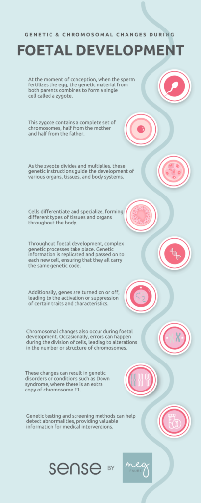
– Uterine hypoplasia, also known as hypoplastic uterus, is a condition where a girl is born with an abnormally small uterus.
– It is a congenital disorder that occurs when the uterus fails to fully develop in the fetus.
– The cause of this abnormal development is unknown.
– Uterine hypoplasia may be associated with Mayer-Rokitansky-Küster-Hauser (MRKH) syndrome, where the girl’s uterus and vagina are absent or underdeveloped.
– Symptoms include primary amenorrhea (failure to start having periods at puberty), abdominal pain, and a small or nonexistent vaginal opening.
– Diagnosis is often made during puberty when a girl fails to start her periods, and it involves a thorough medical history, physical examination, blood tests to check for MRKH syndrome, and imaging tests such as ultrasound and MRI.
– Treatment and care for uterine hypoplasia depend on the individual and their symptoms.

Continue Reading

Gestational Trophoblastic Tumour: Understanding Diagnosis, Treatment, and Recovery

– Gestational trophoblastic tumor is a type of cancer that forms from the cells that would normally develop into the placenta during pregnancy.
– It is rare and usually occurs in women of childbearing age.
– The tumor may be benign or cancerous.
– Symptoms can include abnormal vaginal bleeding, nausea and vomiting, and pelvic pain.
– Treatment options include surgery, chemotherapy, and radiation therapy.
– Clinical trials are being conducted to find new and better ways to treat this type of cancer.
– Gestational trophoblastic disease can occur after any type of pregnancy, including molar pregnancies, pregnancies that end in miscarriage or abortion, and normal pregnancies.
– Gestational trophoblastic disease is rare and can usually be cured with treatment.
– Treatment options for gestational trophoblastic disease include surgery, chemotherapy, and radiation therapy.
– It is important to receive care from a medical team with experience in treating gestational trophoblastic disease.
– The 5-year survival rate for women with low-risk disease is nearly 100%, while the 5-year survival rate for women with high-risk disease is about 90%.
– Gestational trophoblastic disease can sometimes spread to other parts of the body, such as the lungs, making treatment more difficult.

Continue Reading

Unveiling the Enigma: Understanding Endometrioid Tumour Progression

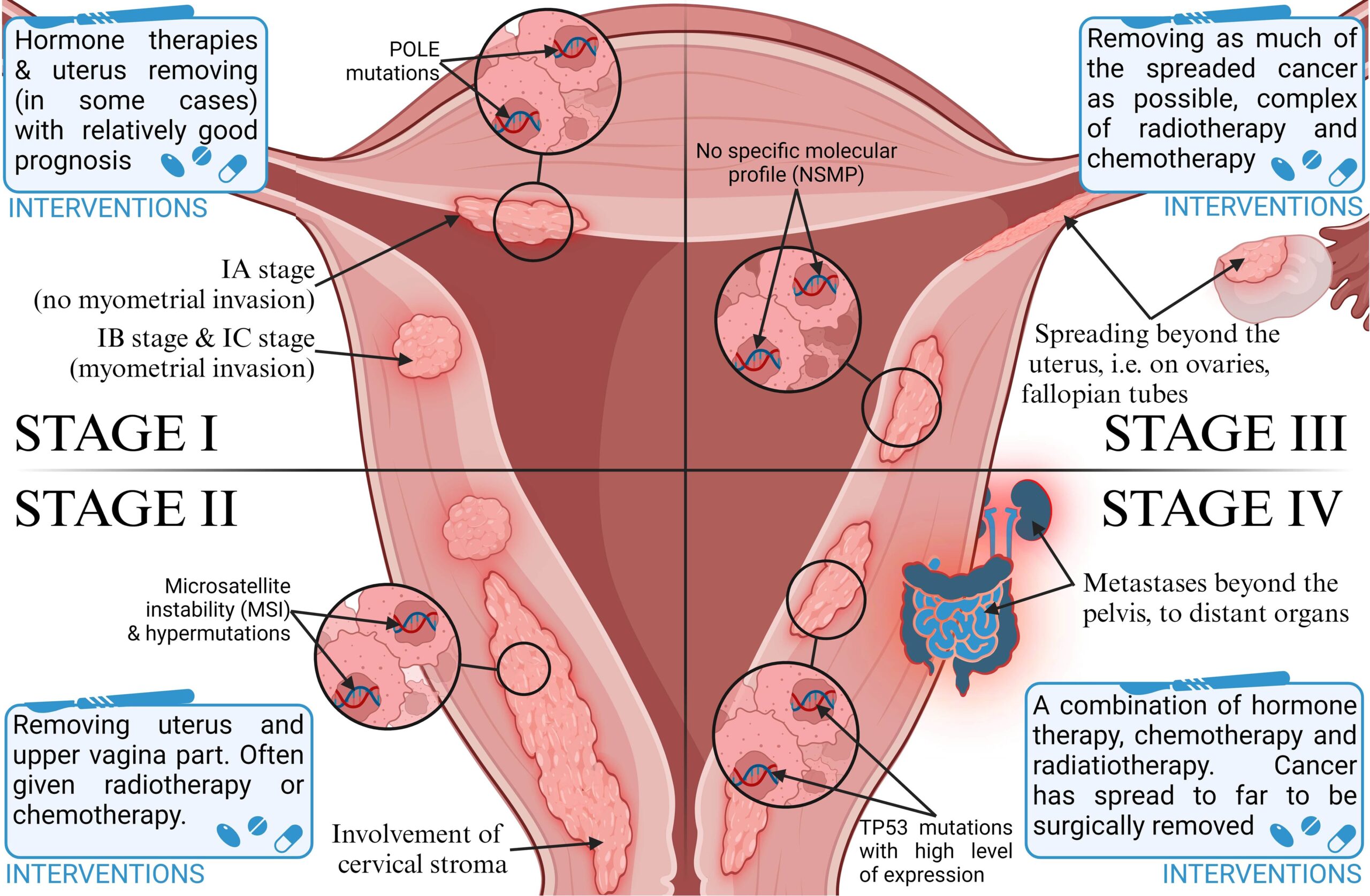
– Endometrioid cancer is the most common type of cancer in the uterus and starts in the inner lining of the uterus (endometrium).
– There are different types of endometrial cancers, including adenocarcinoma (most common), uterine carcinosarcoma, squamous cell carcinoma, small cell carcinoma, transitional carcinoma, serous carcinoma, clear-cell carcinoma, mucinous adenocarcinoma, undifferentiated carcinoma, dedifferentiated carcinoma, and serous adenocarcinoma.
– Endometrioid cancer starts in gland cells and resembles the normal uterine lining.
– Some endometrioid cancers have squamous cells in addition to glandular cells.
– There are several sub-types of endometrioid cancers, including adenocarcinoma (with squamous differentiation), adenoacanthoma, adenosquamous (or mixed cell), secretory carcinoma, ciliated carcinoma, and villoglandular adenocarcinoma.
– The grade of an endometrial cancer is determined by the organization of the cancer cells into gland-like structures.
– Grade 1 tumors of endometrioid cancer have 95% or more of the cancer tissue forming glands.
– Grade 2 tumors have between 50% and 94% of the cancer tissue forming glands.
– Grade 3 tumors have less than half of the cancer tissue forming glands and tend to be aggressive with a worse outlook.
– Type 1 endometrial cancers are usually not aggressive and are caused by too much estrogen. They may develop from atypical hyperplasia.
– Type 2 endometrial cancers are more likely to grow and spread outside the uterus and have a poorer outlook. They are not caused by too much estrogen and include papillary serous carcinoma, clear-cell carcinoma, undifferentiated carcinoma, and grade 3 endometrioid carcinoma.
– Uterine carcinosarcoma (CS) starts in the endometrium and has features of both endometrial carcinoma and sarcoma. It is a type 2 endometrial carcinoma.
– Uterine sarcomas start in the muscle layer or supporting connective tissue of the uterus.
– Cancers that start in the cervix and spread to the uterus are different from cancers that start in the body of the uterus.
– Symptoms of endometrial cancer include vaginal bleeding after menopause, bleeding between periods, and pelvic pain.
– The cause of endometrial cancer is not known, but it is believed that changes in the DNA of cells in the endometrium lead to the growth of cancer cells.
– Early detection of endometrial cancer can lead to successful treatment through surgical removal of the uterus.
– Risk factors for endometrial cancer include changes in hormone balance, certain diseases or conditions, menstruation history, pregnancy history, age, obesity, hormone therapy for breast cancer, and Lynch syndrome.
– Obesity and hormone therapy are notable risk factors for endometrial cancer.
– Lynch syndrome, a genetic syndrome associated with an increased risk of several types of cancer, increases the risk of endometrial cancer.
– Individuals with Lynch syndrome should inquire about appropriate cancer screenings.
– There are no specific facts, stats, or figures provided about endometrioid tumors or the prevalence of Lynch syndrome in the article.

Continue Reading

New Research Revealed: The Shocking Truth About Abruption

– Placental abruption is when a part or all of the placenta separates from the uterus prematurely, causing vaginal bleeding.
– There are two main types of placental abruption: revealed and concealed. Revealed abruption results in visible vaginal bleeding, while concealed abruption involves bleeding that remains within the uterus and may not be visible.
– Major risk factors for placental abruption include previous placental abruption, pre-eclampsia, abnormal lie of the baby, abdominal trauma, smoking or drug use, bleeding in the first trimester, and underlying thrombophilias.
– Clinical features of placental abruption include painful vaginal bleeding, woody and painful uterus on palpation, and the need for systematic assessment and resuscitation.
– General examination involves assessing pallor, distress, peripheral circulation, abdominal tenderness, the feel of the uterus, and the lie and presentation of the fetus/fetuses. The article provides guidance on how to assess bleeding during pregnancy and discusses differential diagnoses for antenatal hemorrhage. It suggests using a cardiotocograph (CTG) at 26 weeks gestation or above to check fetal wellbeing. It advises checking hand-held pregnancy notes for scan reports and looking for signs of placenta praevia. The article also recommends assessing the bleeding externally by looking at pads, avoiding speculum examination until placenta praevia is excluded, and taking triple genital swabs to exclude infection. It warns against performing a digital vaginal examination with known placenta praevia as it could cause massive bleeding. The article mentions placental abruption, placenta praevia, marginal placental bleed, vasa praevia, uterine rupture, and local genital causes as differential diagnoses for antenatal hemorrhage.
– Placental abruption is a common cause of antepartum hemorrhage.
– Investigations that should be performed include hematology (full blood count, Kleihauer test, group and save, cross-match), biochemistry (urea and electrolytes, liver function tests), and fetal wellbeing assessment (cardiotocograph).
– Ultrasound scan should be performed to assess placental abruption, but ultrasound should not be used to exclude abruption.
– Management of placental abruption depends on the health of the fetus: emergency delivery is indicated in the presence of maternal and/or fetal compromise, induction of labor is recommended for hemorrhage at term without compromise, and conservative management is an option for some partial or marginal abruptions without compromise.
– Anti-D should be given within 72 hours of bleeding onset if the woman is rhesus D negative.
– Placental abruption complicates approximately 1% of pregnancies and increases the risk of maternal, fetal, and neonatal morbidity and mortality.
– Risk factors for placental abruption include smoking, alcohol or cocaine use during pregnancy, advanced maternal age, history of maternal hypertension, and preeclampsia.
– Previous placental abruption and multiple gestational pregnancies also increase the risk.
– Trauma from a motor vehicle accident, fall, or blow to the abdomen can cause placental abruption.
– Radiologic imaging, such as ultrasonography, may assist in diagnosing placental abruption.
– Ultrasonography is usually the preferred study due to its benefits of avoiding ionizing radiation, dynamic nature, and availability.
– In severe trauma cases, CT scanning may be required to evaluate for abdominopelvic injuries.
– American College of Radiology guidelines recommend ultrasound FAST scan as a limited bedside adjunct for triage in a pregnant patient with major blunt trauma. The article discusses the use of imaging techniques for diagnosing major blunt trauma in pregnant patients. The American College of Radiology (ACR) states that there are two options for these procedures, but only one should be used to gather clinical information. No other facts, stats, or figures are mentioned in the article.

Continue Reading

Intrauterine Devices: A Contraceptive Option for Safe and Effective Family Planning

Summary:
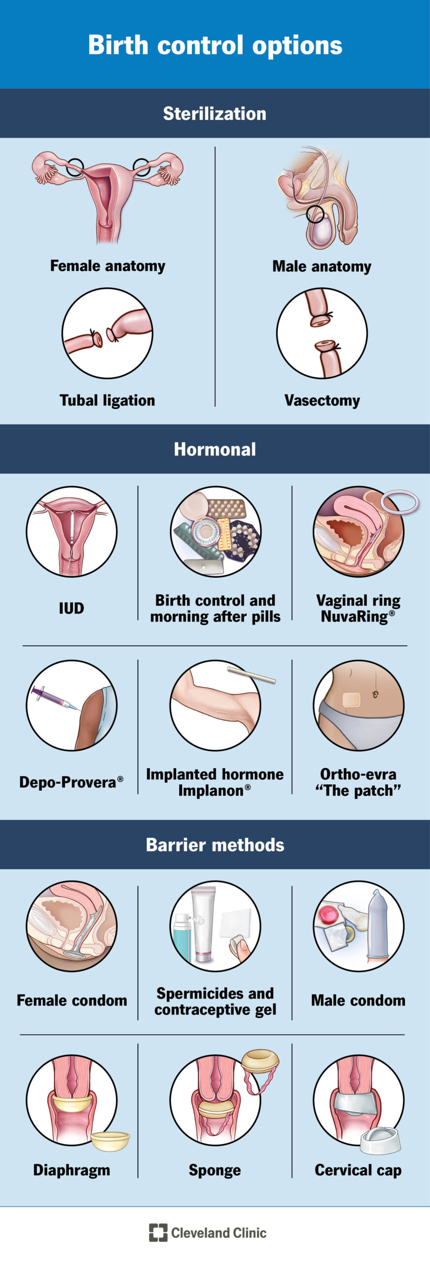
– An intrauterine device (IUD) is a small T-shaped plastic and copper device that is inserted into the womb to prevent pregnancy.
– When inserted correctly, IUDs are over 99% effective and can last for 5 to 10 years.
– IUDs can be inserted at any time during the menstrual cycle if the person is not pregnant.
– IUDs can be removed at any time by a trained medical professional.
– Side effects can include heavier, longer, or more painful periods, spotting or bleeding between periods, and a small risk of infection.
– The IUD does not protect against sexually transmitted infections, so additional protection may be needed.
– The copper in the IUD alters cervical mucus, making it difficult for sperm to reach an egg and preventing the implantation of a fertilized egg.
– IUDs can be left in place until menopause or when contraception is no longer needed for those over 40 years old.
– Before insertion, a GP or nurse will check the position and size of the womb and may test for existing infections.
– The fitting process takes about 20 to 30 minutes, with the IUD being inserted through the cervix into the womb.
– Local anesthesia can be used to minimize discomfort during insertion.
– After having an IUD fitted, period-type cramps and bleeding may occur for a few days.
– A GP may recommend checking the IUD after 3 to 6 weeks.
– If there are any problems or if removal is desired, the GP should be consulted.
– If there is a risk of sexually transmitted infection (STI), it may lead to an infection in the pelvis and should be addressed with a GP.
– IUDs have two thin threads that can be checked by the user to make sure it is still in place.
– If the threads cannot be felt or if the IUD has moved, there may be a risk of pregnancy, and additional contraception should be used until checked by a GP.
– If the partner can feel the IUD during sex, a check-up should be scheduled.
– Additional contraception should be used for 7 days before IUD removal if not replacing the IUD.
– Most individuals with a womb can use an IUD, with exceptions for those who may be pregnant, have untreated STIs or pelvic infections, have womb or cervix issues, or experience unexplained bleeding.
– People who have had an ectopic pregnancy or have an artificial heart valve should consult their GP before getting an IUD.
– IUDs can usually be fitted 4 weeks after giving birth, and alternative contraception should be used until then.
– IUDs have an increased risk of ectopic pregnancy.
– IUDs can be obtained for free from contraception clinics, sexual health or genitourinary medicine (GUM) clinics, GP surgeries, and some young people’s services.
– Contraception services are free and confidential for people under 16.
– Healthcare professionals will not inform parents or caregivers if under 16 seeking contraception, as long as they believe the person fully understands the information and decisions being made.
– Professionals may only disclose information if they believe the individual is at risk of harm, such as abuse.
– An IUD can last between 5 to 10 years, depending on the type.
– Periods may become heavier, longer, or more painful in the first 3 to 6 months after insertion.
– Spotting or bleeding between periods may occur.
– There is a small risk of infection or expulsion of the IUD.
– Previous pelvic infections may make IUDs unsuitable.
– IUDs do not protect against sexually transmitted infections (STIs).
– IUDs release copper into the womb, which alters cervical mucus and makes it difficult for sperm to reach an egg and implant itself.
– Before insertion, a GP or nurse will check the position and size of the womb and test for existing infections.
– The appointment takes about 20 to 30 minutes, with fitting taking no longer than 5 minutes.
– IUD insertion can be uncomfortable or painful, but local anesthesia can be used to help.
– Painkillers can be taken after insertion if needed.
– There is a small chance of getting thrush that keeps coming back after having an IUD fitted.
– If the IUD fails and a woman becomes pregnant, there is an increased risk of an ectopic pregnancy.
– IUDs can be obtained for free from contraception clinics, sexual health or genitourinary medicine (GUM) clinics, GP surgeries, and some young people’s services.
– Contraception services are free and confidential for people under 16.
– Healthcare professionals will not inform parents or carers about a person under 16 seeking contraception, as long as they believe the person understands the information and decisions being made.
– Professionals may disclose information if they believe the person is at risk of harm, such as abuse.

Continue Reading

Cervical Intraepithelial Neoplasm: Understanding Diagnosis, Treatment, and Prevention

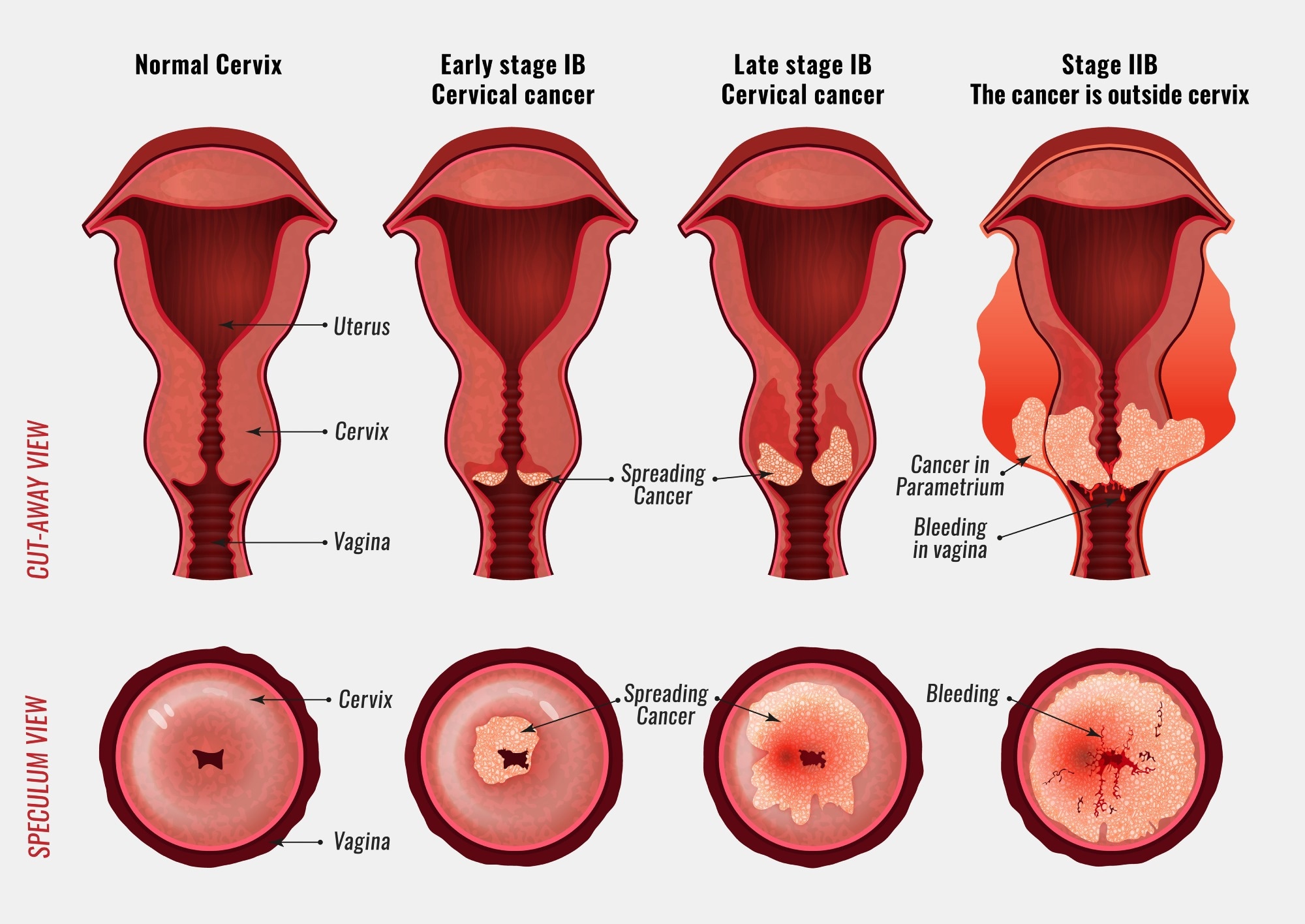
– Cervical dysplasia is a precancerous condition where abnormal cells grow on the surface of the cervix.
– Another name for cervical dysplasia is cervical intraepithelial neoplasia (CIN).
– Most people with cervical dysplasia do not develop cancer.
– Cervical dysplasia is classified on a scale from one to three, with CIN 1 affecting about one-third of the thickness of the epithelium, CIN 2 affecting about one-third to two-thirds of the epithelium, and CIN 3 affecting more than two-thirds.
– Cervical dysplasia primarily affects sexually active individuals assigned female at birth (AFAB) who have a cervix.
– It is most common among women of childbearing age, particularly aged 25 to 35.
– Approximately 250,000 to 1 million cisgender women in the U.S. are diagnosed with cervical dysplasia each year.
– Cervical intraepithelial neoplasia (CIN) is the abnormal growth of cells on the surface of the cervix that could potentially lead to cervical cancer. CIN is graded on a 1-3 scale, with 3 being the most abnormal.
– Human papillomavirus (HPV) infection is necessary for the development of CIN. Many women with HPV infection never develop CIN or cervical cancer. Typically, HPV resolves on its own. However, those with an HPV infection that lasts more than one or two years have a higher risk of developing a higher grade of CIN.
– Most cases of CIN either remain stable or are eliminated by the person’s immune system without the need for intervention. However, a small percentage of cases progress to cervical cancer if left untreated.
– There are no specific symptoms of CIN alone, but signs and symptoms of cervical cancer may include abnormal bleeding, abnormal discharge, changes in bladder or bowel function, pelvic pain, or abnormal appearance or palpation of the cervix.
– The cause of CIN is chronic infection of the cervix with HPV, especially infection with high-risk HPV types 16 or 18.
– Risk factors for developing CIN include infection with high-risk types of HPV, immunodeficiency, poor diet, multiple sex partners, lack of condom use, and cigarette smoking.
– Cervical intraepithelial neoplasia (CIN) is commonly associated with infection by human papillomavirus (HPV).
– Most women with HPV infection do not develop high-grade intraepithelial lesions or cancer.
– There are over 100 different types of HPV, with approximately 40 known to affect the anogenital area.
– The Digene HPV test is a highly accurate test for HPV, serving as both a direct diagnosis and adjuvant to the Pap smear.
– A colposcopy with directed biopsy is the standard for detecting CIN.
– Diagnosis of CIN or cervical carcinoma requires a biopsy for analysis.
– The Bethesda System for Reporting Cervical/Vaginal Cytologic Diagnoses provides a uniform way to describe abnormal epithelial cells.
– CIN is classified into grades: CIN 1 (mild dysplasia), CIN 2 (moderate dysplasia), and CIN 3 (severe dysplasia)
– CIN 3 can also be referred to as cervical carcinoma in situ.
– Locations of CIN findings can be described in terms of quadrants or clock face positions.
– Cervical intraepithelial neoplasm (CIN) is classified as LSIL or HSIL based on its severity.
– Screening for CIN can be done through Pap smear or testing for HPV.
– The accuracy of Pap smear results can vary.
– Abnormal Pap smear results may lead to colposcopy, which involves examining the cervix under magnification and taking a biopsy.
– HPV testing can identify high-risk HPV types responsible for CIN.
– HPV vaccination is the primary prevention method for CIN and cervical cancer, but it does not protect against all types of HPV known to cause cancer.
– Appropriate management and treatment are used as secondary prevention for cervical cancer cases.
– Treatment for CIN 1 is not recommended if it lasts fewer than two years, as it may clear on its own. Instead, close monitoring is advised.
– Treatment for higher-grade CIN involves removal or destruction of the abnormal cells.
– Retinoids may be effective in causing regression of CIN2.
– Therapeutic vaccines are being tested in clinical trials.
– The lifetime recurrence rate of CIN is about 20%.
– Surgical treatment of CIN may increase the risk of infertility or subfertility.
– Women receiving treatment for CIN during pregnancy may have an increased risk of premature birth.
– People with HIV and CIN 2+ should be managed according to general recommendations.
– Most cases of CIN spontaneously regress. Left untreated, about 70% of CIN 1 will regress within one year and 90% within two years. About 50% of CIN 2 cases will regress within two years. Progression to cervical carcinoma in situ (CIS) occurs in approximately 11% of CIN 1 and 22% of CIN 2 cases. Progression to invasive cancer occurs in approximately 1% of CIN 1, 5% of CIN 2, and at least 12% of CIN 3 cases.
– Treatment does not affect the chances of getting pregnant but is associated with an increased risk of miscarriage in the second trimester.
– Between 250,000 and 1 million American women are diagnosed with CIN annually.
– The estimated annual incidence of CIN in the United States is 4% for CIN 1 and 5% for CIN 2 and CIN 3.

Continue Reading

Hypertonic Uterine Contraction: Understanding Causes, Risks, and Management

List of Pertinent Information:

1. Uterine hyperstimulation, also known as hypertonic uterine dysfunction, can occur as a complication of labor induction.
2. It is characterized by frequent contractions (more than five in 10 minutes) or contractions lasting more than two minutes.
3. Uterine hyperstimulation can result in fetal heart rate abnormalities, uterine rupture, or placental abruption.
4. The drug Misoprostol, used for peptic ulcers, can cause uterine hyperstimulation when used to induce labor.
5. Terbutaline is commonly used to treat uterine hyperstimulation.
6. Prostaglandin E2 can be administered before labor to minimize the risk of hyperstimulation and its effects on the fetal heart rate.
7. Tocolytic treatment with β2-adrenergic drugs has been used to stabilize uterine contractions and lower fetal heart rate.
8. Using a balloon catheter for labor induction instead of Prostaglandin E2 can reduce the risk of uterine hyperstimulation and its impact on the fetal heart rate.

Continue Reading