In the realm of medical mysteries lies a condition both rare and perplexing – the partial hydatid mole.
With its enigmatic nature and elusive symptoms, this intriguing anomaly has captured the attention of scientists and doctors alike.
Join us as we embark on a journey to unravel the mysteries surrounding this fascinating medical phenomenon.
partial hydatid mole
A partial hydatid mole refers to a specific type of abnormal pregnancy known as a hydatidiform mole, which occurs when something goes wrong during fertilization and an abnormal mass of tissue develops in the uterus instead of a healthy pregnancy.
A partial hydatid mole is a less common form of this condition, characterized by the presence of both normal and abnormal fetal development.
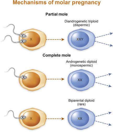
This results in a combination of genetic material from both the mother and the father, leading to a variable clinical presentation and outcomes.
Key Points:
- A partial hydatid mole is an abnormal pregnancy that occurs when fertilization goes wrong and an abnormal mass of tissue develops in the uterus.
- It is a less common form of hydatidiform mole, characterized by both normal and abnormal fetal development.
- The condition results in a combination of genetic material from both the mother and the father.
- A partial hydatid mole has variable clinical presentation and outcomes.
- The abnormal tissue growth replaces a healthy pregnancy in the uterus.
- The condition occurs due to a problem during fertilization.
partial hydatid mole – Watch Video
💡
Pro Tips:
1. A partial hydatid mole is a rare condition in which a fertilized egg develops abnormally, resulting in an abnormal placenta and some fetal tissue.
2. A partial hydatid mole is a molar pregnancy that occurs when two sperm fertilize a single egg, resulting in an extra set of chromosomes (69 instead of the usual 46).
3. Due to its rarity, partial hydatid mole is difficult to diagnose accurately, often being mistaken for other pregnancy complications or miscarriages.
4. Partial hydatid mole tends to have lower levels of the hormone hCG (human chorionic gonadotropin) compared to other types of molar pregnancies, making it harder to detect via routine pregnancy tests.
5. Although prognosis is generally more favorable compared to complete hydatid mole, partial hydatid mole still carries a risk of complications such as persistent trophoblastic disease, requiring careful monitoring and medical intervention.
What Is A Partial Hydatid Mole?
Partial hydatid mole is a rare phenomenon that occurs during pregnancy, specifically in the uterus. It is a type of gestational trophoblastic disease (GTD), which affects the cells that would normally develop into the placenta. In the case of a partial hydatid mole, there is an abnormal growth of these cells, leading to the formation of a mass in the uterus.
The term “partial” indicates that only some of the cells involved in the growth are abnormal, while others are normal. This differentiation is crucial to understanding the characteristics and potential complications associated with partial hydatid mole. Although this condition is rare, it is essential to be aware of its causes, symptoms, and management options.
Symptoms And Signs Of Partial Hydatid Mole
The symptoms of a partial hydatid mole may vary from woman to woman and depend on the stage of pregnancy. Common signs include vaginal bleeding, excessive nausea and vomiting, enlarged uterus, and an abnormal increase in hCG hormone levels. Some women may experience abdominal pain or discomfort, while others may notice the passage of grape-like cysts or vesicles through the vagina.
It is important to note that these signs and symptoms are not exclusive to partial hydatid mole and can be associated with other conditions as well. Therefore, an accurate diagnosis by a healthcare professional is crucial for proper management.
Diagnosing Partial Hydatid Mole
Diagnosing a partial hydatid mole requires a combination of clinical evaluation, medical history, physical examinations, and laboratory tests. A healthcare professional will usually conduct a pelvic exam to check for any abnormalities in the uterus. Additionally, imaging tests such as ultrasounds or MRI may be performed to visualize the uterus and confirm the presence of a partial hydatid mole.
Blood tests, including hCG levels, may also be done as they can provide valuable information about the presence and development of the abnormal growth. Genetic testing of the tissue from the uterus may be recommended to further confirm the diagnosis.
- Clinical evaluation
- Medical history
- Physical examinations
- Laboratory tests
- Pelvic exam
- Imaging tests (ultrasounds or MRI)
- Blood tests (including hCG levels)
- Genetic testing of uterine tissue
Causes And Risk Factors Of Partial Hydatid Mole
The underlying causes of partial hydatid mole are currently not fully comprehended. Nonetheless, it is thought to stem from a genetic anomaly during fertilization, resulting in an excessive amount of genetic material in the developing placenta. The precise risk factors associated with partial hydatid mole have not been well determined, but there are certain factors that may increase a woman’s susceptibility to this condition, such as advanced maternal age or a previous history of GTD.
It is crucial to emphasize that partial hydatid mole is not attributable to any actions or behaviors of the woman. Rather, it is an unexpected abnormality that arises during conception.
Complications Associated With Partial Hydatid Mole
Partial hydatid mole can lead to several complications for both the mother and the developing fetus. One potential complication is the risk of persistent gestational trophoblastic disease, in which the abnormal cells continue to grow even after the mole has been removed. This can require additional treatment and monitoring.
In some cases, partial hydatid mole can also progress to choriocarcinoma, a more aggressive form of GTD. Choriocarcinoma can invade surrounding tissues and potentially spread to other parts of the body.
To summarize:
- Persistent gestational trophoblastic disease can occur after removal of the mole and requires further treatment and monitoring.
- Partial hydatid mole can progress to choriocarcinoma, which is a more aggressive form of GTD.
- Early detection and prompt management are crucial in preventing complications.
“Partial hydatid mole can lead to several complications for both the mother and the developing fetus. One potential complication is the risk of persistent gestational trophoblastic disease, in which the abnormal cells continue to grow even after the mole has been removed. This can require additional treatment and monitoring.”
In some cases, partial hydatid mole can also progress to choriocarcinoma, a more aggressive form of GTD. Choriocarcinoma can invade surrounding tissues and potentially spread to other parts of the body.
Treatment Options For Partial Hydatid Mole
The management of partial hydatid mole typically involves a combination of surgery and chemotherapy. The primary goal of treatment is to remove the abnormal tissues from the uterus and prevent further complications. In most cases, a dilation and curettage (D&C) procedure is performed to remove the partial hydatid mole.
In certain situations, chemotherapy may be recommended to destroy any remaining abnormal cells and reduce the risk of persistent gestational trophoblastic disease or choriocarcinoma. The specific treatment plan will depend on factors such as the stage of pregnancy, the extent of the abnormal growth, and the woman’s overall health.
Prognosis And Long-Term Outcomes
The prognosis for women diagnosed with a partial hydatid mole is generally good. With early detection and appropriate treatment, the majority of women can achieve a complete remission and have successful future pregnancies. Regular monitoring through blood tests and imaging is essential to ensure there is no recurrence or persistent GTD.
It is important for women with a history of partial hydatid mole to consult with a healthcare professional before planning future pregnancies. Close monitoring during subsequent pregnancies can help ensure any potential complications are detected early and managed appropriately.
Prevention And Lifestyle Choices To Reduce The Risk
As the exact causes and risk factors of partial hydatid mole are not well understood, there are no specific preventive measures.
However, maintaining a healthy lifestyle is generally beneficial for overall health and reproductive well-being.
- Regular exercise: Engaging in physical activity helps to maintain a healthy weight and can improve overall well-being.
- Balanced diet: Consuming a nutritious and well-balanced diet provides essential vitamins and minerals needed for a healthy pregnancy. It is important to include a variety of fruits, vegetables, whole grains, lean proteins, and healthy fats.
- Avoiding harmful substances like tobacco or excessive alcohol: It is crucial to refrain from smoking and limit alcohol consumption as these substances can increase the risk of complications during pregnancy.
Additionally, regular prenatal care and early detection of any abnormalities during pregnancy can contribute to better outcomes.
Women should always consult with a healthcare professional for personalized advice regarding their specific risk factors and pregnancy planning.
Remember, early intervention and proactive healthcare can make a significant difference in ensuring a healthy pregnancy.
Support And Resources For Individuals With Partial Hydatid Mole
Facing a diagnosis of partial hydatid mole can be emotionally challenging for women and their families. It is important to seek support from healthcare professionals, support groups, or counseling services specialized in gestational trophoblastic diseases.
Several organizations and online resources provide further information and support for individuals affected by GTD. These resources can help individuals gain a better understanding of the condition, access support networks, and find the most current research and advancements in managing partial hydatid mole.
Research And Advancements In Understanding Partial Hydatid Mole
Although partial hydatid mole is a rare condition, ongoing research and advancements in medical science aim to improve our understanding and management of this phenomenon. Scientists and researchers continue to investigate the genetic and molecular mechanisms underlying partial hydatid mole to develop more accurate diagnostic tools, effective treatment strategies, and potential preventive measures.
In conclusion, partial hydatid mole is a rare but important condition to be aware of, especially for healthcare professionals involved in the management of pregnancy-related disorders. Early diagnosis, appropriate treatment, and regular monitoring can significantly increase the chances of a successful outcome for women affected by partial hydatid mole.
💡
You may need to know these questions about partial hydatid mole
What is the prognosis for a partial hydatidiform mole?
The prognosis for a partial hydatidiform mole is generally less favorable compared to a complete mole. While it may be possible to carry the pregnancy to full term due to the presence of a fetus, there are significant risks involved. These include potential fetal abnormality, hypertension, preterm birth, as well as an increased risk of malignancy or persistence of the molar pregnancy. Therefore, close monitoring and careful management are crucial in these cases to minimize complications and ensure the best possible outcome for both the mother and the fetus.
What are the characteristics of a partial hydatidiform mole?
Partial hydatidiform mole is a rare condition characterized by various microscopic features. Firstly, the presence of two populations of villi is a key characteristic of partial hydatidiform mole. Enlarged villi, measuring at least 3-4 mm, that exhibit central captivation are also typically observed. Additionally, irregular villi with geographic, scalloped borders containing trophoblast inclusions are often present. Finally, trophoblast hyperplasia, which usually manifests as an increase in the number of trophoblast cells, is another common feature of this condition.
In summary, the diagnosis of partial hydatidiform mole is based on the presence of four main microscopic characteristics: two populations of villi, enlarged villi with central captivation, irregular villi with scalloped borders and trophoblast inclusions, as well as trophoblast hyperplasia.
What is the difference between a partial and complete hydatidiform mole?
A complete hydatidiform mole is characterized by the absence of a fetus and any fetal tissue. It is essentially an overgrowth of abnormal placental tissue that can resemble a cluster of grapes. Without any developing fetus, a complete molar pregnancy cannot progress to a viable pregnancy. On the other hand, a partial hydatidiform mole involves the presence of some fetal tissue, although it remains abnormal and cannot survive. While a partial mole may show some signs of fetal development, it is not viable, and the fetus typically cannot survive beyond three months.
Can a baby survive a partial molar pregnancy?
Due to the irregular development of the placenta and fetus in a partial molar pregnancy, it is highly unlikely for a baby to survive. The presence of irregular tissue resembling fluid-filled sacs further hampers the chances of a successful pregnancy. In most cases, unfortunately, a partial molar pregnancy ends in miscarriage, typically occurring early in the pregnancy.
Reference source
https://www.ncbi.nlm.nih.gov/books/NBK459155/
https://www.news-medical.net/health/Molar-Pregnancy-Treatment-and-Prognosis.aspx
https://pubmed.ncbi.nlm.nih.gov/11603213/
https://www.thewomens.org.au/health-information/pregnancy-and-birth/pregnancy-problems/early-pregnancy-problems/hydatidiform-mole


