External Pelvimetry: A Comprehensive Guide to Evaluating Childbirth Potential

– external pelvimetry
– measuring pelvis size and shape
– predicting success of vaginal delivery
– third trimester of pregnancy
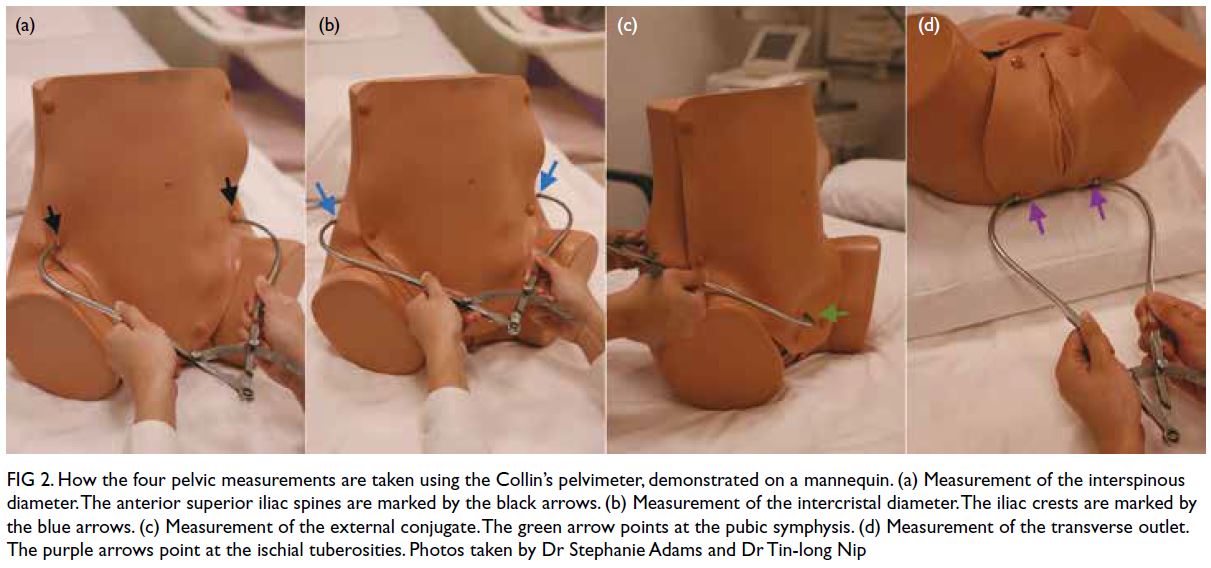
– calipers
– dimensions of the pelvis
– inlet, mid-pelvis, outlet
– limitations of external pelvimetry
– low sensitivity and specificity
– identifying cephalopelvic disproportion (CPD)
– false positive rate
– ultrasound pelvimetry
– clinical assessment
– more reliable methods
– non-invasive method
– evaluating maternal pelvic dimensions
– measuring pelvic landmarks
– calipers or tape measures
– adjunct to traditional methods
– internal pelvimetry
– determining suitability for vaginal delivery
– cesarean section
– limitations of external pelvimetry
– obesity
– fetal position
– pelvic soft tissue
– valuable information
– obstetric care

Continue Reading

Fetal Breath Movement: Understanding Its Significance During Development

Here is the updated list of pertinent information related to the keyword “fetal breath movement” without any duplication:

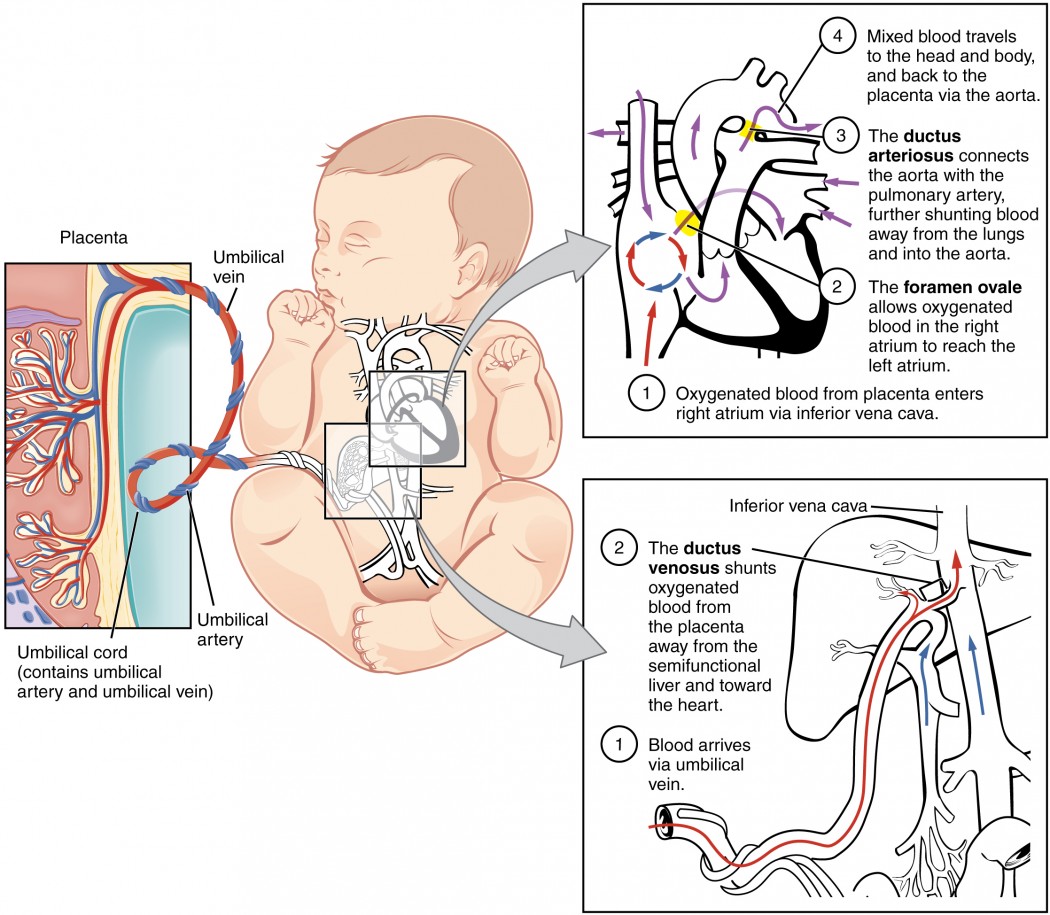
– Babies don’t breathe in the womb in the traditional sense as they don’t have access to air.
– They practice breathing movements called fetal breathing movements.
– Fetal breathing movements help strengthen the muscles involved in breathing and may support the development of air sacs in the lungs.
– These movements start around week 10 of pregnancy but become more frequent after week 20.
– Fetal breathing movements are not necessary for survival and may not happen constantly.
– Oxygen and carbon dioxide exchange in the womb occurs through the umbilical cord and placenta.
– After birth, babies initially rely on the umbilical cord and placenta for oxygen.
– Within about 10 seconds of delivery, babies take their first breath as they respond to the change in environment.
– By week 28 of pregnancy, the lungs of babies are fully developed enough that premature babies can breathe on their own.
– The lungs and circulatory system still need more time to mature.
– By week 37, the lungs have developed more fully and continue to grow and fine-tune until the child is at least 8 years old.
– The lungs begin producing surfactant at around month 6 of pregnancy, which allows them to inflate and deflate.
– Artificial surfactant can be used for babies born extremely preterm, along with breathing assistance from a ventilator, CPAP machine, or small breathing tubes.
– Babies born before week 36 and most born before week 32 may also need breathing support.
– A healthy pregnancy, quitting smoking, following a nutritious diet, avoiding smoke after birth, and encouraging regular exercise can support a baby’s lung development.
– Contractions during delivery squeeze the baby and force amniotic fluid out of the lungs, making it easier for them to breathe.
– As long as the baby is attached to the mother through the placenta and umbilical cord, it is not necessary for them to breathe.
– The baby will take their first breath a few seconds after delivery, without the support of the mother.
– The respiratory system is still growing after birth.
– Alveoli, small air sacs in the lungs, allow the body to transfer oxygen.
– Most newborns have between 20 and 50 million alveoli when born, which can increase to 300 million by the age of eight.
– The ribs surrounding the critical organs will get tougher as the baby develops, making the lungs more secure.
– Newborns may accidentally ingest meconium (first bowel movement) during birth, which can contaminate the lungs if not eliminated promptly.

Continue Reading

The Secretive Phase of Matter: Unlocking Nature’s Mysteries

– Secretory phase of the uterine cycle
– Luteal phase
– Changes in the endometrium
– Preparation of the uterus for implantation
– Days 14 to 28 of the menstrual cycle
– Increase in progesterone levels
– Corpus luteum
– Increased levels of progesterone and estrogen
– Thickening of the endometrium
– Nutrition for the embryo and placenta
– Implantation and hormonal support
– Shedding of the uterine lining
– Symptoms during the secretory phase
– Sore breasts, fatigue, acne breakouts, bloating, mood swings, and uterine cramping
– Abnormal menstrual cycles and hormone imbalance
– Hormone-balancing supplement like Hertime
– Access block on NCBI website
– Possible misuse/abuse situation involving the user’s site
– Lack of understanding on how to use E-utilities efficiently
– Contacting system administrator for access restoration and better site interaction.

Continue Reading

New Research Revealed: The Shocking Truth About Abruption

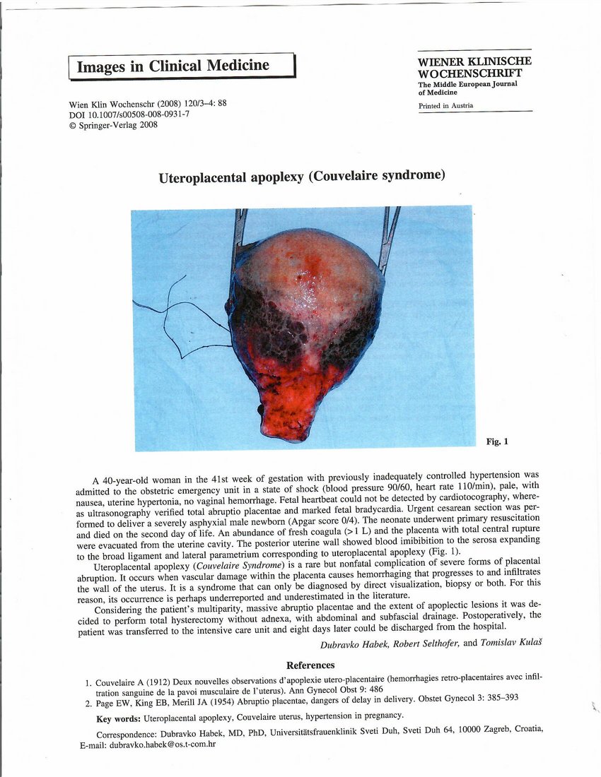
– Placental abruption is when a part or all of the placenta separates from the uterus prematurely, causing vaginal bleeding.
– There are two main types of placental abruption: revealed and concealed. Revealed abruption results in visible vaginal bleeding, while concealed abruption involves bleeding that remains within the uterus and may not be visible.
– Major risk factors for placental abruption include previous placental abruption, pre-eclampsia, abnormal lie of the baby, abdominal trauma, smoking or drug use, bleeding in the first trimester, and underlying thrombophilias.
– Clinical features of placental abruption include painful vaginal bleeding, woody and painful uterus on palpation, and the need for systematic assessment and resuscitation.
– General examination involves assessing pallor, distress, peripheral circulation, abdominal tenderness, the feel of the uterus, and the lie and presentation of the fetus/fetuses. The article provides guidance on how to assess bleeding during pregnancy and discusses differential diagnoses for antenatal hemorrhage. It suggests using a cardiotocograph (CTG) at 26 weeks gestation or above to check fetal wellbeing. It advises checking hand-held pregnancy notes for scan reports and looking for signs of placenta praevia. The article also recommends assessing the bleeding externally by looking at pads, avoiding speculum examination until placenta praevia is excluded, and taking triple genital swabs to exclude infection. It warns against performing a digital vaginal examination with known placenta praevia as it could cause massive bleeding. The article mentions placental abruption, placenta praevia, marginal placental bleed, vasa praevia, uterine rupture, and local genital causes as differential diagnoses for antenatal hemorrhage.
– Placental abruption is a common cause of antepartum hemorrhage.
– Investigations that should be performed include hematology (full blood count, Kleihauer test, group and save, cross-match), biochemistry (urea and electrolytes, liver function tests), and fetal wellbeing assessment (cardiotocograph).
– Ultrasound scan should be performed to assess placental abruption, but ultrasound should not be used to exclude abruption.
– Management of placental abruption depends on the health of the fetus: emergency delivery is indicated in the presence of maternal and/or fetal compromise, induction of labor is recommended for hemorrhage at term without compromise, and conservative management is an option for some partial or marginal abruptions without compromise.
– Anti-D should be given within 72 hours of bleeding onset if the woman is rhesus D negative.
– Placental abruption complicates approximately 1% of pregnancies and increases the risk of maternal, fetal, and neonatal morbidity and mortality.
– Risk factors for placental abruption include smoking, alcohol or cocaine use during pregnancy, advanced maternal age, history of maternal hypertension, and preeclampsia.
– Previous placental abruption and multiple gestational pregnancies also increase the risk.
– Trauma from a motor vehicle accident, fall, or blow to the abdomen can cause placental abruption.
– Radiologic imaging, such as ultrasonography, may assist in diagnosing placental abruption.
– Ultrasonography is usually the preferred study due to its benefits of avoiding ionizing radiation, dynamic nature, and availability.
– In severe trauma cases, CT scanning may be required to evaluate for abdominopelvic injuries.
– American College of Radiology guidelines recommend ultrasound FAST scan as a limited bedside adjunct for triage in a pregnant patient with major blunt trauma. The article discusses the use of imaging techniques for diagnosing major blunt trauma in pregnant patients. The American College of Radiology (ACR) states that there are two options for these procedures, but only one should be used to gather clinical information. No other facts, stats, or figures are mentioned in the article.

Continue Reading
Hypertonic Uterine Contraction: Understanding Causes, Risks, and Management

List of Pertinent Information:

1. Uterine hyperstimulation, also known as hypertonic uterine dysfunction, can occur as a complication of labor induction.
2. It is characterized by frequent contractions (more than five in 10 minutes) or contractions lasting more than two minutes.
3. Uterine hyperstimulation can result in fetal heart rate abnormalities, uterine rupture, or placental abruption.
4. The drug Misoprostol, used for peptic ulcers, can cause uterine hyperstimulation when used to induce labor.
5. Terbutaline is commonly used to treat uterine hyperstimulation.
6. Prostaglandin E2 can be administered before labor to minimize the risk of hyperstimulation and its effects on the fetal heart rate.
7. Tocolytic treatment with β2-adrenergic drugs has been used to stabilize uterine contractions and lower fetal heart rate.
8. Using a balloon catheter for labor induction instead of Prostaglandin E2 can reduce the risk of uterine hyperstimulation and its impact on the fetal heart rate.

Continue Reading

Partial Placenta Praevia: Causes, Symptoms, and Management Strategies

List:

– Placenta previa
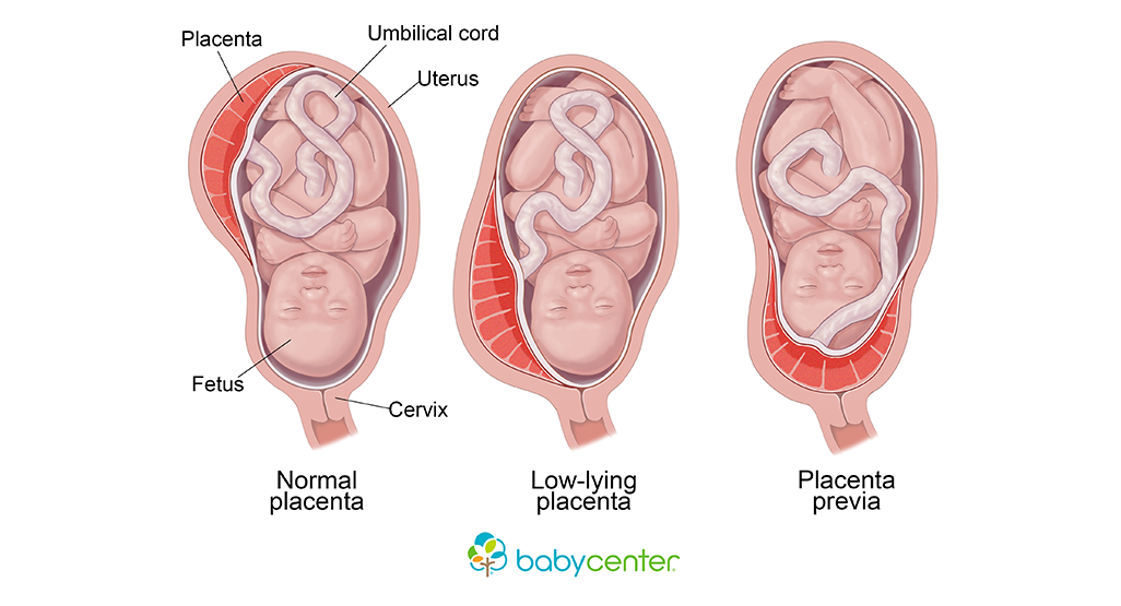
– Condition where the placenta implants at the bottom of the uterus, covering the cervix
– Painless vaginal bleeding after 20 weeks of pregnancy
– 1 in every 200 pregnancies affected by placenta previa
– Other causes of vaginal bleeding during pregnancy
– Thinning and spreading of the bottom part of the uterus can cause bleeding if the placenta is anchored to the bottom
– Sexual intercourse can cause bleeding
– Complications of placenta previa: major bleeding, shock, fetal distress, premature labor or delivery, health risks to the baby, emergency cesarean delivery, hysterectomy, blood loss for the baby, and death
– Causes and risk factors for placenta previa: low implantation of the fertilized egg, abnormalities of the uterine lining, scarring of the uterine lining, abnormalities of the placenta, and multiple pregnancies
– Tests used to diagnose placenta previa: ultrasound scans, feeling the mother’s belly
– Differentiating between placenta previa and placental abruption
– Life-threatening condition for both the mother and baby
– Diagnosis involves ultrasound scan and gentle speculum vaginal examination
– Treatment options depend on factors: type and location of the placenta, amount of blood loss, gestational age of the baby, and the health of both the baby and mother
– Medical treatment during pregnancy: bed rest, hospitalization, close monitoring, blood transfusion, avoiding activities that trigger contractions
– Delivery usually done through a caesarean section
– Postpartum monitoring for complications such as postpartum bleeding
– Baby closely monitored for health issues related to prematurity or lack of oxygen during delivery
– Prompt medical attention needed if experiencing vaginal bleeding during pregnancy
– Treatment options depend on factors such as the type and location of the placenta, amount of blood lost, gestational age of the baby, and the health of both the baby and mother
– Treatment during pregnancy may include bed rest, hospitalization, close monitoring, blood transfusion, and avoiding activities that trigger contractions
– Caesarean section usually performed once the baby is old enough
– Tests to check the mother’s blood cell counts and clotting ability needed.

Continue Reading

The Shocking Truth: Premature Separation of Normally Implanted Placenta

– Placental abruption is a condition in which the placenta separates from the wall of the uterus before birth.
– It can happen partially or completely, leading to a lack of oxygen and nutrients for the baby.
– Symptoms include vaginal bleeding, pain, contractions, discomfort, and tenderness.
– It occurs in about 1 in 100 pregnant individuals.
– Placental abruption is more likely to occur in the third trimester, but can happen after 20 weeks of pregnancy.
– Mild cases can lead to complete separation and may require close monitoring.
– Placental abruption is related to about 1 in 10 premature births.
– Premature babies are at higher risk for health problems, disabilities, and death.
– Placental abruption can cause anemia and life-threatening complications for the pregnant individual and the baby.
– Immediate diagnosis and treatment are necessary to prevent hemorrhage and blood clotting complications.
– Delivery by cesarean birth may be required.
– The causes of placental abruption may include previous abruption, high blood pressure, smoking, cocaine use, physical trauma, age 35 or older, infection in the uterus, preterm labor, early water breaking, issues with the uterus or umbilical cord, excess fluid around the baby, carrying multiples, asthma, family history of abruption, previous c-section, and exposure to air pollution.
– If a person has previously experienced placental abruption, they have a 10% chance of it occurring again in a later pregnancy.
– Measures to reduce the risk of placental abruption include closely monitoring and treating high blood pressure, avoiding smoking or using street drugs, always wearing a seatbelt when in a car, and taking prenatal vitamins with folic acid.

Continue Reading