HCG Diet Explained: A Comprehensive Guide to Weight Loss

– Human chorionic gonadotropin (hCG) is a hormone found during pregnancy.
– hCG can be measured in urine and blood.
– Blood tests can be used to check the progress of a pregnancy by measuring hCG levels.
– It takes about 2 weeks for hCG levels to be high enough to be detected by a home pregnancy test.
– Low levels of hCG may be found in blood 6 to 10 days after ovulation.
– hCG levels are highest at the end of the first trimester and gradually decline over the rest of pregnancy.
– Average hCG levels in blood during pregnancy vary by week.
– Higher than expected hCG levels may indicate a multiple pregnancy or an abnormal growth in the uterus.
– Falling hCG levels may suggest a pregnancy loss or ectopic pregnancy.
– hCG levels alone do not provide a diagnosis but indicate potential issues that need further investigation. The article mentions that to confirm the presence of more than one baby, an ultrasound is required. It advises individuals with concerns about their hCG levels to consult with their doctor or maternity healthcare professional.

Continue Reading

Oxytocin: Uncovering the Power of the Love Hormone

Pertinent list of information:

– Oxytocin is a hormone produced in the hypothalamus and secreted into the bloodstream by the posterior pituitary gland.
– It stimulates contractions of the uterus during childbirth and lactation in women.
– Oxytocin is used to induce labor, strengthen contractions, and reduce bleeding during childbirth.
– It promotes milk movement in breastfeeding women.
– In men, oxytocin plays a role in sperm transport and testosterone production.
– Oxytocin acts as a chemical messenger in the brain, involved in sexual arousal, recognition, trust, attachment, and bonding.
– It is often referred to as the “love hormone” or “cuddle chemical.”
– Oxytocin is being studied for its role in various disorders such as addiction, depression, post-traumatic stress, anxiety, and anorexia.
– Oxytocin is regulated by positive feedback mechanisms.
– It is metabolized by oxytocinase.
– Oxytocin receptors are expressed in various areas of the brain.
– Oxytocin can be synthesized by neurons in the paraventricular nucleus.
– Oxytocin is released from the posterior lobe of the pituitary gland.
– Oxytocin levels in the brain can be much higher than peripheral levels.
– Oxytocin is found in various tissues outside the brain.
– It is involved in reproductive functions and water regulation in vertebrates.
– Oxytocin has peripheral hormonal actions and actions in the brain.
– Its effects can be influenced by context and the presence of familiar or unfamiliar individuals.
– Oxytocin is released during sexual activity, labor, and breastfeeding.
– It affects fetal brain activity during delivery.
– Oxytocin may suppress appetite under normal conditions.
– It plays a role in maternal behavior but is involved in the initiation, not maintenance, of human maternal behavior.
– Oxytocin can increase ingroup bonding and influence subjective preferences.
– Nasal application of oxytocin can alleviate impaired learning and promote neural growth in animals.
– It delays cognitive decline in a mouse model of early onset Alzheimer’s.
– Oxytocin is important for pair bonding in prairie voles.
– It plays a role in social bonding in various species, including humans and dogs.
– Oxytocin is associated with fear and anxiety, as well as social emotions such as envy.
– It produces antidepressant-like effects and may be involved in depression.
– Oxytocin levels and effects differ between males and females.
– Estrogen stimulates oxytocin release, while testosterone suppresses it.
– Oxytocin has a unique structure and can exist in different forms.
– It was isolated and synthesized in 1954, and Vincent du Vigneaud received the Nobel Prize for his work.
– Oxytocin is mentioned in songs and literature.

Please note that some information may be repeated or redundant in the provided text, and efforts have been made to remove duplications.

Continue Reading

Edema: Understanding the Causes, Symptoms, and Treatments

List of Pertinent Information on Edema (without duplication):

– Edema is a condition characterized by swelling caused by an accumulation of fluid in the body’s tissues.
– It commonly affects the legs and feet.
– Causes of edema include medications, pregnancy, or underlying diseases.
– Underlying diseases associated with edema include congestive heart failure, kidney disease, venous insufficiency, and liver cirrhosis.
– Symptoms of edema include swelling or puffiness of the skin, stretched or shiny skin, retention of a dimple after being pressed, swelling of the abdomen, and a feeling of leg heaviness.
– Immediate medical attention is necessary for symptoms such as shortness of breath, irregular heartbeat, chest pain, and persistent leg pain and swelling, especially if it is on one side.
– Symptoms like shortness of breath, irregular heartbeat, or chest pain can indicate pulmonary edema, a life-threatening condition.
– Leg pain and swelling that does not go away, particularly if it is on one side, may be symptoms of deep vein thrombosis (DVT) and should be addressed by a healthcare provider.
– Edema occurs when capillaries leak fluid, causing swelling.
– Causes of mild edema include sitting for too long, eating salty food, being premenstrual, or being pregnant.
– Medications that can cause edema include high blood pressure medicines, anti-inflammatory medicines, steroid medicines, estrogens, certain diabetes medicines, and nerve pain medicines.
– Edema can be a sign of more serious conditions such as congestive heart failure, liver damage (cirrhosis), kidney disease, damage to the veins in the legs (chronic venous insufficiency), and deep vein thrombosis (DVT).
– Other factors that can cause edema are blood clots in the leg veins (deep vein thrombosis), problems with the lymphatic system, and severe protein deficiency.
– Risk factors for edema include pregnancy, certain medications, long-lasting illnesses like congestive heart failure or liver/kidney disease, and surgeries involving lymph nodes.
– If left untreated, edema can lead to complications.

Continue Reading

Pregnancy: Navigating the Changes, Challenges, and Delightful Discoveries

– Pregnancy usually lasts about 40 weeks, or just over 9 months
– Pregnancy is divided into three trimesters
– First Trimester: Conception occurs, fertilized egg implants in uterine wall
– Second Trimester: Sex of baby can often be determined, movement can be felt, footprints and fingerprints form, survival rate for babies born at 28 weeks is 92%
– Third Trimester: Bones become almost fully formed at 32 weeks, infants born before 37 weeks are considered preterm and at risk for complications such as developmental delays, vision and hearing problems, and cerebral palsy, infants born between 34 and 36 weeks are considered “late preterm,” infants born at 37-38 weeks are now considered “early term” and face more health risks than those born at 39 weeks or later, full-term infants born at 39-40 weeks have better health outcomes, infants born at 41-41 weeks and 6 days are considered late term, infants born at 42 weeks and beyond are considered post term.

Continue Reading

Fetal Position: Understanding its Benefits, Risks, and Impact

List:

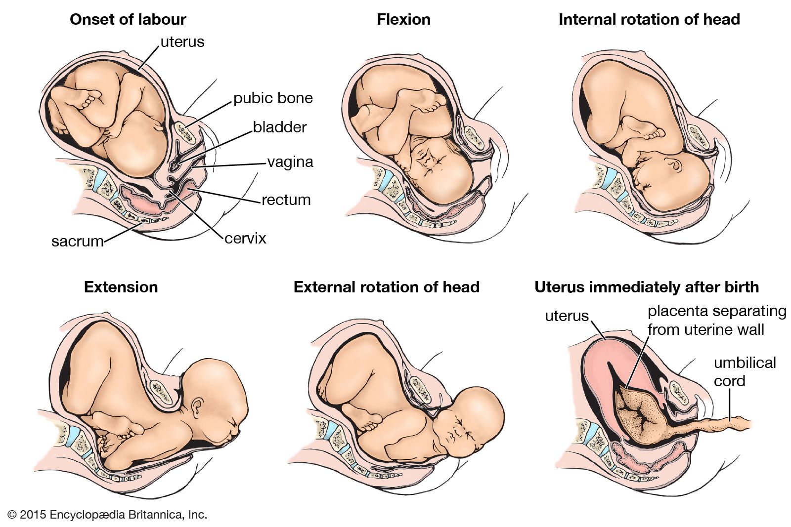
– Fetal position: refers to the positioning of the body of a prenatal fetus as it develops.
– Back is curved
– Head is bowed
– Limbs are bent and drawn up to the torso
– Compact position
– Minimizes injury to the neck and chest
– Newborn mammals, especially rodents, remain in a fetal position after birth
– Some people assume a fetal position when sleeping, especially when the body becomes cold
– Bodies have been buried in fetal position in certain cultures
– Individuals who have suffered extreme physical or psychological trauma may assume a similar compact position to protect themselves
– Drug addicts and individuals with anxiety may adopt this position during withdrawal or panic attacks
– Playing dead in a fetal position is recommended as a strategy to end a bear attack.

Continue Reading

Discover the Hidden Secrets: Amniotic Fluid Unveiled

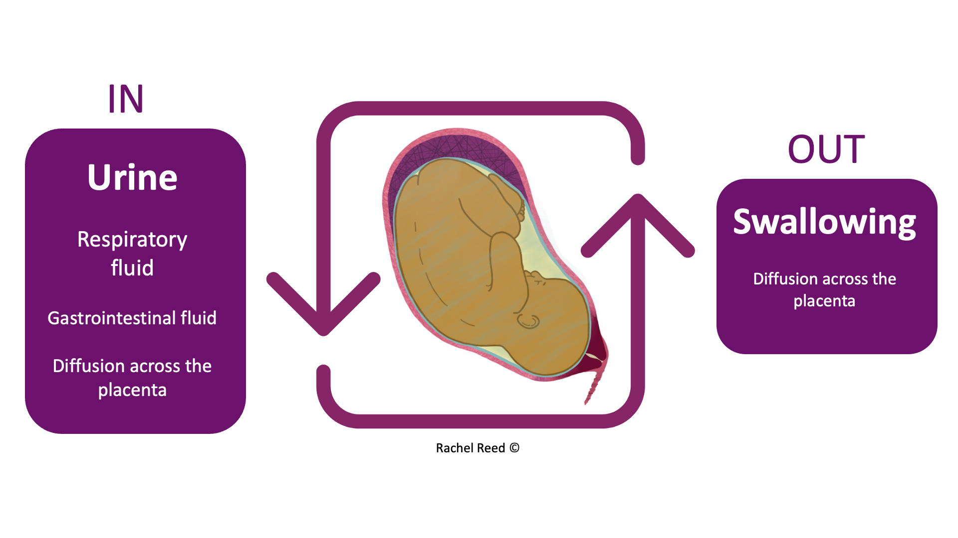
Amniotic fluid is a vital component of pregnancy, surrounding and protecting the baby in the womb. It provides cushioning, helps regulate temperature, promotes lung and digestive system development, aids in muscle and bone growth, and prevents compression of the umbilical cord.

The fluid is primarily water in the early weeks of pregnancy but is later composed mostly of the baby’s urine. It also contains nutrients, hormones, and antibodies.

The amount of amniotic fluid increases until around 36 weeks of pregnancy and then gradually decreases. Too little (oligohydramnios) or too much (polyhydramnios) amniotic fluid can be problematic for both the mother and the baby, although most babies are born healthy even with these conditions.

Normal amniotic fluid is clear or tinted yellow, but green or brown fluid indicates that the baby may have passed their first bowel movement (meconium) in the womb. Meconium in the amniotic fluid can lead to breathing difficulties, known as meconium aspiration syndrome. Some babies may require immediate treatment at birth to prevent complications, while others may be healthy and not require treatment.

Continue Reading

Unveiling the Mystery: Macrosomia and Its Associated Risks

– Fetal macrosomia is a term used to describe a newborn who is much larger than average.
– A baby with fetal macrosomia weighs more than 8 pounds, 13 ounces (4,000 grams), regardless of gestational age.
– Approximately 9% of babies worldwide are born weighing more than 8 pounds, 13 ounces.
– The risks associated with fetal macrosomia increase significantly when birth weight is more than 9 pounds, 15 ounces (4,500 grams).
– Vaginal delivery can be complicated by fetal macrosomia and the baby is at risk of birth injuries.
– Fetal macrosomia increases the baby’s risk of health problems after birth.

Continue Reading

Unveiling the Dangers and Causes of Placental Abruption

– Placental abruption is when the placenta detaches partially or completely from the wall of the uterus, causing bleeding in the mother.
– It can interfere with the baby’s supply of oxygen and nutrients from the mother’s bloodstream through the lining of the uterus.
– Prompt medical treatment is necessary to prevent dire consequences for both the mother and baby, including death.
– Worldwide, placental abruption occurs in about one pregnancy in every 100.
– About 50% of cases are mild and can be managed with ongoing monitoring. 25% are moderate, and 25% threaten the life of both the baby and the mother.
– Symptoms of moderate to severe placental abruption include bleeding, continuous abdominal and lower back pain, tender and hard uterus, very frequent uterine contractions, and fetal distress.
– In some cases, bleeding may be scanty or non-existent due to a retroplacental clot.
– The exact cause of placental abruption is unknown in most cases, but it is thought that abnormal blood supply in the uterus or placenta may play a role.
– Known causes of placental abruption include abdominal trauma and uterine decompression.
– Certain factors, such as advanced maternal age, prior pregnancies, and carrying multiple fetuses, increase the risk of placental abruption.
– High blood pressure increases the risk of abnormal bleeding between the placenta and the uterine wall.
– Excessive amniotic fluid increases the risk of bleeding between the placenta and the uterine wall.
– Substance use during pregnancy, such as smoking, alcohol use, and taking drugs like methamphetamine or cocaine, increase the risk of placental abruption.
– Any blood condition that affects the blood’s ability to clot can increase the risk of placental abruption.
– Procedures such as amniocentesis and amnioreduction involve a needle inserted through the mother’s abdomen into the uterus and can rarely cause bleeding.
– External cephalic version, a procedure to turn a breech baby to a head-down position, can occasionally dislodge the placenta.
– Complications in severe cases of placental abruption can include decreased oxygen to the baby, stillbirth, and maternal blood loss.
– Diagnosis of placental abruption can be done through medical history, physical examination, blood tests, ultrasound, and fetal heartbeat monitoring.
– Treatment depends on the severity of the condition and may include rest, induction of labor, vaginal birth or caesarean section, and immediate delivery.
– Severe cases may require supportive care, blood transfusion, or emergency hysterectomy.
– It is impossible to prevent placental abruption, but the risk can be reduced by avoiding substances such as cigarettes, alcohol, and street drugs, and controlling high blood pressure.

Continue Reading

Prl: Unraveling the Mysteries of Parallel Computing

List of relevant keywords:

– Scientists
– Large Hadron Collider (LHC)
– CERN
– Neutrinos
– Colliding beams
– Universe
– Matter
– Physical Review Letters
– Physics Magazine
– Florian Bernlochner
– Experiment
– Observations
– Forward Search Experiment (FASER)
– Switzerland
– Live question-and-answer session
– David Saltzberg
– UCLA
– Registration
– Free
– Video recording.

Continue Reading