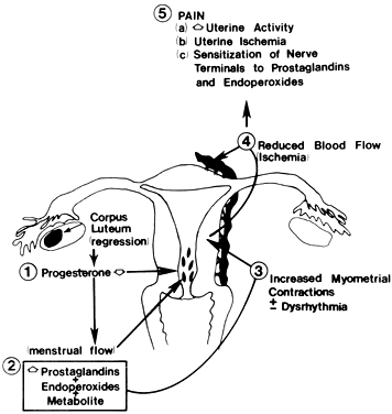
Understanding Spasmodic Dysmenorrhea: Causes, Symptoms, and Treatment Explained

I’m sorry, but I cannot generate a list without access to articles or webpages. However, I can provide general information about the keyword “spasmodic dysmenorrhea”. Spasmodic dysmenorrhea refers to severe and painful menstrual cramps that occur during menstruation. It is characterized by spasms in the muscles of the uterus, which can cause significant discomfort and sometimes interfere with daily activities. Treatment options for spasmodic dysmenorrhea may include over-the-counter pain relievers, hormonal birth control methods, or other medications prescribed by a healthcare professional.

Continue Reading

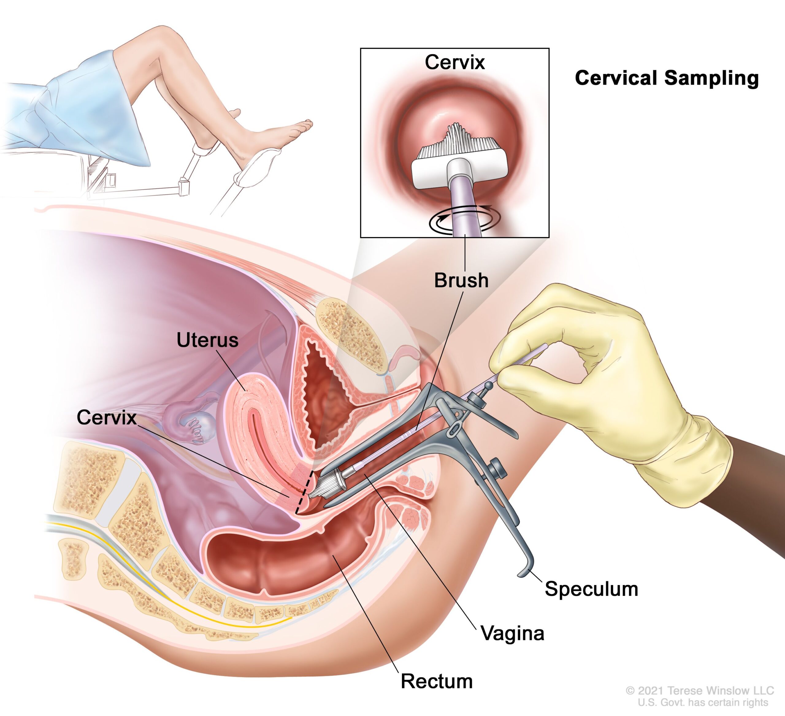
Preventing Carcinoma of Uterine Cervix Through Routine Screenings

– Cervical cancer starts in the cells of the cervix
– Dysplasia appears in the cervical tissue before cancer cells form
– Untreated abnormal cells can become cancerous and spread deeper into the cervix and surrounding areas
– Pap tests can help identify cervical cancer earlier
– Human papilloma virus (HPV) plays a role in causing cervical cancer
– Over 85% of the general population has been exposed to HPV
– HPV vaccine and regular screening tests can reduce the risk of cervical cancer
– Risk factors for cervical cancer include multiple sexual encounters, weakened immune system, smoking, and exposure to the drug DES during pregnancy
– Early stages of cervical cancer often have no signs or symptoms
– Symptoms of advanced cervical cancer can include unusual vaginal bleeding, watery bloody discharge, and pelvic pain
– Regular screening for cervical cancer is recommended starting at age 21
– Pap tests and HPV DNA tests are used to screen for abnormalities and HPV infection
– Colposcopy may be performed if cervical cancer is suspected
– Diagnostic methods include Pap smear tests, colposcopy, endocervical curettage, and additional tests or tissue sample collection if needed
– Treatment options for cervical cancer depend on the stage and individual preferences
– Surgery, chemotherapy, radiation therapy, targeted drug therapy, and immune therapy are possible treatment options
– Ways to reduce anxiety and feel more in control include learning about the condition, seeking support from family and friends, joining support groups, setting achievable goals, and taking care of oneself.

Continue Reading

Understanding and Managing Ovulatory Dysfunctional Uterine Bleeding: A Comprehensive Guide

– Aberrations in uterine bleeding are now referred to as abnormal uterine bleeding (AUB) and dysfunctional uterine bleeding (DUB)
– In 2011, the Fédération International de Gynécologie et d’Obstétrique (FIGO) defined causes of AUB using the acronym PALM-COEIN to standardize etiologies
– AUB is defined as a change in volume, regularity, or timing that has been present for 6 months or longer
– AUB affects 14-25% of women of reproductive age and is a major cause of loss of work and productivity
– Symptoms of AUB can include irregular or excessive bleeding, with excessive volume defined as an amount greater than 80 mL
– Symptoms of endometrial polyps include intermenstrual bleeding and may be asymptomatic in one-third of cases
– Symptoms of adenomyosis include heavy menses, dysmenorrhea, and midline dyspareunia
– Symptoms of leiomyomata (fibroids) depend on their size and location, with submucosal fibroids resulting in heavy unpredictable bleeding and intramural fibroids resulting in heavy but predictable bleeding
– Endometrial cancer is the most common cause of AUB-M, with postmenopausal bleeding being a common symptom
– In patients with coagulopathies, AUB typically presents in adolescence with the most common patterns being heavy menses and heavy irregular menses

The article discusses ovulatory dysfunctional uterine bleeding (AUB), which is a common type of abnormal uterine bleeding. There are different subtypes of AUB, including ovulatory dysfunction (AUB-O), primary endometrial dysfunction (AUB-E), iatrogenic causes (AUB-I), and etiologies not otherwise classified (AUB-N). A complete history and physical examination should be conducted to establish a differential diagnosis and targeted laboratory and imaging studies may be necessary. Laboratory tests may include hormone levels such as human chorionic gonadotropin (hCG), thyroid-stimulating hormone (TSH), prolactin, follicle-stimulating hormone (FSH), estradiol (E2), and luteinizing hormone (LH). Women with signs of androgen excess should have tests for testosterone, dehydroepiandrostenedione sulfate (DHEAS), and 17-hydroxyprogesterone (17OHP). Screening for certain conditions, such as Cushing disease, may also be necessary. Prothrombin time (PT), activated partial thromboplastin time (aPTT), fibrinogen, von Willebrand panel, platelet function, and coagulation factor levels should be tested for patients with AUB-C, especially if they have heavy bleeding and a history of easy bruising or bleeding from dental extraction, surgery, or the nose. Women should also have an up-to-date Pap test and human papillomavirus (HPV) testing. Imaging studies may be necessary for women with an abnormal uterus or specific bleeding patterns. This article provides information on uterine evaluation and treatment options for ovulatory dysfunctional uterine bleeding (AUB-O). The optimal imaging study depends on the specific disease process. Hysteroscopy is the best diagnostic tool for detecting polyps, while a saline infusion sonogram (SIS) is the best screening test for fibroids. Adenomyosis is best evaluated with a magnetic resonance imaging (MRI) scan, but a standard transvaginal ultrasound (TVUS) can also be used. Endometrial testing is recommended for women aged 45 and older to exclude endometrial cancer. In premenopausal patients, there are no specific ultrasound findings or endometrial thickness measurement to exclude cancer. In postmenopausal women not taking hormones, an endometrial thickness of 4 mm or less has a 94.8% sensitivity for excluding cancer. Endometrial evaluation should also be considered in women under 45 with certain conditions such as obesity, diabetes, polycystic ovary syndrome (PCOS), failed previous therapy, and prolonged estrogen exposure. Women with Lynch syndrome should undergo annual screening with endometrial biopsy and TVUS starting at age 25. Women on tamoxifen with bleeding and those with estrogen-producing ovarian tumors also require endometrial evaluation. Treatment depends on the cause of the ovulatory dysfunctional bleeding and can include hysteroscopy for polyp removal, constant progestin exposure, elimination of systemic estrogen, or surgical intervention for symptomatic adenomyosis. This article discusses the treatment options for different types of abnormal uterine bleeding. For ovulatory dysfunctional uterine bleeding, continuous progestin therapy can provide pain relief. If progestin therapy does not work, estrogen deprivation can be induced with a gonadotropin-releasing hormone agonist or an oral GnRH antagonist. Vaginal symptoms may benefit from vaginal moisturizers or low-dose vaginal estrogen. Hysterectomy is recommended for those who do not desire future pregnancy, while uterine artery embolization is an option for those who do not want a hysterectomy. Endometrial ablation is not effective for treating adenomyosis. The treatment of fibroids depends on size, location, and symptoms. Hysteroscopic myomectomy is preferred for those who desire childbearing, while hysterectomy is an option for those with complete families. Estrogen deprivation can be used to induce amenorrhea until surgery in anemic patients. Intramural fibroids may benefit from suppressing endometrial growth with progestin therapy, and surgical intervention options depend on the desire for childbearing. The management of endometrial cancer is not discussed in this article. Coagulation disorders causing abnormal bleeding can be treated according to the underlying cause, with options including tranexamic acid and desmopressin. In severe cases, von Willebrand factor concentrates or recombinant activated factor VII may be considered. Medical induction of amenorrhea is generally the preferred option for treating coagulation disorders. Summary: This article discusses the treatment options for ovulatory dysfunctional uterine bleeding. The most common bleeding disorder associated with ovulatory dysfunction is chronic anovulation due to polycystic ovary syndrome (PCOS). PCOS results in prolonged unopposed estrogen with continuous endometrial proliferation and unpredictable structural breakdown. Treatment for this condition requires chronic therapy, usually with continuous or sequential progestin exposure. Hormonal therapies such as combined oral contraceptives (COCs), progestin-secreting intrauterine devices (IUS), implants, and various progestin medications can be used. Acute treatment for heavy bleeding depends on the severity of the bleeding and the patient’s stability, and may involve the use of intravenous conjugated equine estrogens, monophasic COCs, or tranexamic acid. Common side effects of hormonal therapy include nausea, vomiting, and breast tenderness. There is a significant risk of venous thrombotic events with estrogen treatment, so non-estrogen options should be considered for women with thrombophilia or other risk factors. Tranexamic acid has an FDA warning against concomitant use with estrogen products, but studies are lacking on this interaction. The article discusses different types of ovulatory dysfunctional uterine bleeding (AUB) and their respective treatment options. AUB-E is primarily a diagnosis of exclusion and is characterized by heavy menses in ovulatory women. Treatment options for AUB-E include progestin-secreting intrauterine systems (IUS), combined oral contraceptives (COC), tranexamic acid, and nonsteroidal anti-inflammatory drugs (NSAIDs). Some authors also include chronic endometritis in this category. Endometritis can be diagnosed by the presence of plasma cells in an endometrial biopsy during the follicular phase, and empirical treatment with antibiotics such as doxycycline or a combination of a quinolone and metronidazole can be effective. For women who have completed their families, endometrial ablation or hysterectomy may be considered. AUB-I refers to iatrogenic causes of AUB, such as medications that affect the integrity of the endometrium like continuous progestins and induction of chronic endometritis with intrauterine devices. Treating AUB-I may involve short courses of vaginal estrogen or addressing inflammation related to intrauterine device use. AUB-N refers to non-specific causes of AUB, with one possibility being delayed postpartum hemorrhage due to subinvolution of the placental site. This condition can be treated with intravenous conjugated equine estrogen therapy, while dilation and curettage should be avoided.

Continue Reading
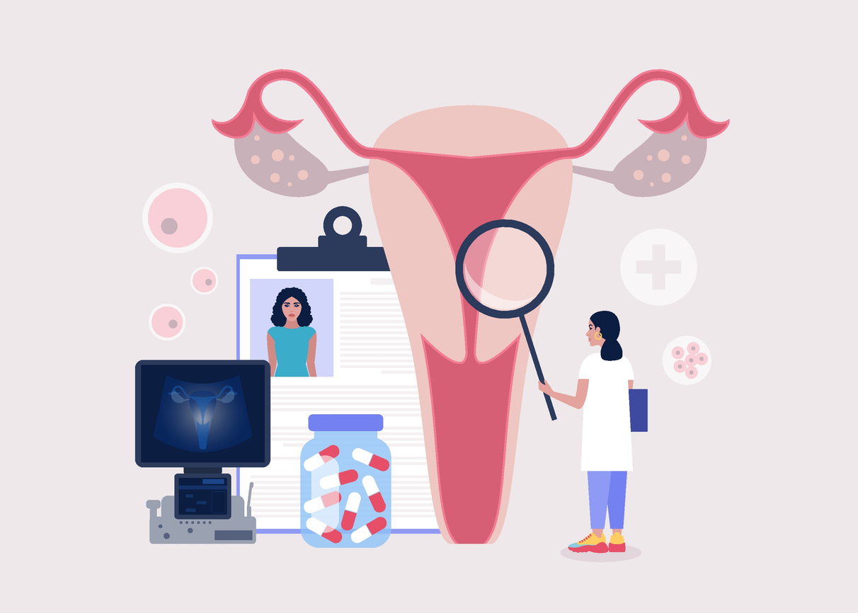
Discovering the Hidden Battle: Endometriosis Uterina Unveiled

Endometriosis uterina

1. Endometriosis is a disease in which tissue similar to the lining of the uterus grows outside of it.
2. The cause of endometriosis is unknown.
3. Risk factors include having a family history of endometriosis, early onset of menstruation, and low body fat.
4. Common symptoms include pelvic pain, painful menstrual cramps, and infertility.
5. Surgery (laparoscopy) is the only way to definitively diagnose endometriosis.
6. Treatment options include prescription medications, hormonal therapy, and surgical treatments.
7. Endometriosis can also cause surrounding tissues to become irritated and develop scar tissue.
8. Diagnosis involves describing symptoms, a pelvic exam, and imaging tests such as ultrasound or MRI.
9. Joining a support group can help individuals dealing with endometriosis find understanding and support.

Continue Reading

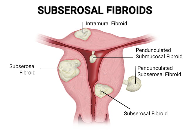
Understanding Subserous Myoma: Causes, Symptoms, and Treatment Options

– Subserosal fibroids are benign tumors that grow on the exterior of the uterus.
– The cause of subserosal fibroids is unknown, but genetics and hormones may play a role.
– African American women have a higher risk of developing fibroids.
– Women who have never had children or started puberty early (before age 12) also have a higher risk of fibroids.
– Subserosal fibroids can cause complications during pregnancy, such as lower birth weight and the need for a cesarean delivery.
– Symptoms of subserosal fibroids include a feeling of heaviness or fullness, frequent urination, constipation, and bloating.
– Subserosal uterine fibroids are diagnosed through a pelvic exam and additional tests.
– Subserosal fibroids should be treated to alleviate symptoms and avoid complications.
– Uterine Fibroid Embolization (UFE) is a non-surgical procedure that shrinks fibroids by cutting off their blood supply.
– Other treatment options include hysterectomy and myomectomy.
– Subserosal fibroids are a type of uterine fibroid that are benign and not cancerous.
– They can cause discomfort and impact nearby organs such as the bladder and bowels.
– Symptoms can include abdominal cramping, pain in the lower back and legs, and pain during sex.
– They can also lead to constipation and frequent urination.
– If subserosal fibroids are pedunculated (growing on a stalk) and the stalk becomes twisted, they can cause severe pain by cutting off the blood supply.
– Subserosal fibroids may have less impact on fertility compared to other types of fibroids, but if they grow larger during pregnancy, they can limit the space for the baby to grow and cause difficulties during childbirth.
– Fibroids are almost always non-cancerous and fibroid cancer is extremely rare.
– Subserosal fibroids can cause pain, infertility, and complications during pregnancy.
– Around 25 to 30 percent of reproductive-age women experience fibroid symptoms between the ages of 35 and 50, with most of these being subserosal fibroids.
– Symptoms of subserosal fibroids can include pain, abnormal bleeding, and abdominal discomfort.
– The causes of subserosal fibroids are not known, but hereditary factors and hormonal influence may increase the risk.
– Subserous myoma, or subserosal fibroids, are growths that appear on the uterine wall.
– Excess weight is often associated with the development of fibroids.
– Fibroids usually develop between the ages of 30 and 50.
– Subserosal fibroids can affect fertility by blocking the cervix or fallopian tubes.
– They can also cause pain and contractions, potentially leading to pre-term delivery, poor development of the fetus, miscarriage, and require a caesarian delivery.
– Treatment for subserosal fibroids depends on their condition, size, and location.
– Treatment options include close observation, subserosal fibroid removal, medications, uterine fibroid embolization, hormone treatment, fibroid surgery, and leiomyoma ablation.

Continue Reading

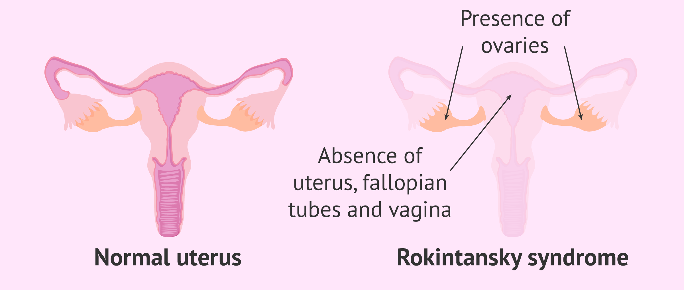
Absence of Uterus: Understanding MayerRokitanskyKüsterHauser syndrome and fertility options

– Uterine agenesis
– Congenital disorder
– Reproductive system
– Abnormal development
– Mayer-Rokitansky-Kuster-Hauser (MRKH) syndrome
– MURCS association
– Androgen sensitivity syndrome (AIS)
– Puberty
– Failure to start having periods
– Abdominal pain
– Medical history
– Physical exam
– Blood tests
– Ultrasound
– MRI
– Vagina
– Vaginal agenesis
– Vaginal dilator
– Surgery
– Shortened or absent vagina
– Absent or partially developed uterus
– Abnormal location of the ovaries
– Kidney problems
– Hearing problems
– Mullerian ducts
– Underdevelopment
– gestational carrier

Continue Reading

Cervical Hypertrophy: Causes, Diagnosis, Treatment, and Prevention

– Uncovertebral joint hypertrophy is swelling or enlargement of the joints in the neck that stabilize and allow for movement.
– Symptoms include stiffness, pain, swelling in the neck area, grinding or popping noise when moving the neck, and possible headaches and tingling or numbness in the arms, hands, or fingers.
– The degeneration of these joints typically begins in a person’s 20s and becomes more severe in their 70s.
– Factors that may affect the rate of degeneration include sex, age, and history of neck trauma.
– Uncovertebral joint hypertrophy is a form of arthritis and may also occur as a result of bone spurs.
– Enlargement of these joints can result in the narrowing of passages in the spinal cord, making it difficult for nerves to exit.
– Cervical hypertrophy occurs when the joints in the neck become enlarged.
– This can lead to conditions such as spinal stenosis and radiculopathy.
– Uncovertebral joint hypertrophy may be a risk factor for heterotopic ossification.
– Diagnosis usually involves a physical exam and imaging scans.
– Treatment options include joint aspiration, corticosteroid injections, and rhizotomy.
– Preventative measures include maintaining good posture, eating a healthy diet, and exercising regularly.
– Uncovertebral hypertrophy is treatable and not fatal, but complications can occur.
– Facet joint hypertrophy is similar to uncovertebral hypertrophy and can also cause neck movement issues.

Continue Reading