Epithelial Tumour of Ovary: Understanding Diagnosis, Treatment, and Prognosis

List pertinent to the keyword ‘epithelial tumour of ovary’:

1. Epithelial ovarian tumors are classified as benign, borderline, or malignant.
2. Benign tumors are non-cancerous.
3. Borderline tumors may become harmful if allowed to grow.
4. Malignant tumors, called carcinomas, are invasive cancers that can spread to other areas of the body.
5. The four main types of epithelial ovarian cancer are serous carcinoma, mucinous carcinoma, endometrioid carcinoma, and clear cell carcinoma.
6. Serous carcinomas are the most common, accounting for 52% of cases.
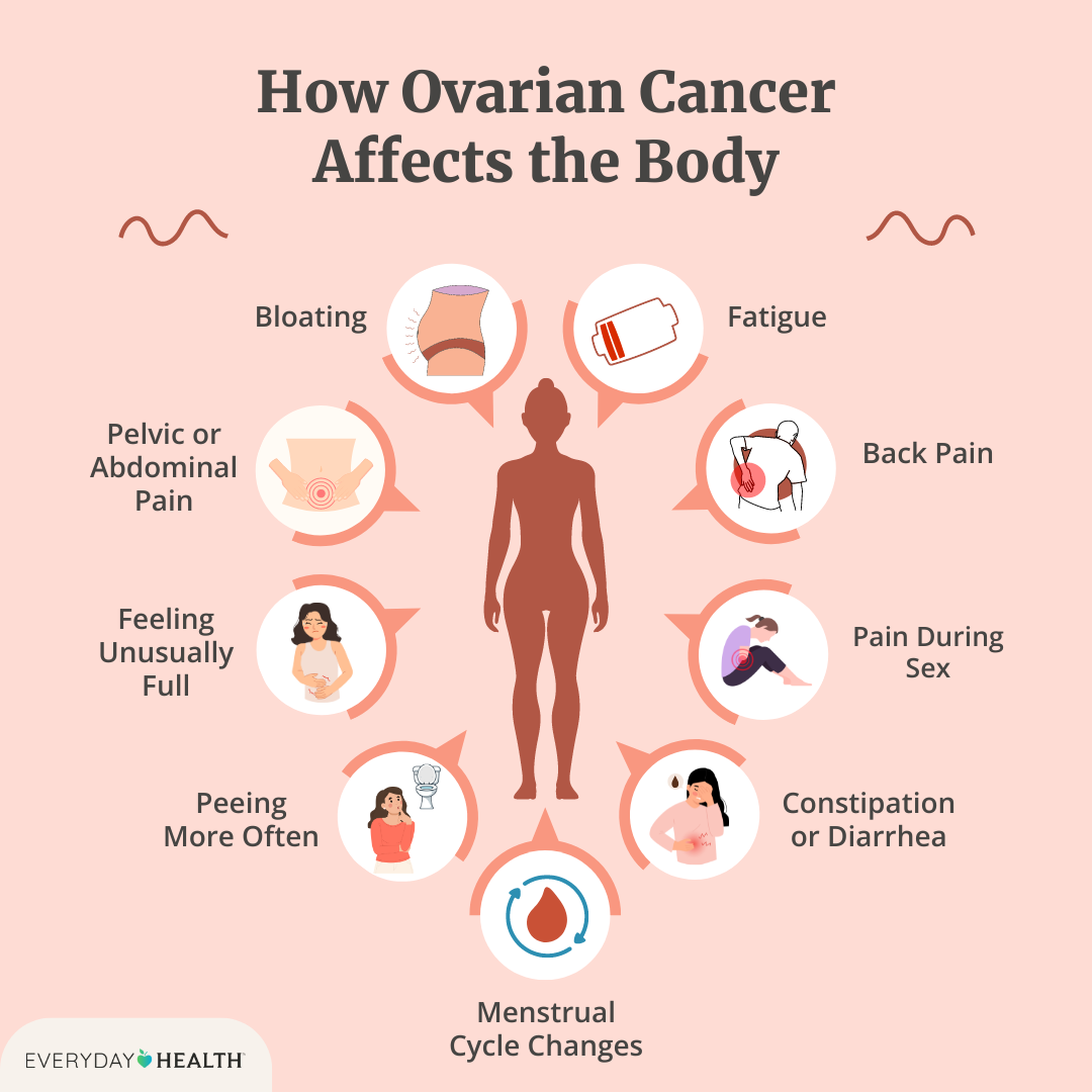
7. Symptoms of epithelial ovarian cancer include feeling full quickly after eating, loss of appetite, bloating, abdominal and pelvic pain, ascites, urinary symptoms, vaginal bleeding, and digestive issues.
8. Gene mutations, such as BRCA1 and BRCA2, can increase the risk of ovarian cancer.
9. Around 20-25% of ovarian cancers are inherited.
10. People with a personal or family history of breast cancer have an increased risk of ovarian cancer.
11. Common risk factors for ovarian cancer include age, family history of ovarian cancer, breast cancer, or colorectal cancer.
12. Epithelial ovarian cancer cannot be detected by one specific test.
13. CA-125 blood test may be used to detect signs of ovarian cancer.
14. Imaging tests such as transvaginal ultrasound, computed tomography, or magnetic resonance imaging may be requested to examine reproductive organs.
15. Laparoscopy may be used as a surgical diagnostic test to examine reproductive organs and take tissue samples for biopsy.
16. A biopsy is the only way to determine if a mass is cancerous or benign.
17. Ascites formation in the abdomen may help in diagnosing ovarian cancer through a procedure called paracentesis.
18. Treatment for ovarian cancer is personalized based on individual needs and goals.
19. There are four stages of ovarian cancer.
20. Surgery is performed to remove as much of the cancer as possible.
21. Chemotherapy is commonly used in the treatment of epithelial ovarian cancer.
22. Targeted therapy, which attacks proteins in cells, is sometimes used in combination with surgery and chemotherapy.
23. Hormone therapy is rarely used for epithelial ovarian cancer.
24. Palliative care aims to help manage emotional stress, medication side effects, pain, and symptoms that affect the patient’s quality of life.

Continue Reading

Combined Chemotherapy: Maximizing Treatment Effectiveness for Cancer Patients

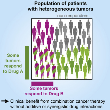
– Combination chemotherapy is the use of more than one medication at a time to treat cancer.
– Using a combination of drugs increases the chance of eliminating all cancer cells, but it also increases the risk of drug interactions.
– Combination chemotherapy was inspired by the approach to treating tuberculosis using a combination of antibiotics.
– Combination chemotherapy has been found to be more effective for treating certain cancers such as acute lymphocytic leukemia and Hodgkin’s lymphoma.
– Combination chemotherapy is more effective than single drug therapy and sequential chemotherapy.
– Targeted therapies, which block specific pathways in cancer cells, have been used in combination with chemotherapy.
– Combination chemotherapy is used for various types of solid tumors, such as non-small cell lung cancer and breast cancer.
– Combination chemotherapy is also used for certain leukemias and Hodgkin lymphoma.
– Chemotherapy used in combination with immunotherapy can enhance the effectiveness of both treatments.
– Immunotherapy drugs help the immune system recognize and attack cancer cells.
– The abscopal effect, where chemotherapy helps the immune system target abnormal cells, can occur when chemotherapy is combined with immunotherapy.
– Combination chemotherapy has increased survival rates for many diseases.
– Combination chemotherapy involves using multiple chemotherapy medications at the same time.
– There are several advantages to using combination chemotherapy, including decreased resistance of tumors to treatment, earlier administration of medications, targeting multiple processes in cancer growth, increased effectiveness, lower doses of medications, and synergetic effects of drug combinations.
– Combination chemotherapy has been found to improve survival or result in a better response to treatment, especially as adjuvant treatment after surgery.
– However, there are also disadvantages to combination chemotherapy, including an increased risk of side effects.
– The article discusses the use of multiple chemotherapy drugs in combination treatment.
– It states that when multiple drugs are used, side effects of both drugs may compound.
– For example, if two drugs that cause a low white blood cell count are used, the risk of chemotherapy-induced neutropenia is increased.
– If a person develops a side effect while taking multiple medications, it may be difficult to determine which medication is responsible.
– In these cases, all medications may need to be discontinued if the side effect is serious.
– Sometimes side effects occur due to interactions between medications, not because of a specific medication.
– The more medications a person is taking, the greater the chance of an interaction occurring.
– Combination chemotherapy can extend life, reduce the risk of cancer recurrence, and improve the results of immunotherapy.
– However, adding more medications can increase side effects and the intensity of treatment.
– Advances in managing side effects have been made, such as anti-nausea drugs reducing or eliminating nausea, and drugs increasing white blood cell count allowing for higher and more effective doses of chemotherapy.

Continue Reading

Hypoplasia of uterus: Understanding its causes, symptoms, and treatment

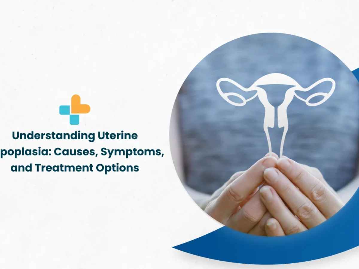
– Uterine hypoplasia, also known as hypoplastic uterus, is a condition where a girl is born with an abnormally small uterus.
– It is a congenital disorder that occurs when the uterus fails to fully develop in the fetus.
– The cause of this abnormal development is unknown.
– Uterine hypoplasia may be associated with Mayer-Rokitansky-Küster-Hauser (MRKH) syndrome, where the girl’s uterus and vagina are absent or underdeveloped.
– Symptoms include primary amenorrhea (failure to start having periods at puberty), abdominal pain, and a small or nonexistent vaginal opening.
– Diagnosis is often made during puberty when a girl fails to start her periods, and it involves a thorough medical history, physical examination, blood tests to check for MRKH syndrome, and imaging tests such as ultrasound and MRI.
– Treatment and care for uterine hypoplasia depend on the individual and their symptoms.

Continue Reading

Gestational Trophoblastic Tumour: Understanding Diagnosis, Treatment, and Recovery

– Gestational trophoblastic tumor is a type of cancer that forms from the cells that would normally develop into the placenta during pregnancy.
– It is rare and usually occurs in women of childbearing age.
– The tumor may be benign or cancerous.
– Symptoms can include abnormal vaginal bleeding, nausea and vomiting, and pelvic pain.
– Treatment options include surgery, chemotherapy, and radiation therapy.
– Clinical trials are being conducted to find new and better ways to treat this type of cancer.
– Gestational trophoblastic disease can occur after any type of pregnancy, including molar pregnancies, pregnancies that end in miscarriage or abortion, and normal pregnancies.
– Gestational trophoblastic disease is rare and can usually be cured with treatment.
– Treatment options for gestational trophoblastic disease include surgery, chemotherapy, and radiation therapy.
– It is important to receive care from a medical team with experience in treating gestational trophoblastic disease.
– The 5-year survival rate for women with low-risk disease is nearly 100%, while the 5-year survival rate for women with high-risk disease is about 90%.
– Gestational trophoblastic disease can sometimes spread to other parts of the body, such as the lungs, making treatment more difficult.

Continue Reading

Unveiling the Enigma: Understanding Endometrioid Tumour Progression

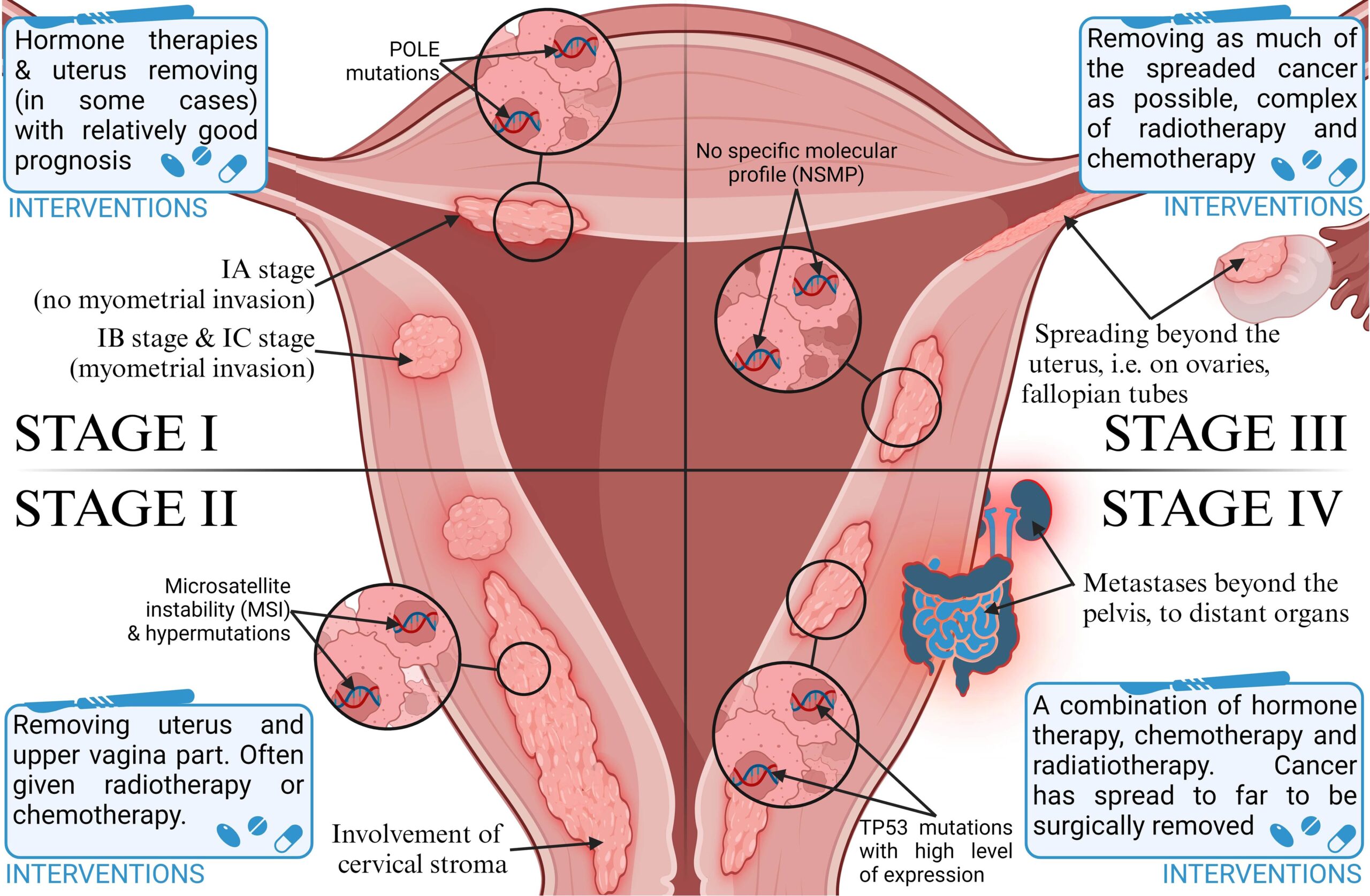
– Endometrioid cancer is the most common type of cancer in the uterus and starts in the inner lining of the uterus (endometrium).
– There are different types of endometrial cancers, including adenocarcinoma (most common), uterine carcinosarcoma, squamous cell carcinoma, small cell carcinoma, transitional carcinoma, serous carcinoma, clear-cell carcinoma, mucinous adenocarcinoma, undifferentiated carcinoma, dedifferentiated carcinoma, and serous adenocarcinoma.
– Endometrioid cancer starts in gland cells and resembles the normal uterine lining.
– Some endometrioid cancers have squamous cells in addition to glandular cells.
– There are several sub-types of endometrioid cancers, including adenocarcinoma (with squamous differentiation), adenoacanthoma, adenosquamous (or mixed cell), secretory carcinoma, ciliated carcinoma, and villoglandular adenocarcinoma.
– The grade of an endometrial cancer is determined by the organization of the cancer cells into gland-like structures.
– Grade 1 tumors of endometrioid cancer have 95% or more of the cancer tissue forming glands.
– Grade 2 tumors have between 50% and 94% of the cancer tissue forming glands.
– Grade 3 tumors have less than half of the cancer tissue forming glands and tend to be aggressive with a worse outlook.
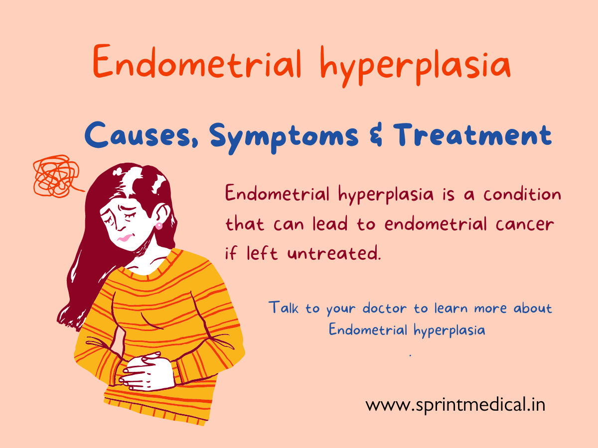
– Type 1 endometrial cancers are usually not aggressive and are caused by too much estrogen. They may develop from atypical hyperplasia.
– Type 2 endometrial cancers are more likely to grow and spread outside the uterus and have a poorer outlook. They are not caused by too much estrogen and include papillary serous carcinoma, clear-cell carcinoma, undifferentiated carcinoma, and grade 3 endometrioid carcinoma.
– Uterine carcinosarcoma (CS) starts in the endometrium and has features of both endometrial carcinoma and sarcoma. It is a type 2 endometrial carcinoma.
– Uterine sarcomas start in the muscle layer or supporting connective tissue of the uterus.
– Cancers that start in the cervix and spread to the uterus are different from cancers that start in the body of the uterus.
– Symptoms of endometrial cancer include vaginal bleeding after menopause, bleeding between periods, and pelvic pain.
– The cause of endometrial cancer is not known, but it is believed that changes in the DNA of cells in the endometrium lead to the growth of cancer cells.
– Early detection of endometrial cancer can lead to successful treatment through surgical removal of the uterus.
– Risk factors for endometrial cancer include changes in hormone balance, certain diseases or conditions, menstruation history, pregnancy history, age, obesity, hormone therapy for breast cancer, and Lynch syndrome.
– Obesity and hormone therapy are notable risk factors for endometrial cancer.
– Lynch syndrome, a genetic syndrome associated with an increased risk of several types of cancer, increases the risk of endometrial cancer.
– Individuals with Lynch syndrome should inquire about appropriate cancer screenings.
– There are no specific facts, stats, or figures provided about endometrioid tumors or the prevalence of Lynch syndrome in the article.

Continue Reading
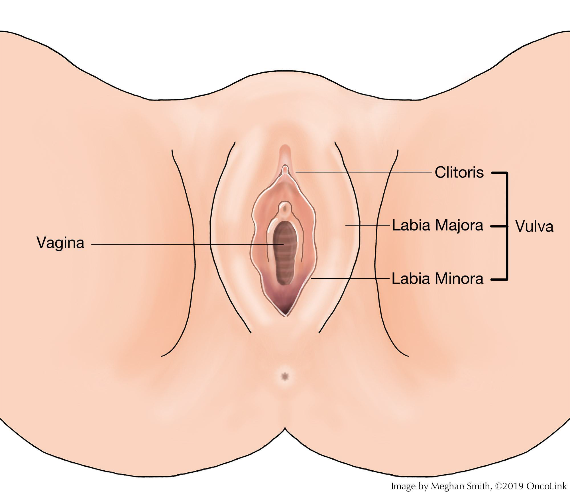
Primordial Uterus: Unveiling the Origins of Life

– Development of paramesonephric ducts in the female reproductive system
– Role of anti-Müllerian hormone (AMH) in the regression of paramesonephric ducts in males
– Persistence of paramesonephric ducts in males with mutations in AMH or AMH receptor genes
– Persistent Mullerian duct syndrome and its manifestations
– Abnormalities and complications associated with paramesonephric duct anomalies
– Difficulty in diagnosing paramesonephric duct anomalies
– Surgical advances improving the sexual function, fertility, and obstetric outcomes for women with these anomalies
– Assisted reproductive technology for women with paramesonephric duct anomalies
– Johannes Peter Müller and his discovery of paramesonephric ducts

Continue Reading

Abactus Venter: Unveiling the Hidden World of Gut Microbes

Here is a list of pertinent keywords related to “abactus venter”:

1. Latin phrase
2. Legal context
3. Medical context
4. Woman’s belly
5. Childbirth
6. Abortion
7. Uterus
8. Normal size and position
9. Condition after childbirth or abortion
10. Physical state
11. Involution
12. Womb
13. Pre-pregnancy state
14. Contraction of uterine muscles
15. Blood clots
16. Placental remnants
17. Duration and progression
18. Health
19. Hormonal levels
20. Type of delivery or abortion
21. Natural process
22. Reducing the size of the uterus.

Continue Reading

Abdominal Stalk: Unveiling the Fascinating Science of Digestion

– Body-stalk anomaly is a rare abdominal wall defect in which the abdominal organs develop outside of a baby’s abdominal cavity and remain attached directly to the placenta.
– This condition is accompanied by a short or non-existent umbilical cord and is almost always fatal for the fetus.
– The cause of body-stalk anomaly is unknown, but theories include early rupture of the amnion or amniotic band constriction due to that rupture, disruption of the embryo’s vascular system, or abnormalities in the fertilized egg.
– Body-stalk anomaly has been associated with cocaine usage and younger mothers but is mostly considered to occur randomly and is not believed to be genetic.
– Diagnosis is usually made through prenatal ultrasound in either the first 10-14 weeks or 16-20 weeks of pregnancy, and abnormalities in abdominal structures, head, arms, and legs can be seen.
– Early detection allows parents to have the option of early termination.
– There is no known treatment for body-stalk anomaly.
– The focus of treatment for this condition is counseling and support for the expectant mother and family, as well as allowing the option to terminate the pregnancy or let it proceed naturally, knowing the baby will live for only a short time after delivery.

Continue Reading