Atresia of Vagina: Causes, Symptoms, Diagnosis, and Treatment

List:

1. Vaginal Atresia
2. Bardet-Biedl syndrome
3. Fraser syndrome
4. Mayer-Rokitansky-Küster-Hauser (MRKH) syndrome
5. Vision loss
6. Obesity
7. Kidney problems
8. Intellectual disorders
9. Skin-covered eyes
10. Joined fingers and toes
11. Abnormalities of the urinary tract
12. Underdeveloped or absent vagina and uterus
13. Kidney anomalies
14. Cloacal malformation
15. Surgical treatment
16. Vaginal replacement techniques

Continue Reading

Exploring the Intricate Cavity of the Uterus: Unveiling Mysteries

List of pertinent information about the cavity of the uterus (no duplications):

– The uterine cavity is the inside portion of the uterus.
– It is triangular in shape and formed by the internal surface of the body of the uterus.
– The base of the cavity is located between the openings of the fallopian tubes.
– The apex is the internal opening of the uterus that connects to the cervix.
– The part of the uterine cavity that enters the openings of the fallopian tubes is a narrow, flattened area.
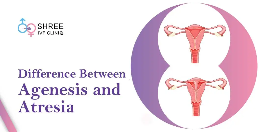
– Abnormalities of the womb or congenital uterine abnormalities refer to women who have a womb that is different in shape or size from the norm.
– These abnormalities can be discovered during an ultrasound scan or if a woman experiences miscarriage, bleeding, or difficulties conceiving.
– Women with womb abnormalities may have an increased risk of miscarriage, preterm birth, or fertility problems depending on the shape of the womb.
– Women with bicornuate wombs have a slightly higher risk of miscarriage and preterm birth.
– Women with a unicornate womb have half the size of a normal womb and an increased risk of ectopic pregnancy, late miscarriage, or preterm birth.
– Women with a didelphic womb, which is split in two, may have a small increased risk of preterm birth.
– Women with a septate/subseptate womb may have an increased risk of miscarriage, preterm birth, or fertility problems.
– Septate wombs may cause difficulties with conception.
– Septate wombs have an increased risk of early miscarriage and preterm birth.
– Babies in later pregnancy with septate wombs may not be in a head-down position, leading to a higher likelihood of needing a C-section.
– Many women with congenital uterine abnormalities, like a septate uterus, are not aware of their condition.
– Surgery to resect the septum before pregnancy is common for women with a septate uterus, but it was not recommended by the consultant in this case.
– Despite having a complete septate uterus, the woman in the case was able to carry her baby to term but had an elective C-section because the baby was breech.
– An arcuate womb has a dip at the top but resembles a normal womb.
– Having an arcuate womb does not increase the risk of preterm birth or early miscarriage.
– An arcuate womb may increase the risk of late miscarriage.
– Babies in later pregnancy with an arcuate womb may not be in a head-down position, increasing the likelihood of needing a C-section.

Continue Reading

Repair of Vesicovaginal Fistula: A LifeChanging Procedure

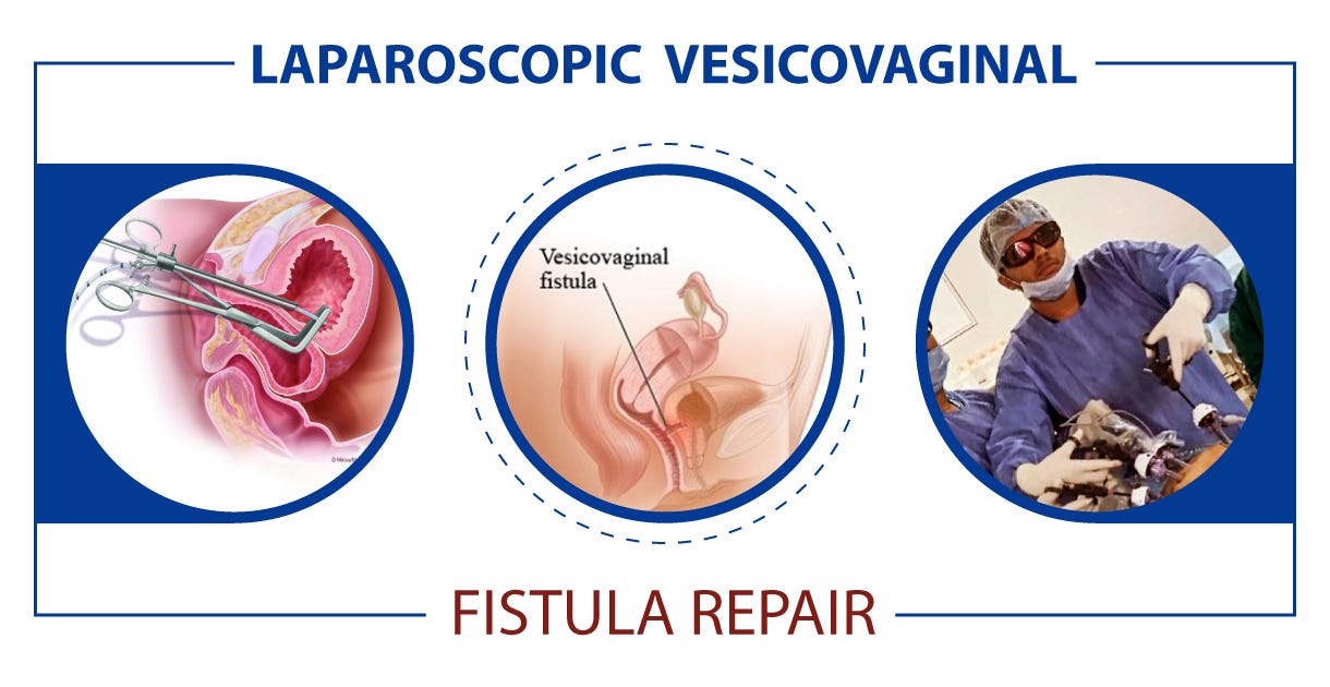
– Repair of vesicovaginal fistula is a surgical procedure to close or remove a fistula between the bladder and vagina.
– Before surgery, patients may be instructed to fast and shower with a specific soap to prevent infection.
– General anesthesia is administered during the surgery.
– Stents may be inserted in the ureters to protect them during surgery.
– The fistula is either excised or closed with stitches, and the incision is closed with stitches.
– Antibiotic-soaked bandages may be placed in the vagina to prevent infection.
– In some cases, an incision may be made through the abdomen, and a suprapubic catheter may be inserted into the bladder for urine drainage.
– Risks of the surgery include excessive bleeding, infection, damage to the ureters, bladder spasms, vaginal bleeding, bladder stones, incomplete bladder emptying, smaller bladder, shortened vaginal canal, and the possibility of a new fistula forming after surgery.
– Repair success is achieved by performing the repair in a single layer, using a non-absorbable suture material.
– Some surgeons use absorbable sutures for the vaginal closure to avoid suture-induced granulomas.
– Repair of vesicovaginal fistula in radiation patients may require additional techniques like omentoplasty or myocutaneous flap interposition.
– Different surgical techniques for VVF repair include vaginal and abdominal approaches.
– The success rates for VVF repair are high if certain surgical principles are followed.
– Vascularized flaps or grafts may be used for repairing large or radiotherapy-related fistulas.
– Different incisions such as the Dührssen and Schuchardt incisions can be used to improve exposure during surgery.

Continue Reading

Epidermal Carcinoma: Causes, Symptoms, Stages, Treatment Options Explained

– SCCs (Squamous Cell Carcinomas) can manifest as scaly red patches, open sores, rough or thickened skin, wart-like skin, or raised growths with a central depression.
– Sometimes, SCCs may crust over, itch, or bleed.
– These lesions usually occur in sun-exposed areas of the body but can also appear in other body parts, including the genitals.
– The appearance of SCCs can vary from person to person.
– Squamous cell carcinoma of the skin is a type of cancer that starts as a growth of cells on the skin.
– It usually starts in squamous cells, which are found in the middle and outer layers of the skin.
– Squamous cell carcinoma is a common type of skin cancer and is usually not life-threatening if treated.
– Most cases of squamous cell carcinoma are caused by excessive exposure to ultraviolet (UV) radiation from sunlight, tanning beds, or lamps.
– The cancer can occur anywhere on the skin, but in people who sunburn easily, it is often found in sun-exposed areas.
– In people with Black and brown skin, squamous cell carcinomas are more likely to appear in areas not exposed to the sun, such as the genitals.
– Symptoms of squamous cell carcinoma include firm bumps on the skin, flat sores with a scaly crust, new sores or raised areas on old scars or sores, rough patches on the lips or inside the mouth, and raised patches or wartlike sores on or in the anus or genitals.
– It is recommended to see a doctor if a sore or scab doesn’t heal in about two months or if there is a flat patch of scaly skin that won’t go away.
– Epidermal carcinoma, specifically squamous cell carcinoma of the skin, is more common in individuals with low levels of melanin.
– The risk of squamous cell carcinoma is highest in people with blond or red hair, light-colored eyes, freckles, and easily sunburned skin.
– Factors that increase the risk of this type of carcinoma include excessive sun exposure, both from natural sunlight and tanning beds.
– A history of sunburns, especially during childhood or adolescence, is also a risk factor.
– Having precancerous skin lesions, a previous history of skin cancer, a weakened immune system, or a rare genetic disorder called xeroderma pigmentosum can increase the risk.
– Infection with human papillomavirus (HPV) can also increase the risk of developing squamous cell carcinoma.
– Untreated squamous cell carcinoma can destroy healthy tissue, spread to lymph nodes or other organs, and potentially be fatal.
– Factors that may increase the risk of the cancer spreading include large or deep growth, involvement of mucous membranes (such as the lips), and a weakened immune system.
– Most cases of squamous cell carcinoma can be prevented.

Continue Reading

Hypoplasia of the uterus: Causes, symptoms, and treatments

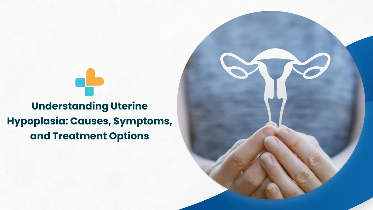
– Uterine hypoplasia is a condition where a girl is born with a small uterus
– It is a congenital disorder present at birth
– The cause of abnormal fetal development leading to uterine hypoplasia is unknown
– Uterine hypoplasia may be a symptom of Mayer-Rokitansky-Küster-Hauser (MRKH), which involves underdeveloped or absent uterus and vagina
– Symptoms may include failure to start periods, abdominal pain, and a small or no vaginal opening
– Diagnosis is often not made until puberty when a girl fails to start having periods and visits a doctor
– Diagnosis involves a medical history, physical exam, pelvic exam, blood tests, ultrasound, and MRI
– Treatment depends on the individual and her symptoms

Continue Reading

Ovarian Tumour: Understanding Symptoms, Diagnosis, Treatment, and Support

– Ovarian tumors can be noncancerous (benign) or cancerous (malignant).
– Symptoms of ovarian tumors include stomach bloating, increased belly size, stomach or pelvic pain, constipation, difficulty urinating or urinating frequently, feeling full more quickly, painful cramps during menstruation, lower back pain, nausea or vomiting, pain during sex, and vaginal bleeding after menopause.
– The causes of ovarian tumors are still being studied.
– There are two broad categories of ovarian tumors: benign and malignant.
– Benign tumors can develop into malignant tumors if left untreated.
– Types of ovarian tumors include surface epithelial tumors, stromal tumors, and germ cell tumors.
– Ovarian tumors can be classified into four stages if they are malignant: Stage I, Stage II, Stage III, and Stage IV.
– Risk factors for ovarian tumors include age, family history, genetic mutations (BRCA1 or BRCA2), and obesity.
– Women who have never conceived and carried a pregnancy or have done so after age 35 may have an increased risk of ovarian cancer.
– Continuous use of estrogen after menopause can also increase the risk of ovarian cancer.
– The reason for ovarian tumors forming is unknown, so prevention methods are currently unknown.
– To lower the risk of ovarian cancer, steps that can be taken include eating a healthy diet, exercising regularly, maintaining a healthy weight, not smoking, and taking birth control pills.
– The information in the article is for educational purposes only and should not replace advice from a healthcare provider.

Continue Reading

The Mysterious Claustrum Virginale: Unveiling Its Secrets

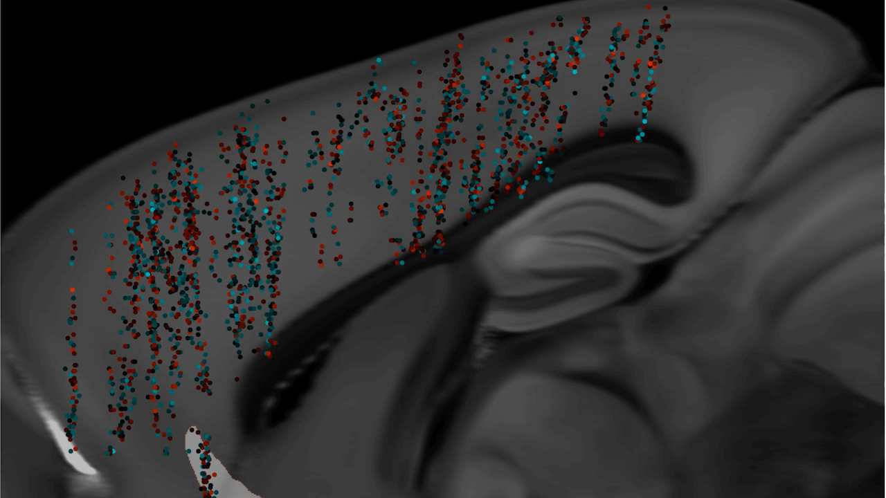
The claustrum is a thin sheet of neurons and supporting glial cells in the brain that connects to the cerebral cortex and subcortical regions. It is located between the insular cortex and putamen. The claustrum is considered to be the most densely connected structure in the brain and is hypothesized to integrate various sensory inputs into one experience. It may also play a role in salience processing and attention. The claustrum is made up of different cell types, with the principal cell type being the Golgi type I neuron. It is believed to synchronize activity in different parts of the brain and support consciousness. The claustrum may also differentiate between relevant and irrelevant information. Its precise boundaries are still debated.

The claustrum is a brain structure that has extensive connections to both cortical and subcortical regions. It is highly connected to thalamic nuclei, the basal ganglia, and various regions of the cortex. The claustrum is the most highly connected structure per regional volume in the brain and may serve as a hub to coordinate activity of cerebral circuits. It is involved in processing sensory information and the physical and emotional state of an animal. Inputs to the claustrum are organized by modality, including prefrontal, visual, auditory, and somatomotor processing areas. The claustrum possesses a distinct topological organization for each sensory modality and has dense connectivity with frontal cortices. Local connectivity within the claustrum is dominated by feed-forward disynaptic inhibition. Excitatory claustrum neurons form synapses across the anteroposterior axis and are biased toward neurons that do not share projection targets. Overall, these findings suggest that the claustrum is capable of performing local transformations of diverse input information from across the brain.

The claustrum is a part of the brain composed of various cell types. Excitatory cells in the claustrum project to different brain regions. Inhibitory neurons make up 10%-15% of the claustrum and express parvalbumin, somatostatin, or vasoactive intestinal peptide. The claustrum can be identified by its prominent plexus of parvalbumin-positive fibers. Different methods, such as electrophysiological, morphological, genetic, and connectomic approaches, have been used to study the claustrum in mice. The claustrum has widespread connectivity with cortical components associated with consciousness and sustained attention. It acts as a “conductor” in coordinating the function of all connections. The claustrum has reciprocal connections to the prefrontal cortex, visual, auditory, sensory, and motor regions. It is proposed that the claustrum functions in the gating of selective attention, selectively controlling input from different modalities. The claustrum may also implement resistance to certain inputs to prevent distraction. The claustrum integrates various sensory and motor modalities from different parts of the cortex to facilitate consciousness.

Functional imaging studies show dampened activity in the claustrum when anesthetized versus awake in rats. The claustrum has strong functional connections with the contralateral hemisphere’s claustrum, the mediodorsal thalamus, the medial prefrontal cortex, and surrounding and distant cortical areas.

The claustrum is a region in the brain that plays a role in integrating different modalities, such as sensory and motor functions. It has been shown to have connections with various parts of the cortex and is involved in processes such as spatial navigation and slow-wave sleep. The claustrum also has the ability to select between task relevant and irrelevant information for directed attention. It receives input from visual and auditory centers and can be activated by unexpected stimuli. Electrical stimulation of the claustrum can cause inhibition, leading to a blank stare and unresponsiveness. Salvinorin A, a hallucinogenic compound, can bind to Kappa Opioid Receptors in the claustrum, inducing a loss of awareness and synesthesia. High frequency stimulation of the claustrum in cats and humans has been shown to induce a decrease in awareness and consciousness.

MRI studies have also shown increased signal intensity within the claustrum. The claustrum has been associated with status epilepticus, where epileptic seizures occur without recovery of consciousness in between events. Increased signal intensity in the claustrum is associated with Focal dyscognitive seizures, which impair awareness or consciousness without convulsions. Studies have shown that the claustrum is active during REM sleep and may play a role in spatial memory consolidation. Damage to the claustrum is associated with duration of loss of consciousness in traumatic brain injuries. Stimulation of the extreme capsule, near the claustrum, can disrupt consciousness in a case study. Decreased grey matter volume in the left claustrum is associated with greater delusions in schizophrenia. Lesions to both claustrums would be needed to cause total loss of function. Electrical stimulation of the claustrum did not disrupt consciousness in a study of five patients. Damage to the claustrum may mimic other diseases or mental disorders.

Summary:

The claustrum, a region in the brain, has been found to be involved in various neurological conditions. In autism, a decrease in grey matter volume in the claustrum and insula is associated with an increase in positive symptoms. For epilepsy, increased claustral signal intensity has been observed in MRI scans of diagnosed individuals. Electrical stimulation of the claustrum has been found to disrupt consciousness, while lesions in the claustrum can lead to a loss of consciousness. However, a recent study found no disruption of consciousness with electrical stimulation. Artificial activation of the claustrum can silence brain activity across the cortex. Lesions in the claustrum have been identified as the likely origin of parkinsonism across different conditions. In mice, suppression of the claustrum attenuates anxiety and stress and increases chronic stress-resistance. The claustrum has extensive connections throughout the cortex and with sensory and motor regions in animals. The article discusses the anatomy and function of the claustrum in cats, rodents, and monkeys. In cats, high-frequency stimulation of the claustrum can alter motor activity and induce changes in awareness. The claustrum in cats responds to sensory stimuli and is connected to the motor, somatosensory, visual, and auditory cortex. Sensory input in cats is segregated based on modalities, with a preference for peripheral sensory information. In rodents, the claustrum has distinct patterning of connectivity with different cortical areas and plays specialized roles in cortical processing. In mice, inhibitory interneurons strongly modulate the activity of the claustrum and synchronize activity of claustrocortical projections to influence brain rhythms and coordinated activity of different cortical regions. In monkeys, the claustrum has widespread connections. The claustrum has connections with various cortical regions including the frontal lobe, visual cortex, temporal cortex, parieto-occipital cortex, and somatosensory areas. It also projects towards subcortical areas such as the amygdala, caudate nucleus, and hippocampus. The claustrum may have bi-directional connections with motor structures in the cortex. 70% of movement neurons in the dorsocaudal claustrum are non-selective and can fire for various types of movements, while the remaining neurons are more specific and only fire for one type of movement.

Continue Reading
The Importance of Arteria Ovarica in Female Reproduction:

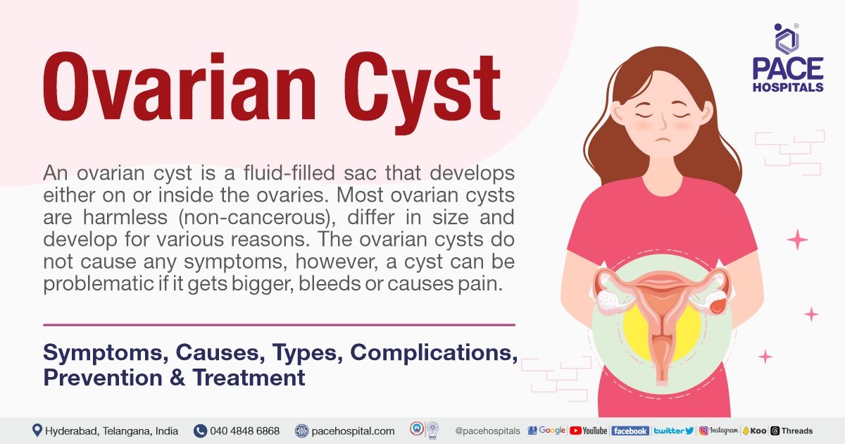
1. The ovarian artery is an artery that supplies oxygenated blood to the ovary in females.
2. It arises from the abdominal aorta below the renal artery.
3. It can be found within the suspensory ligament of the ovary, anterior to the ovarian vein and ureter.
4. The ovarian arteries are paired structures that arise from the abdominal aorta, usually at the level of L2.
5. After emerging from the aorta, the artery travels within the suspensory ligament of the ovary and enters the mesovarium.
6. The ovarian arteries may anastomose with the uterine artery in the broad ligament.
7. Small branches are given to the ureter and the uterine tube.
8. One branch passes on to the side of the uterus and unites with the uterine artery.
9. Other offsets are continued on the round ligament of the uterus, through the inguinal canal, to the integument of the labium majus and groin.
10. In 20% of cases, the ovarian arteries arise from the renal arteries.
11. They may also arise from adrenal, lumbar, or internal iliac arteries.
12. The ovarian artery supplies blood to the ovary and uterus.
13. The ovarian arteries swell during pregnancy, in order to increase the uterine blood supply.

Continue Reading