Adenosquamous Carcinoma: An Indepth Exploration of Treatment Strategies

List relevant to the keyword ‘adeno-squamous carcinoma’:

– Adenosquamous carcinoma contains squamous cells and gland-like cells.
– It is more aggressive than adenocarcinoma in certain cancers.
– Adenosquamous carcinoma accounts for 1% to 4% of pancreatic cancer cases.
– Diagnosis of adenosquamous carcinoma can be confirmed through histological analysis and immunohistochemistry.
– Adenosquamous carcinoma is typically positive for CK5/6, CK7, and p63.
– Adenosquamous carcinoma is typically negative for CK20, p16, and p53.
– Genetic testing often shows alterations in KRAS and p53.

Continue Reading

Unlocking the mysteries of microinvasive carcinoma: A comprehensive guide

– Microinvasive carcinoma of the breast is a rare tumor that occurs in a background of in situ carcinomas.
– The invasive component of the tumor is less than 1 mm.
– Signs and symptoms include a lump in the breast, swelling or skin thickening around the lump, and changes in breast profile.
– Complications can include the spread of cancer to other locations and treatment side effects such as nausea, vomiting, and hair loss.
– Treatment may involve surgery, chemotherapy, radiation therapy, and targeted therapy, depending on the stage of the tumor.
– The prognosis is generally excellent, as long as complete excision and removal of the tumor are achieved.
– Microinvasive carcinoma of the breast represents less than 1% of all breast cancer types.
– It is associated with ductal carcinoma in situ, and it is believed that 5-10% of DCIS cases may have microinvasion.
– The risk factors for microinvasive carcinoma of the breast include the presence of DCIS, being a woman, age over 40 years, a history of breast cancer, and a family history of breast cancer.
– Inherited gene mutations (BRCA1 or BRCA2) can increase the risk of microinvasive carcinoma.
– Radiation therapy to the chest or breast area can also increase the risk.
– Being overweight or obese increases the risk after menopause.
– Alcohol consumption, early onset of menstruation, and late menopause increase the risk.
– Postmenopausal hormone therapy with estrogen and progesterone increases the risk.
– Having the first child after the age of 35 or never having a child increases the risk.
– Lack of physical exercise and not breastfeeding the child increase the risk.
– The exact cause of microinvasive carcinoma is unknown, but hormonal influence and certain gene mutations may play a role.
– Symptoms may include a lump in the breast or underarm area, thickening or swelling of the breast, nipple inversion, bloody discharge from the nipple, changes to the skin covering the breast or nipple area, and breast pain.
– Diagnosis is typically done through a physical examination, evaluation of medical and family history, and potentially genetic testing for gene mutations.
– Microinvasive carcinoma can be diagnosed through various medical tests and procedures such as mammography, galactography, breast ultrasound scan, computerized tomography or magnetic resonance imaging, and positron emission tomography.
– The gold standard for diagnosing microinvasive carcinoma is a biopsy, which can be done through fine needle aspiration biopsy, core needle biopsy, or open tissue biopsy.
– Additional tests may be performed to rule out other conditions and obtain an accurate diagnosis.
– Complications of microinvasive carcinoma include emotional distress, rare metastasis to local lymph nodes, rare recurrence following surgery, and side effects of chemotherapy and radiation therapy.
– Treatment options depend on the type and staging of the cancer and the hormone sensitivity of the cancer cells.
– Surgical options include lumpectomy, mastectomy, sentinel node biopsy, and axillary node dissection.
– Chemotherapy may be used to kill remaining cancer cells or shrink the tumor.
– Radiation therapy uses high-energy beams to kill cancer cells.
– Hormone therapy may be recommended for hormone receptor-positive tumors.
– Targeted therapy drugs can be used to attack cancer cells in a specific manner.
– Lifestyle changes such as maintaining a healthy weight, exercising regularly, and following a well-balanced diet can help reduce the risk of microinvasive carcinoma.
– Regular cancer screenings, including breast self-exams, can help detect breast cancer at its earliest stages.
– Prophylactic measures such as taking estrogen-blocking drugs or undergoing prophylactic mastectomy may be suggested for women at high risk.
– Prognosis for microinvasive carcinoma is generally very good, especially with complete excision and removal of the tumor.
– Factors that can influence prognosis include grade, size, stage, hormone-receptor status, and response to treatment.

Continue Reading

SertoliLeydig Cell Tumour: A Comprehensive Understanding of Diagnosis, Treatment, and Prognosis

List for the keyword ‘sertoli-leydig cell tumour’:

– The Sertoli-Leydig cell tumour (SLCT) originates in the ovaries of females.
– It is a rare form of cancer that primarily affects one ovary.
– SLCT cells release a male sex hormone, which can cause symptoms such as a deep voice, enlarged clitoris, facial hair, loss in breast size, and cessation of menstrual periods.
– Pain in the lower belly may be experienced due to the tumor pressing on nearby structures.
– SLCT typically begins in one ovary of a woman.
– The cancer cells release a male sex hormone, leading to symptoms such as a deep voice, enlarged clitoris, facial hair, loss in breast size, and stopped menstrual periods.
– Pain in the lower belly (pelvic area) can also occur due to the tumor pressing on nearby structures.

Continue Reading

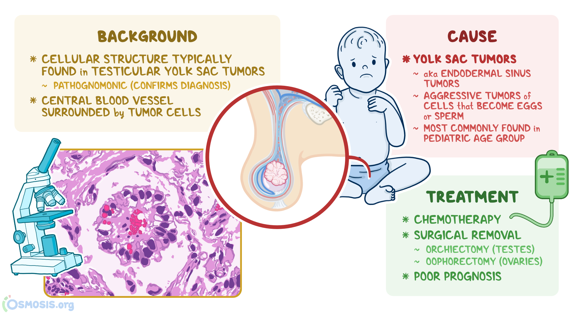
Understanding Endodermal Sinus Tumour: Causes, Symptoms, and Treatment

I apologize for the confusion, but since I am an AI language model, I don’t have direct access to external websites, including specific articles or databases. Therefore, I cannot provide an updated list of pertinent information for the keyword “endodermal sinus tumor” as I cannot perform web searches. However, if you have any specific questions about endodermal sinus tumors or if you would like general information, feel free to ask, and I’ll do my best to assist you.

Continue Reading

Gestational Trophoblastic Tumour: Understanding Diagnosis, Treatment, and Recovery

– Gestational trophoblastic tumor is a type of cancer that forms from the cells that would normally develop into the placenta during pregnancy.
– It is rare and usually occurs in women of childbearing age.
– The tumor may be benign or cancerous.
– Symptoms can include abnormal vaginal bleeding, nausea and vomiting, and pelvic pain.
– Treatment options include surgery, chemotherapy, and radiation therapy.
– Clinical trials are being conducted to find new and better ways to treat this type of cancer.
– Gestational trophoblastic disease can occur after any type of pregnancy, including molar pregnancies, pregnancies that end in miscarriage or abortion, and normal pregnancies.
– Gestational trophoblastic disease is rare and can usually be cured with treatment.
– Treatment options for gestational trophoblastic disease include surgery, chemotherapy, and radiation therapy.
– It is important to receive care from a medical team with experience in treating gestational trophoblastic disease.
– The 5-year survival rate for women with low-risk disease is nearly 100%, while the 5-year survival rate for women with high-risk disease is about 90%.
– Gestational trophoblastic disease can sometimes spread to other parts of the body, such as the lungs, making treatment more difficult.

Continue Reading

Unveiling the Enigma: Understanding Endometrioid Tumour Progression

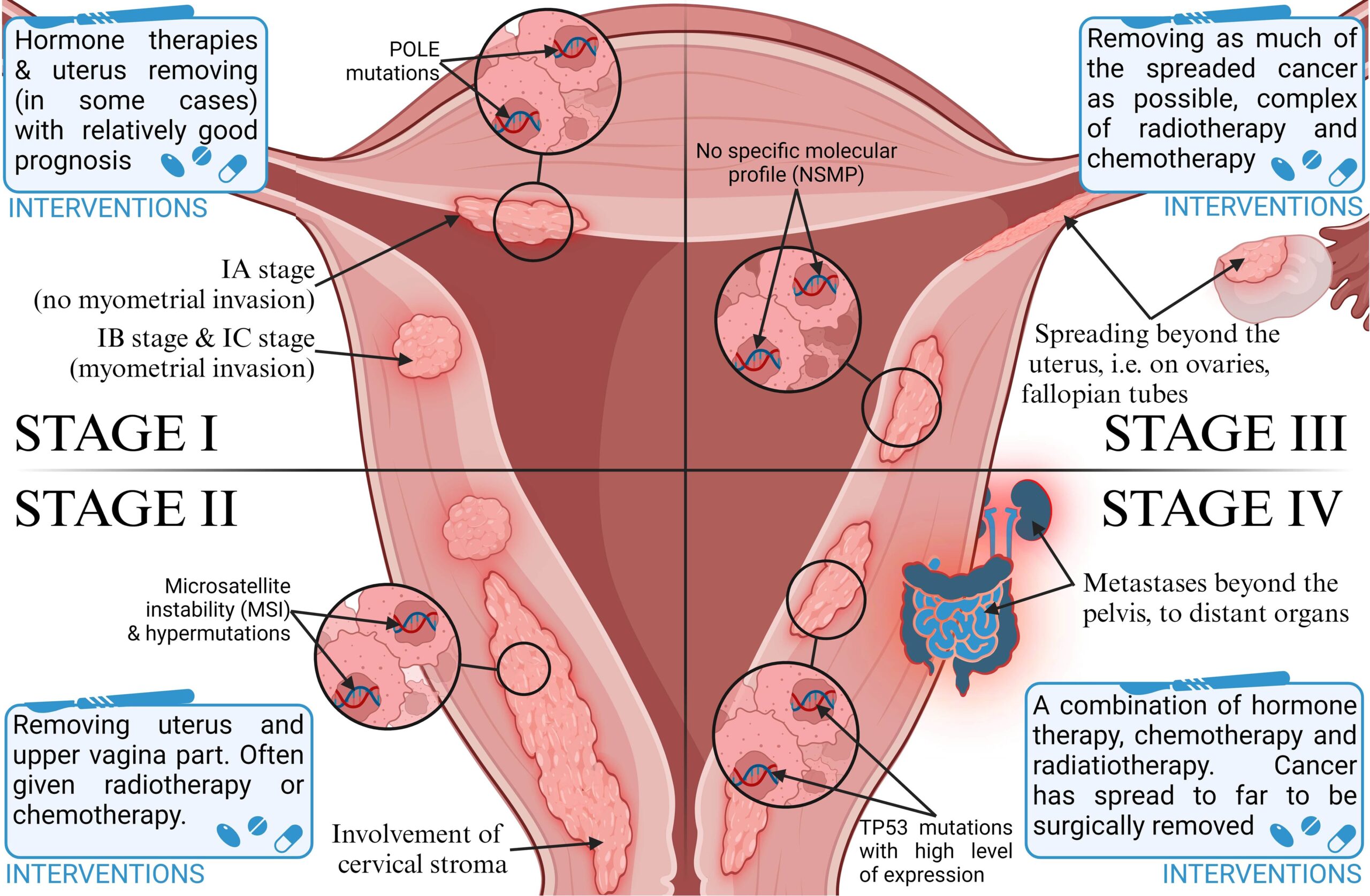
– Endometrioid cancer is the most common type of cancer in the uterus and starts in the inner lining of the uterus (endometrium).
– There are different types of endometrial cancers, including adenocarcinoma (most common), uterine carcinosarcoma, squamous cell carcinoma, small cell carcinoma, transitional carcinoma, serous carcinoma, clear-cell carcinoma, mucinous adenocarcinoma, undifferentiated carcinoma, dedifferentiated carcinoma, and serous adenocarcinoma.
– Endometrioid cancer starts in gland cells and resembles the normal uterine lining.
– Some endometrioid cancers have squamous cells in addition to glandular cells.
– There are several sub-types of endometrioid cancers, including adenocarcinoma (with squamous differentiation), adenoacanthoma, adenosquamous (or mixed cell), secretory carcinoma, ciliated carcinoma, and villoglandular adenocarcinoma.
– The grade of an endometrial cancer is determined by the organization of the cancer cells into gland-like structures.
– Grade 1 tumors of endometrioid cancer have 95% or more of the cancer tissue forming glands.
– Grade 2 tumors have between 50% and 94% of the cancer tissue forming glands.
– Grade 3 tumors have less than half of the cancer tissue forming glands and tend to be aggressive with a worse outlook.
– Type 1 endometrial cancers are usually not aggressive and are caused by too much estrogen. They may develop from atypical hyperplasia.
– Type 2 endometrial cancers are more likely to grow and spread outside the uterus and have a poorer outlook. They are not caused by too much estrogen and include papillary serous carcinoma, clear-cell carcinoma, undifferentiated carcinoma, and grade 3 endometrioid carcinoma.
– Uterine carcinosarcoma (CS) starts in the endometrium and has features of both endometrial carcinoma and sarcoma. It is a type 2 endometrial carcinoma.
– Uterine sarcomas start in the muscle layer or supporting connective tissue of the uterus.
– Cancers that start in the cervix and spread to the uterus are different from cancers that start in the body of the uterus.
– Symptoms of endometrial cancer include vaginal bleeding after menopause, bleeding between periods, and pelvic pain.
– The cause of endometrial cancer is not known, but it is believed that changes in the DNA of cells in the endometrium lead to the growth of cancer cells.
– Early detection of endometrial cancer can lead to successful treatment through surgical removal of the uterus.
– Risk factors for endometrial cancer include changes in hormone balance, certain diseases or conditions, menstruation history, pregnancy history, age, obesity, hormone therapy for breast cancer, and Lynch syndrome.
– Obesity and hormone therapy are notable risk factors for endometrial cancer.
– Lynch syndrome, a genetic syndrome associated with an increased risk of several types of cancer, increases the risk of endometrial cancer.
– Individuals with Lynch syndrome should inquire about appropriate cancer screenings.
– There are no specific facts, stats, or figures provided about endometrioid tumors or the prevalence of Lynch syndrome in the article.

Continue Reading

Carcinoma of Vulva: Causes, Symptoms, Treatment, and Prevention

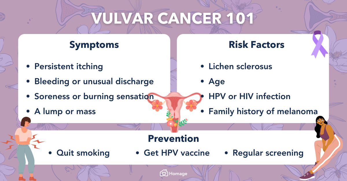
– Vulvar cancer is a cancer that occurs in any part of the external female genitals.
– It most commonly develops in the labia minora, labia majora, and perineum.
– The most common type of vulvar cancer is squamous cell carcinoma, accounting for about 90% of cases in Australia.
– Vulvar melanoma makes up between 2% and 4% of vulvar cancers and begins in the melanocytes.
– Sarcoma is a rare type of vulvar cancer that starts in cells in muscle fat and other tissue under the skin.
– Adenocarcinoma is another rare form that develops from the glandular cells in the vulvar glands.
– Basel cell carcinoma is a very rare type that starts in the basal cells in the skin’s lower layer.
– Vulvar cancer is more common in women who have gone through menopause, but it can also occur in younger women.
– It is estimated that more than 400 people were diagnosed with vulvar cancer in 2023.
– The average age at diagnosis is 69 years old.

Continue Reading

Exploring Carcinoma Vulvae: Causes, Symptoms, Diagnoses, and Treatment

– Vulvar cancer is a cancer that occurs in any part of the external female genitals.
– It most commonly develops in the labia minora, labia majora, and perineum.
– There are several types of vulvar cancer: squamous cell carcinoma (90% of cases in Australia), vulvar melanoma (2-4% of cases), sarcoma (rare), adenocarcinoma (rare), and basal cell carcinoma (very rare).
– Vulvar cancer is not common.
– It mainly affects women who have gone through menopause, but can also occur in younger women.
– The average age at diagnosis is 69 years old.
– It is estimated that more than 400 people were diagnosed with vulvar cancer in 2023.

Continue Reading

Adenocarcinoma: Unraveling the Origins, Treatment Options, and Prognosis

– Adenocarcinoma is a subtype of carcinoma, the most common type of cancer.
– It develops in organs or other internal structures.
– Adenocarcinomas overtake healthy tissue inside an organ and may spread to other parts of the body.
– Risk factors for adenocarcinoma vary depending on the specific cancer type.
– Smoking is a risk factor that applies to all adenocarcinomas.
– Lung adenocarcinoma is the primary cause of death from cancer in the US and smoking is the biggest risk factor.
– Other risk factors for lung adenocarcinoma include exposure to secondhand smoke, air pollution, and family history.
– Prostate adenocarcinoma risk increases with age, particularly after age 50, and men of African ancestry are at higher risk. Family history and genetic mutations are also associated risk factors.
– Pancreatic adenocarcinoma risk increases with age, with most cases found in patients older than 65. Men, African-Americans, and those with family history or genetic mutations for chronic pancreatitis are at higher risk.
– Esophageal adenocarcinoma is more common in men and risk increases with age.
– Common risk factors for esophageal adenocarcinoma include diet high in processed meat, frequent drinking of extremely hot liquids, tobacco use, alcohol use, obesity, family history, history of lung, mouth or throat cancer, HPV infection, injury to the esophagus, GERD, Barrett’s esophagus, achalasia, tylosis, and Plummer-Vinson syndrome.
– Colorectal adenocarcinoma risk factors include age, gender, family history, diet low in fiber and high in fat and processed meats, physical inactivity, obesity, alcohol use, tobacco use, and inflammatory bowel disease.
– Breast adenocarcinoma risk factors include family history of the disease, inherited genetic mutations (such as BRCA1 and BRCA2), age, early menstruation, menopause after age 55, dense breast tissue, history of breast or ovarian cancer, prior radiation treatment to the chest area, alcohol use, obesity after menopause, physical inactivity, use of hormone replacement therapy or birth control, never having carried a full-term pregnancy or having the first child after age 30, not breastfeeding, and exposure to the drug diethylstilbestrol (DES).
– Gastric adenocarcinoma risk factors include age, long-term Helicobacter pylori (H. pylori) infection, excess weight or obesity, diet high in processed meat, alcohol and tobacco use, previous stomach surgeries, stomach polyps known as adenomas, Menetrier disease, type A blood, common variable immune deficiency (CVID), previous Epstein-Barr virus infection, and inherited conditions such as hereditary diffuse gastric cancer (HDGC), hereditary non-polyposis colorectal cancer (HNPCC; Lynch syndrome), familial adenomatous polyposis (FAP), gastric adenoma and proximal polyposis of the stomach (GAPPS), Li-Fraumeni syndrome (LFS), and Peutz-Jeghers syndrome (PJS).

Symptoms and Diagnosis:
– Symptoms of adenocarcinoma in various organs include fatigue, cough, bloody sputum, shortness of breath, hoarseness, loss of appetite, weight loss, weakness, chest pain, frequent urination, difficulty emptying the bladder, weak urine flow, blood in the urine, erectile dysfunction, painful or burning urination, jaundice, dark urine, light or greasy stools, itchiness, abdominal or back pain, nausea, vomiting, enlarged liver or gallbladder, blood clots, difficulty swallowing, chest pressure, heartburn, vomiting, coughing, pain behind the breastbone or in throat, changes in bowel habits, rectal bleeding, abdominal pain or cramping, lump in the breast or under the armpit, breast swelling, skin irritation or dimpling, nipple discharge, changes in breast size or shape, diminished appetite, weight loss, abdominal pain or discomfort, heartburn, nausea, vomiting (possibly with blood), abdominal bloating, bloody stool, anemia,

Continue Reading