Primordial Uterus: Unveiling the Origins of Life

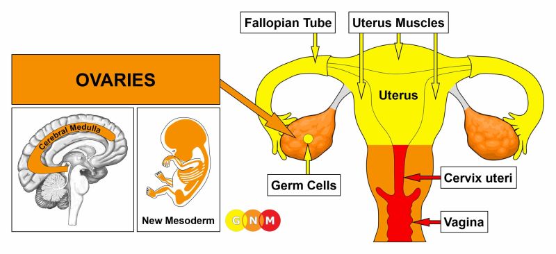
– Development of paramesonephric ducts in the female reproductive system
– Role of anti-Müllerian hormone (AMH) in the regression of paramesonephric ducts in males
– Persistence of paramesonephric ducts in males with mutations in AMH or AMH receptor genes
– Persistent Mullerian duct syndrome and its manifestations
– Abnormalities and complications associated with paramesonephric duct anomalies
– Difficulty in diagnosing paramesonephric duct anomalies
– Surgical advances improving the sexual function, fertility, and obstetric outcomes for women with these anomalies
– Assisted reproductive technology for women with paramesonephric duct anomalies
– Johannes Peter Müller and his discovery of paramesonephric ducts

Continue Reading

Fetal Posture: Unlocking the Secrets of Optimal Development

– Fetal presentation before birth refers to the position of the baby in the uterus right before delivery.
– The most common position is cephalic occiput anterior, where the baby’s head is down and face down.
– Another position is cephalic occiput posterior, where the baby’s head is down but face up. This can make labor longer and may require manual rotation or assisted delivery.
– Breech presentation occurs when the baby’s feet or buttocks are in place to come out first during birth. This happens in about 3% to 4% of babies.
– The most common type of breech presentation is frank breech, where the baby’s knees aren’t bent and the feet are close to the baby’s head.
– A procedure called external cephalic version can be performed to try to move the baby into a head-down position if they are in a frank breech position.
– If the procedure is not successful or the baby moves back into a breech position, the delivery options should be discussed with the healthcare team.
– A complete breech presentation is when the baby has both knees bent and both legs pulled close to the body.
– An incomplete breech presentation is when one or both of the legs are not pulled close to the body, and one or both of the feet or knees are below the baby’s buttocks.
– If a baby is in a complete or incomplete breech presentation after 36 weeks of pregnancy, the health care professional may try to move the baby into a head-down position using external cephalic version.
– If the procedure is not successful or if the baby moves back into a breech position, alternative delivery options should be discussed with the health care team.
– A transverse lie is when the baby is lying horizontally across the uterus.
– If the baby is in a transverse lie at week 37 of pregnancy, the health care professional may try to move the baby into a head-down position using external cephalic version.
– If the procedure is not successful or if the baby moves back into a transverse lie, alternative delivery options should be discussed.
– If pregnant with twins and only one twin is head down, the health care provider may deliver the first twin vaginally and then suggest delivering the second twin in the breech position or try to move the second twin into a head-down position using external cephalic version.
– Delivery by C-section may be suggested for the second twin.

Continue Reading

Fetal Tone: Understanding the Importance of Prenatal Development

– A biophysical profile (BPS or BPP) is a test performed after 32 weeks of pregnancy to evaluate the fetus’s health.
– It is typically done for high-risk pregnancies or when there are irregular test results.
– The test uses ultrasound to evaluate four areas: fetal body movement, muscle tone, breathing movements, and amniotic fluid volume.
– A scoring system is used to rate the fetus in each area.
– If one or more of the areas are not met, a nonstress test may be needed to assess the fetus’s heart rate.
– A modified biophysical profile combines a nonstress test with an ultrasound assessment of amniotic fluid.
– The purpose of a biophysical profile is to assess the fetus’s well-being.
– It may be ordered for high-risk pregnancies or pregnancies that go beyond 40 weeks.
– The five parameters of a biophysical profile include a nonstress test and four ultrasound assessments.
– A biophysical profile is a test used in the third trimester of pregnancy to assess the unborn baby’s overall health.
– It combines a nonstress test, which checks the baby’s heart rate and contractions, with an ultrasound evaluation.
– Five areas are assessed during the profile: body movements, muscle tone, breathing movements, amniotic fluid, and heartbeat.
– Each area is given a score of either 0 (abnormal) or 2 (normal), and the total score ranges from 0 to 10.
– A score of 8 or 10 is considered normal, while 6 is borderline and below 6 indicates possible problems.
– The test can help determine if the baby needs to be born early.
– Reasons for needing a biophysical profile include concerns about the baby’s health, decreased fetal movement, fetal growth problems, or pregnancy going past 42 weeks.
– The procedure is safe and painless, involving sensors attached to belts for the nonstress test and an ultrasound wand with gel for the ultrasound.
– The test poses very little risk to the mother and baby, and concerns about ultrasounds over a long period of time have not been proven.
– Mothers should discuss any concerns with their healthcare provider.
– A biophysical profile is a test done during pregnancy to assess the health of the fetus.
– It is typically done after 32 to 34 weeks of pregnancy.
– The test involves a nonstress test and an ultrasound.
– The results of the test are scored based on the baby’s body movements, muscle tone, breathing movements, amniotic fluid, and heartbeat.
– Depending on the score, the healthcare provider may suggest inducing labor or delivering the baby by C-section if the score is low, while if the score is normal, the pregnancy can continue as usual.
– There are no special precautions to take after the test.
– The article advises individuals to discuss the test and any instructions with their healthcare provider before agreeing to it.

Continue Reading

Fetal Heart Sound: Understanding the Rhythmic Melodies

– Fetal heart monitoring measures the heart rate and rhythm of the baby during late pregnancy and labor.
– The average fetal heart rate is between 110 and 160 beats per minute, with a variation of 5 to 25 beats per minute.
– Abnormal fetal heart rate may indicate oxygen deprivation or other problems.
– There are two methods of fetal heart monitoring: external and internal.
– External monitoring uses a Doppler ultrasound device to listen to and record the baby’s heartbeat through the mother’s abdomen.
– Internal monitoring involves placing a thin wire electrode on the baby’s scalp, connected to a monitor.
– Fetal heart monitoring is especially helpful in high-risk pregnancies or when monitoring the effects of preterm labor medicines.
– Fetal heart rate can be affected by factors such as uterine contractions, pain medicines or anesthesia, tests done during labor, and pushing during the second stage of labor.
– Risks of fetal heart rate monitoring include discomfort caused by the transducer belts and slight discomfort during internal monitoring.
– The accuracy of fetal heart rate monitoring may be affected by factors such as the mother’s obesity, position of the baby or mother, and certain conditions like polyhydramnios.
– The article advises patients to discuss any concerns with their healthcare provider.
– Before the procedure, patients may be asked to sign a consent form and may need to follow specific instructions.
– The amniotic sac must be broken and the cervix dilated for internal monitoring.
– The test can be done at a healthcare provider’s office or as part of a hospital stay.
– Fetal heartbeat can be detected by a vaginal ultrasound as early as 5 1/2 to 6 weeks gestation.
– Embryonic cardiac activity begins approximately 22 days after conception.
– The earliest the baby’s cardiac activity can be detected is between five and six weeks gestation.
– At this stage, the heartbeat is seen through an ultrasound image rather than heard through a Doppler.
– It may still be too early to detect a heartbeat if it is not seen on the ultrasound screen at the first visit.
– Transvaginal ultrasounds are commonly used early on to detect the heartbeat.
– Fetal Dopplers can detect the heartbeat as early as 8 weeks, but other factors may make it difficult.
– Most fetal heart tones can be heard by 10-12 weeks.
– A baby’s heartbeat can be heard with a stethoscope starting at 18 to 20 weeks.

Continue Reading

Fetal Position: Understanding its Benefits, Risks, and Impact

List:

– Fetal position: refers to the positioning of the body of a prenatal fetus as it develops.
– Back is curved
– Head is bowed
– Limbs are bent and drawn up to the torso
– Compact position
– Minimizes injury to the neck and chest
– Newborn mammals, especially rodents, remain in a fetal position after birth
– Some people assume a fetal position when sleeping, especially when the body becomes cold
– Bodies have been buried in fetal position in certain cultures
– Individuals who have suffered extreme physical or psychological trauma may assume a similar compact position to protect themselves
– Drug addicts and individuals with anxiety may adopt this position during withdrawal or panic attacks
– Playing dead in a fetal position is recommended as a strategy to end a bear attack.

Continue Reading

Discover the Hidden Secrets: Amniotic Fluid Unveiled

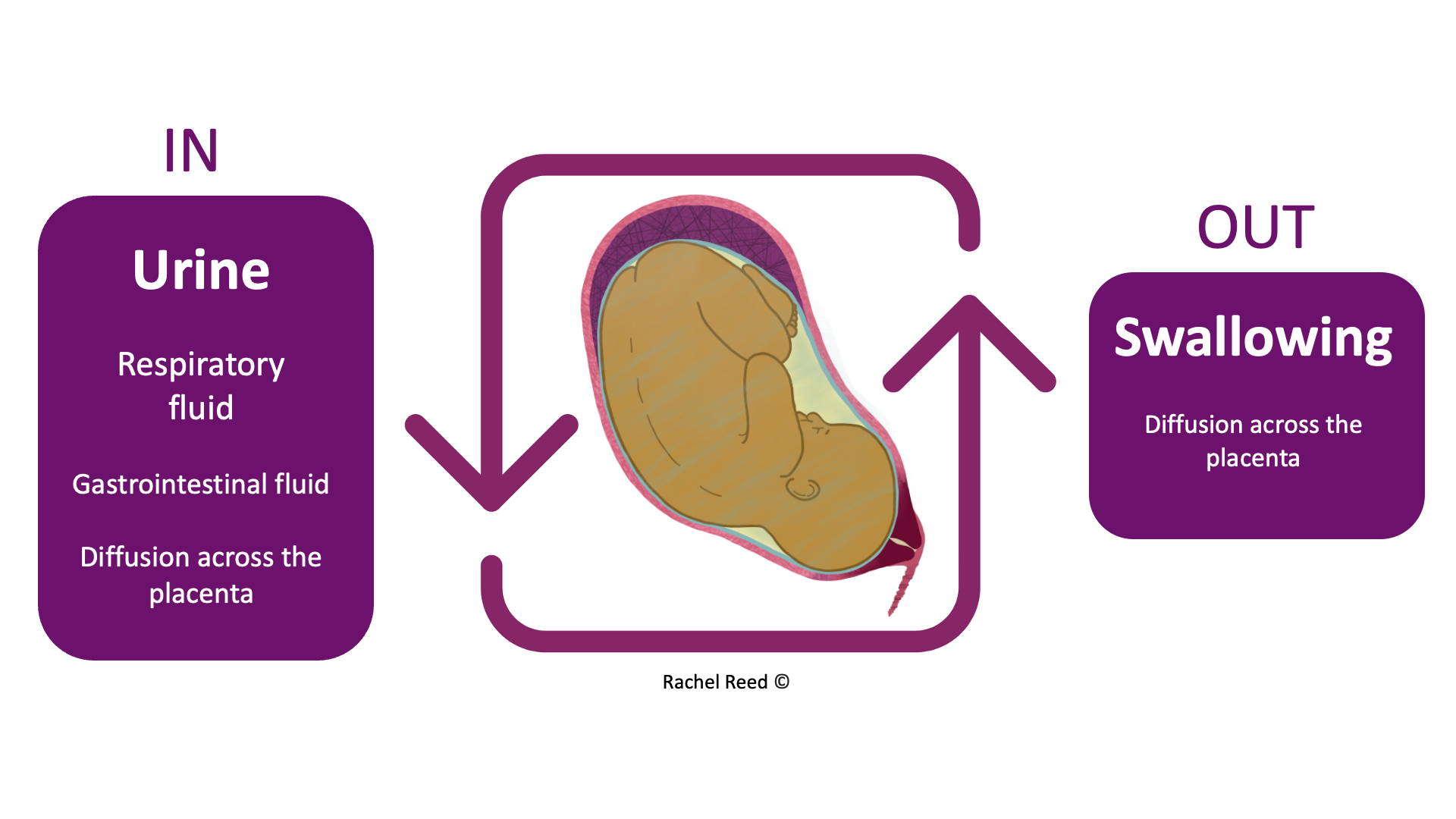
Amniotic fluid is a vital component of pregnancy, surrounding and protecting the baby in the womb. It provides cushioning, helps regulate temperature, promotes lung and digestive system development, aids in muscle and bone growth, and prevents compression of the umbilical cord.

The fluid is primarily water in the early weeks of pregnancy but is later composed mostly of the baby’s urine. It also contains nutrients, hormones, and antibodies.

The amount of amniotic fluid increases until around 36 weeks of pregnancy and then gradually decreases. Too little (oligohydramnios) or too much (polyhydramnios) amniotic fluid can be problematic for both the mother and the baby, although most babies are born healthy even with these conditions.

Normal amniotic fluid is clear or tinted yellow, but green or brown fluid indicates that the baby may have passed their first bowel movement (meconium) in the womb. Meconium in the amniotic fluid can lead to breathing difficulties, known as meconium aspiration syndrome. Some babies may require immediate treatment at birth to prevent complications, while others may be healthy and not require treatment.

Continue Reading