Unlocking the Secrets of Persistent Occipitotransverse Position: A Comprehensive Guide

– Persistent occipitotransverse position
– Malpresentations and malpositions in obstetrics
– Vertex presentation
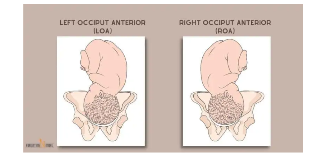
– Left occipito-anterior position
– Right occipito-anterior position
– Defects in the powers
– Pendulous abdomen
– Defects in the passages
– Contracted pelvis
– Uterine anomalies
– Defects in the passenger
– Preterm fetus
– Multiple pregnancy
– Signs suggesting malpresentations
– Nonengagement of the presenting part
– Premature rupture of membranes
– Delay in descent of presenting part
– Complications of malpresentations
– Cord presentation and prolapse
– Prolonged labor
– Obstructed labor
– Instrumental and operative delivery
– Trauma to genital tract
– Postpartum hemorrhage
– Puerperal infection
– Perinatal mortality
– Occipito-posterior position
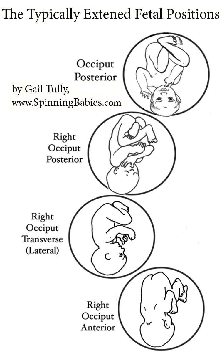
– Right occipito-posterior
– Left occipito-posterior
– Shape of the pelvis
– Anthropoid pelvis
– Android pelvis
– Maternal kyphosis
– Anterior insertion of placenta
– Placenta previa
– Diagnosis of occipito-posterior position
– Ultrasonography
– Lateral view x-ray
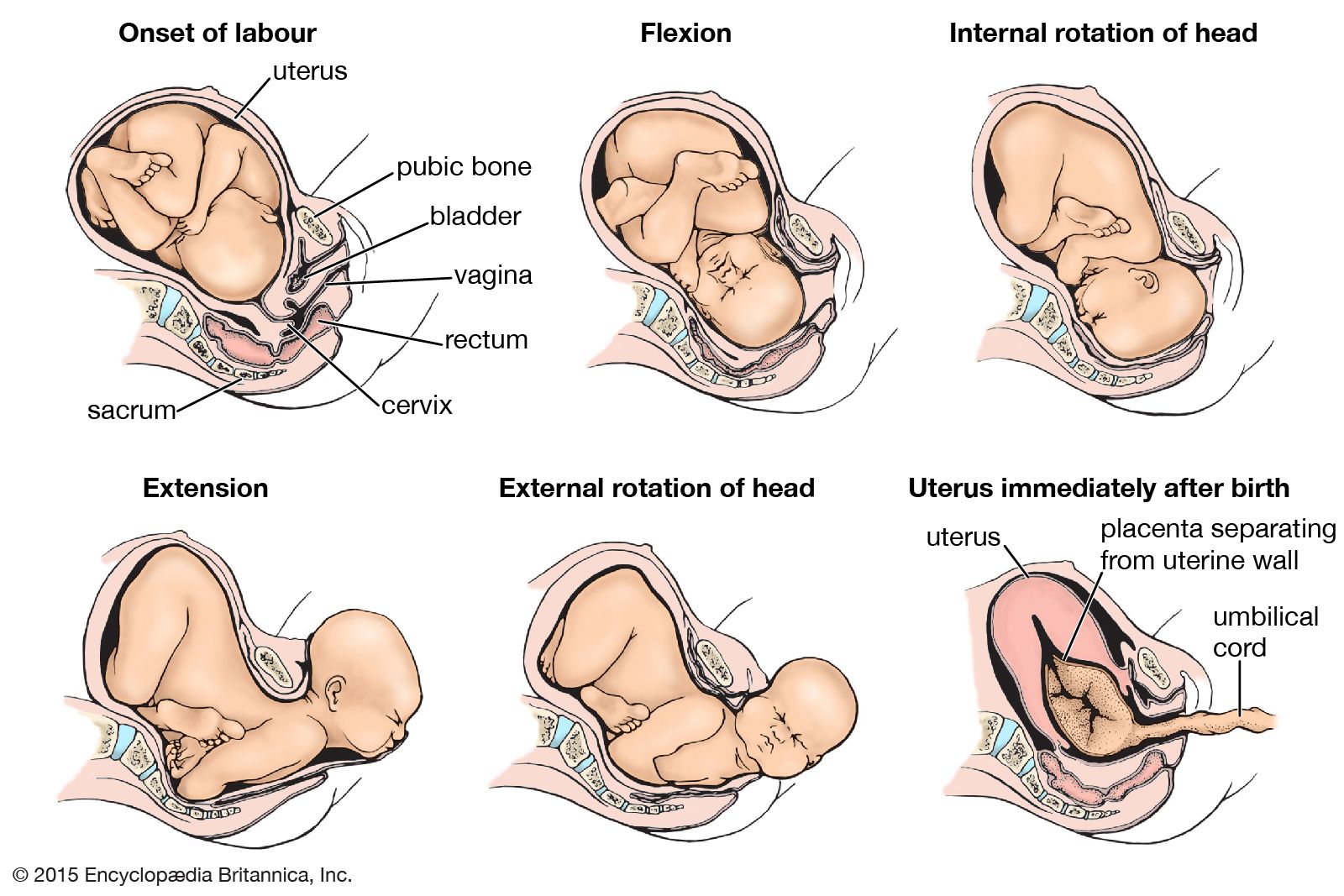
– Mechanism of labor
– Biparietal diameter
– Occipito-frontal diameter
– Deflexion of the occiput
– Normal mechanism of labor
– Abnormal mechanisms
– Deep transverse arrest
– Direct occipito-posterior
– Factors favoring long anterior rotation
– Well-flexed head
– Good uterine contractions
– Roomy pelvis
– Good pelvic floor
– Failure of long anterior rotation
– Uterine inertia
– Contracted pelvis
– Lax or rigid pelvic floor
– Management of labor
– Contracted pelvis
– Presentation or prolapse of cord
– Oxytocin
– Analgesia
– Premature rupture of membranes
– Second stage of labor
– Waiting for 60-90 minutes
– Observing mother and fetus
– Methods for management of persistent occipitotransverse position
– Internal rotation
– Direct occipito-posterior
– Deep transverse arrest
– Vacuum extraction
– Manual rotation
– Forceps
– Kielland’s forceps
– Barton’s forceps
– Scanzoni double application
– Caesarean section
– Craniotomy
– Preferred methods in modern obstetrics
Continue Reading

The Left Mentoanterior Position: Understanding Fetal Presentation during Labor

– Face presentation is a cephalic presentation in which the head is completely extended.
– The incidence of face presentation is about 1 in 300 labours.
– Primary face presentation occurs during pregnancy and may be caused by factors such as anencephaly, loops of the cord around the neck, foetal neck tumors, hypertonicity of neck muscles, dolicocephaly (long antero-posterior diameter of the head), dead or premature foetus, or idiopathic causes.
– Secondary face presentation occurs during labour and may be due to factors such as contracted pelvis, pendulous abdomen, marked lateral obliquity of the uterus, further deflexion of brow or occipito-posterior positions, or other malpresentations such as polyhydramnios and placenta praevia.
– Left mento-anterior (LMA) and right mento-anterior (RMA) are more common positions of face presentation.
– Diagnosis during pregnancy is difficult, but the back is difficult to feel and the limbs may be felt more prominently in mento-anterior position. Ultrasound or X-ray can confirm the diagnosis.
– Diagnosis during labour is done through vaginal examination, which shows identifying features such as supra-orbital ridges, malar processes, nose, mouth, and chin.
– Late in labour, the face may become oedematous (tumefaction), which can be misdiagnosed as a buttock (breech presentation). Differentiating factors include the formation of a triangle with foetal mouth and malar processes as apexes, anus on the same line as ischial tuberosities, feeling of a hard gum through the mouth, and no hard object through the anus.
– The mechanism of labour in mento-anterior position involves descent.
– Engagement by submento-bregmatic diameter: 9.5 cm
– Submental region hinges below the symphysis in flexion position
– Submento-vertical diameter: 11.5 cm
– Biparietal diameter does not pass the plane of pelvic inlet until the chin is below the level of the ischial spines and the face begins to distend the perineum
– In about 2/3 of cases, long anterior rotation of 3/8 circle occurs during mento-posterior position
– In about 1/3 of cases, deep transverse arrest of the face, persistent mento-posterior, or direct mento-posterior occur during mento-posterior position
– Direct mento-posterior cannot be delivered due to obstruction caused by the length of the sacrum and neck
– Management of labour includes excluding foetal anomalies and contracted pelvis
– Spontaneous delivery usually occurs in mento-anterior position during second stage of labour
– Forceps delivery and episiotomy may be indicated in prolonged second stage of labour in mento-anterior position
– Wait for long anterior rotation of 3/8 circle in mento-posterior position during second stage of labour
– Oxytocin is used to compete inertia during this period if there are no contraindications
– Caesarean section is the safest option if long anterior rotation fails or there is foetal or maternal distress
– Manual rotation and forceps extraction or rotation and extraction by Kielland forceps are alternative methods, but are hazardous and not commonly used
– Craniotomy may be performed if the foetus is dead
– Complications may occur, refer to complications of malpresentations and malposition for more information.
– There is an increased risk of trauma to the baby in face presentation, so internal manipulation, vacuum extractors, and manual extraction should be avoided.
– Abnormalities in fetal heart rate are more common in face presentation. Monitoring is crucial during labor.
– Complications of face presentation include prolonged labor, facial trauma, facial edema, skull molding, respiratory distress, spinal cord injury, abnormal fetal heart rate patterns, and low Apgar scores.
– Informed consent should be obtained from the mother, and failure to do so is considered negligence.
– Forceps and oxytocin used during labor can put a baby at risk of complications. Forceps can cause head injuries and oxytocin can deprive the baby of oxygen due to strong contractions.
– Mothers should be given the option of a C-section if facing complications.
– Face presentation babies should be closely monitored and delivered by an experienced physician.
– If negligent practices cause injury to the baby, it can be considered medical malpractice. ABC Law Centers specialize in birth injury cases and offer free legal consultations.

Continue Reading

Discover the Fascinating World of Left Occipitotransverse Birth

– Left Occiput Anterior (LOA)
– Left Occiput Posterior (LOP)
– Left Occiput Transverse (LOT)
– Occiput Anterior (OA)
– Occiput Posterior (OP)
– Left Mentum Anterior (LMA)
– Right Mentum Anterior (RMA)
– Left Frontum Anterior (LFA)
– Right Frontum Anterior (RFA)
– Right Mentum Posterior (RMP)
– Right Mentum Transverse (RMT)
– Left Mentum Anterior (LMA)
– Left Mentum Posterior (LMP)
– Left Mentum Transverse (LMT)
– Mentum Anterior (MA)
– Mentum Posterior (MP)
– Complete Breech
– Frank Breech
– Single or Double Footling Breech
– Kneeling Breech
– Transverse Position
– Sacrum Anterior
– Sacrum Posterior

Continue Reading

The Persistent Occipitoposterior Position: A Guide to Delivery

– Occipito-posterior position is a malposition in which the baby’s back is directed posteriorly during a vertex presentation
– Occipito-posterior position occurs in approximately 10% of labors
– Right occipito-posterior (ROP) is more common than left occipito-posterior (LOP)
– Causes of occipito-posterior position include the shape of the pelvis (anthropoid and android pelvises are common causes), maternal kyphosis, anterior insertion of the placenta, and other malpresentations such as placenta previa, pelvic tumors, pendulous abdomen, polyhydramnios, and multiple pregnancy
– Diagnosis during pregnancy can be done through inspection, palpation, and auscultation
– Complications of occipito-posterior position include premature rupture of membranes, cord presentation and prolapse, prolonged labor, obstructed labor, increased incidence of instrumental and operative delivery, trauma to the genital tract, postpartum hemorrhage, and perinatal mortality
– The persistent occipitoposterior position occurs in about 3% of cases
– Long internal rotation, occurring in about 90% of cases, allows for delivery to proceed as in normal labor
– Direct occipitoposterior position occurs in about 6% of cases and can be managed by spontaneous delivery or with the aid of outlet forceps
– Deep transverse arrest occurs in 1% of cases and requires vacuum extraction or manual rotation and extraction with forceps
– Vacuum extraction can be used for rotation of the head, while manual rotation and extraction with forceps is done under general anesthesia
– Different types of forceps, such as Kielland’s forceps and Barton’s forceps, can be used for rotation and extraction of the head in persistent occipitoposterior position
– The Scanzoni double application method, which is considered hazardous, involves applying forceps twice for rotation and extraction
– If other methods fail, a Caesarean section may be necessary. Other indications for a C-section include contracted pelvis, placenta previa, prolapsed pulsating cord, and elderly primigravida
– Craniotomy may be performed if the fetus is dead
– Vacuum extraction and Caesarean section are the commonly used methods in modern obstetrics for managing persistent occipitoposterior position
Continue Reading

Left Occipitoanterior: A Guide to Fetal Positioning and Birth

– LOA: Left Occiput Anterior. Occiput (back of the baby’s head) is on the left side of the pelvis, baby’s back is closest to the mother’s belly.
– LOP: Left Occiput Posterior. Occiput is on the left side of the pelvis, baby’s back is closest to the mother’s spine.
– LOT: Left Occiput Transverse. Occiput is on the left side of the pelvis, baby’s back is on the left side of the mother’s body.
– OA: Occiput Anterior. Occiput is in the central portion of the pelvis, baby’s back is lined up directly central on the mother’s belly.
– OP: Occiput Posterior. Occiput is in the central portion of the pelvis, baby’s back is lined up against mother’s spine.
– RMA: Right Mentum Anterior. Mentum (chin) is on the right side of the pelvis, back is closest to the mom’s belly.
– RMP: Right Mentum Posterior. Mentum is on the right side of the pelvis, back is closest to the mom’s spine.
– RMT: Right Mentum Transverse. Mentum is on the right side of the pelvis, back is on mom’s right side.
– LMA: Left Mentum Anterior. Mentum is on the left side of the pelvis, back is closest to the mom’s belly.
– LMP: Left Mentum Posterior. Mentum is on the left side of the pelvis, back is closest to the mom’s spine.
– LMT: Left Mentum Transverse. Mentum is on the left side of the pelvis, back is on mom’s left side.
– MA: Mentum Anterior. Mentum is in the central portion of the pelvis, back is directly lined up the center of mom’s abdomen.
– MP: Mentum Posterior. Mentum is in the central portion of the pelvis, back is directly lined up against mom’s spine.
– RFA: Right Frontum Anterior. Frontum (forehead) is on the right side of the pelvis, back is closest to mom’s belly.
– RFP: Right Frontum Posterior. Frontum is on the right side of the pelvis, back is closest to mom’s spine.
– Complete Breech: Baby is sitting cross-legged in the pelvis, sacrum is presenting part.
– Frank Breech: Baby is in a pike position with legs extended towards the face, sacrum is presenting part.
– Single or Double Footling Breech: Baby has one or both feet lower in the pelvis than the rest of the body.
– Kneeling Breech: Baby is kneeling, knees enter the pelvis first.
– Right sacrum anterior: Sacrum is on the right side of the pelvis, back is closest to the mother’s belly.
– Right sacrum posterior: Sacrum is on the right side of the pelvis, back is closest to the mother’s spine.
– Right sacrum transverse: Sacrum is on the right side of the pelvis, back is on the mother’s right side.
– Left sacrum anterior: Sacrum is on the left side of the pelvis, back is closest to the mother’s belly.
– Left sacrum posterior: Sacrum is on the left side of the pelvis, back is closest to the mother’s spine.
– Left sacrum transverse: Sacrum is on the left side of the pelvis, back is on the mother’s left side.
– Sacrum anterior: Sacrum is in a central portion of the pelvis, baby’s back is directly in the center of the mother’s abdomen.
– Sacrum posterior: Sacrum is in a central portion of the pelvis, baby’s back is lined up against the mother’s spine.
– Transverse position: Baby’s spine and mother’s spine make a right angle, presenting part is usually the shoulder.
– Optimal position for birth: Baby is head down and anterior, with the back close to the mother’s belly.

Continue Reading

Fetal Posture: Unlocking the Secrets of Optimal Development

– Fetal presentation before birth refers to the position of the baby in the uterus right before delivery.
– The most common position is cephalic occiput anterior, where the baby’s head is down and face down.
– Another position is cephalic occiput posterior, where the baby’s head is down but face up. This can make labor longer and may require manual rotation or assisted delivery.
– Breech presentation occurs when the baby’s feet or buttocks are in place to come out first during birth. This happens in about 3% to 4% of babies.
– The most common type of breech presentation is frank breech, where the baby’s knees aren’t bent and the feet are close to the baby’s head.
– A procedure called external cephalic version can be performed to try to move the baby into a head-down position if they are in a frank breech position.
– If the procedure is not successful or the baby moves back into a breech position, the delivery options should be discussed with the healthcare team.
– A complete breech presentation is when the baby has both knees bent and both legs pulled close to the body.
– An incomplete breech presentation is when one or both of the legs are not pulled close to the body, and one or both of the feet or knees are below the baby’s buttocks.
– If a baby is in a complete or incomplete breech presentation after 36 weeks of pregnancy, the health care professional may try to move the baby into a head-down position using external cephalic version.
– If the procedure is not successful or if the baby moves back into a breech position, alternative delivery options should be discussed with the health care team.
– A transverse lie is when the baby is lying horizontally across the uterus.
– If the baby is in a transverse lie at week 37 of pregnancy, the health care professional may try to move the baby into a head-down position using external cephalic version.
– If the procedure is not successful or if the baby moves back into a transverse lie, alternative delivery options should be discussed.
– If pregnant with twins and only one twin is head down, the health care provider may deliver the first twin vaginally and then suggest delivering the second twin in the breech position or try to move the second twin into a head-down position using external cephalic version.
– Delivery by C-section may be suggested for the second twin.

Continue Reading

Transverse Arrest: Causes, Symptoms, and LifeSaving Interventions

– The study will be conducted in hospitals in Australia that have 2,000 or more deliveries per year.
– The intervention will be performed by experienced obstetricians or midwives.
– The inclusion criteria for the study include being at least 18 years old, having a singleton pregnancy, being at least 37 weeks gestation, planning a vaginal birth, having a cephalic presentation, and having confirmed occiput transverse position.
– The exclusion criteria include clinical suspicion of cephalopelvic disproportion, previous caesarean section, brow or face presentation, pathologic CTG, fetal scalp abnormalities, chorioamnionitis, intrapartum hemorrhage, maternal diabetes, suspected fetal bleeding disorder, and major fetal abnormalities.
– The intervention, called manual rotation, is performed when the woman is at full cervical dilatation and the fetal position is occiput transverse. The technique used will be at the discretion of the operator and may involve applying pressure to the lambdoid suture or flexion and rotation of the fetal head.
– The comparator is the standard practice of waiting until full dilatation is reached before performing any intervention.
– The primary outcome measure is operative delivery (vacuum, forceps, or caesarean section).
– Secondary outcomes include the rate of caesarean section, serious maternal morbidity or mortality, and serious perinatal and neonatal morbidity and mortality.
– Prolonged second stage of labour is defined differently based on parity and use of epidural analgesia.
– Other outcomes measured include length of second stage, time from randomization to delivery, estimated blood loss, perineal or vaginal trauma requiring suturing, length of hospital stay, and outcomes for operative delivery.
– Secondary outcomes assessed include breastfeeding status, satisfaction with birth, depression, health-related quality of life, and pelvic floor function.
– The sample size for the study is 416 participants, based on power calculations and previous studies.
– The study aims to evaluate the effectiveness of manual rotation in reducing adverse outcomes during transverse arrest.
– The primary outcome measure is serious morbidity and/or mortality, which includes factors such as neonatal injury, low Apgar score, abnormal cord pH levels, birth trauma, seizures, ventilation, tube feeding, NICU admission, and neonatal jaundice.
– Data collection will occur at three possible time points: antenatal, latent phase of labor, or active phase of the first stage of labor.
– Informed consent will be obtained, and participants will be informed of the potential risks of manual rotation.
– An ultrasound will be performed at full dilatation, and the fetal position will be confirmed by a second ultrasound.
– The treatment allocation is recorded on a randomization sheet kept by the investigator.
– The findings are recorded by the investigator.
– The data will be stored securely and checked for accuracy.
– The analysis will be done according to specific guidelines and will include variables such as maternal factors, gestation, and neonatal gender.
– Subgroup analyses will also be performed based on different techniques of manual rotation and operator ability.
– A Data and Safety Monitoring Committee has been established to ensure the safety of the trial participants.
– Any adverse events will be reported to this committee, and serious complications will be referred to them as well.
– The study has received approval from the Ethics Review Committee of the Sydney Local Health District in Sydney, Australia, with the protocol number X110410.

Continue Reading

Fetal Position: Understanding its Benefits, Risks, and Impact

List:

– Fetal position: refers to the positioning of the body of a prenatal fetus as it develops.
– Back is curved
– Head is bowed
– Limbs are bent and drawn up to the torso
– Compact position
– Minimizes injury to the neck and chest
– Newborn mammals, especially rodents, remain in a fetal position after birth
– Some people assume a fetal position when sleeping, especially when the body becomes cold
– Bodies have been buried in fetal position in certain cultures
– Individuals who have suffered extreme physical or psychological trauma may assume a similar compact position to protect themselves
– Drug addicts and individuals with anxiety may adopt this position during withdrawal or panic attacks
– Playing dead in a fetal position is recommended as a strategy to end a bear attack.

Continue Reading