Understanding Annexitis: Causes, Symptoms, and Effective Treatment Options

– Adnexitis is inflammation of the ovaries or fallopian tubes.
– It can cause female infertility.
– Adnexitis is caused by ascending bacterial infections, such as gonorrhea, chlamydia, and bacterial vaginosis, which are sexually transmitted diseases.
– Common symptoms of adnexitis include irregular and painful menstruation, vaginal discharge with strong or unpleasant odor, bleeding after intercourse, discomfort when urinating, high fever, and bleeding between periods.
– Complications of adnexitis can include chronic pelvic pain, ectopic pregnancies, and female infertility.
– Treatment for adnexitis usually involves antibiotics, and it is important to complete the treatment even if symptoms improve.
– Prevention methods include using condoms, regular gynecological check-ups, tests for STDs, practicing good intimate and sexual hygiene, and abstaining from sexual intercourse.
– Bacteria reach the fallopian tubes and ovaries through the vagina.
– Young, sexually active women are at higher risk, especially during the menstrual period and with frequent partner swapping.
– Gynaecological procedures like using a spiral increase the risk.
– Symptoms include fever, lower abdominal pain, malaise, nausea, vomiting, and foul-smelling vaginal discharge.
– Diagnosis is done through a gynaecological examination, with pain on palpation being typical.
– Chlamydia bacteria can be detected in vaginal smears.
– Ultrasound and blood tests may also be used.
– Treatment includes antibiotics, pain-relievers, and anti-inflammatory medication.
– Laparoscopy may be necessary if there are abscesses or peritoneal infection.
– If the inflammation is caused by an infected spiral, it must be removed.
– Untreated or late-treated inflammation can lead to infertility and growths in the fallopian tubes, increasing the risk of ectopic pregnancy.

Continue Reading

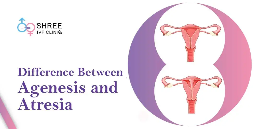
Atresia of Vagina: Causes, Symptoms, Diagnosis, and Treatment

List:

1. Vaginal Atresia
2. Bardet-Biedl syndrome
3. Fraser syndrome
4. Mayer-Rokitansky-Küster-Hauser (MRKH) syndrome
5. Vision loss
6. Obesity
7. Kidney problems
8. Intellectual disorders
9. Skin-covered eyes
10. Joined fingers and toes
11. Abnormalities of the urinary tract
12. Underdeveloped or absent vagina and uterus
13. Kidney anomalies
14. Cloacal malformation
15. Surgical treatment
16. Vaginal replacement techniques

Continue Reading

Understanding Subserous Myoma: Causes, Symptoms, and Treatment Options

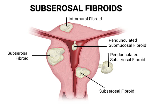
– Subserosal fibroids are benign tumors that grow on the exterior of the uterus.
– The cause of subserosal fibroids is unknown, but genetics and hormones may play a role.
– African American women have a higher risk of developing fibroids.
– Women who have never had children or started puberty early (before age 12) also have a higher risk of fibroids.
– Subserosal fibroids can cause complications during pregnancy, such as lower birth weight and the need for a cesarean delivery.
– Symptoms of subserosal fibroids include a feeling of heaviness or fullness, frequent urination, constipation, and bloating.
– Subserosal uterine fibroids are diagnosed through a pelvic exam and additional tests.
– Subserosal fibroids should be treated to alleviate symptoms and avoid complications.
– Uterine Fibroid Embolization (UFE) is a non-surgical procedure that shrinks fibroids by cutting off their blood supply.
– Other treatment options include hysterectomy and myomectomy.
– Subserosal fibroids are a type of uterine fibroid that are benign and not cancerous.
– They can cause discomfort and impact nearby organs such as the bladder and bowels.
– Symptoms can include abdominal cramping, pain in the lower back and legs, and pain during sex.
– They can also lead to constipation and frequent urination.
– If subserosal fibroids are pedunculated (growing on a stalk) and the stalk becomes twisted, they can cause severe pain by cutting off the blood supply.
– Subserosal fibroids may have less impact on fertility compared to other types of fibroids, but if they grow larger during pregnancy, they can limit the space for the baby to grow and cause difficulties during childbirth.
– Fibroids are almost always non-cancerous and fibroid cancer is extremely rare.
– Subserosal fibroids can cause pain, infertility, and complications during pregnancy.
– Around 25 to 30 percent of reproductive-age women experience fibroid symptoms between the ages of 35 and 50, with most of these being subserosal fibroids.
– Symptoms of subserosal fibroids can include pain, abnormal bleeding, and abdominal discomfort.
– The causes of subserosal fibroids are not known, but hereditary factors and hormonal influence may increase the risk.
– Subserous myoma, or subserosal fibroids, are growths that appear on the uterine wall.
– Excess weight is often associated with the development of fibroids.
– Fibroids usually develop between the ages of 30 and 50.
– Subserosal fibroids can affect fertility by blocking the cervix or fallopian tubes.
– They can also cause pain and contractions, potentially leading to pre-term delivery, poor development of the fetus, miscarriage, and require a caesarian delivery.
– Treatment for subserosal fibroids depends on their condition, size, and location.
– Treatment options include close observation, subserosal fibroid removal, medications, uterine fibroid embolization, hormone treatment, fibroid surgery, and leiomyoma ablation.

Continue Reading
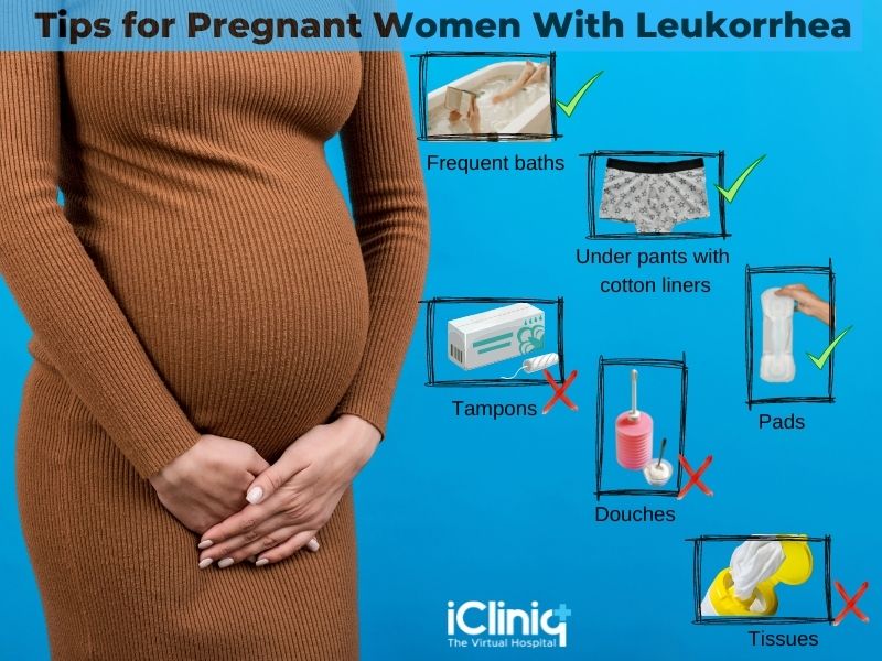
Fluor Albus: Understanding Causes, Symptoms, and Treatments

List relevant to the keyword ‘fluor albus’:

– Leukorrhea, also known as fluor albus or “the whites,” is a thick, whitish, yellowish or greenish vaginal discharge.
– It is a non-pathological symptom usually caused by inflammatory conditions of the vagina or cervix.
– Leukorrhea can be confirmed by finding >10 WBC per high-power field under a microscope when examining vaginal fluid.
– Yellowish or odorous discharge may indicate a bacterial infection or STD.
– Leukorrhea can also be caused by parasitic protozoan called Trichomonas vaginalis, which causes symptoms such as burning sensation, itching, and frothy discharge.
– Treatment includes antibiotics, such as metronidazole, clindamycin, or tinidazole, depending on the underlying cause.
– Leucorrhoea, also known as white discharge, is a common condition characterized by thick, yellowish or white vaginal discharge.
– It can be a sign of various gynaecological conditions and should be evaluated and treated promptly.
– Conventional treatment for leucorrhoea offers short-term relief but can have mild side effects such as burning, itching, and vaginal discomfort.
– Homeopathy has proven to be effective in treating leucorrhoea, with a case study showing positive results and reduced chances of recurrence.
– Homeopathic treatment is personalized based on the patient’s specific symptoms and aims to address the root cause of the condition.
– Dr Batra’s® has over 35 years of experience in providing treatment for leucorrhoea, with a success rate of 96% in treating women’s health problems including leucorrhoea.
– Homeopathy is said to help manage physical, mental, and emotional symptoms, and it is recommended for both acute and chronic cases.

Continue Reading

Understanding Adnexal Disease: Causes, Symptoms, and Treatment Options

List:
1. Adnexal cysts, including ovarian cysts, may resolve on their own without treatment.
2. Hormonal contraceptives may be prescribed to prevent the formation of new cysts.
3. Fallopian tube cysts may be monitored using ultrasound if they are asymptomatic.
4. Larger or symptomatic paraovarian or paratubal cysts may require surgical removal.
5. Surgeons must take caution during the procedure to avoid damaging the fallopian tube.
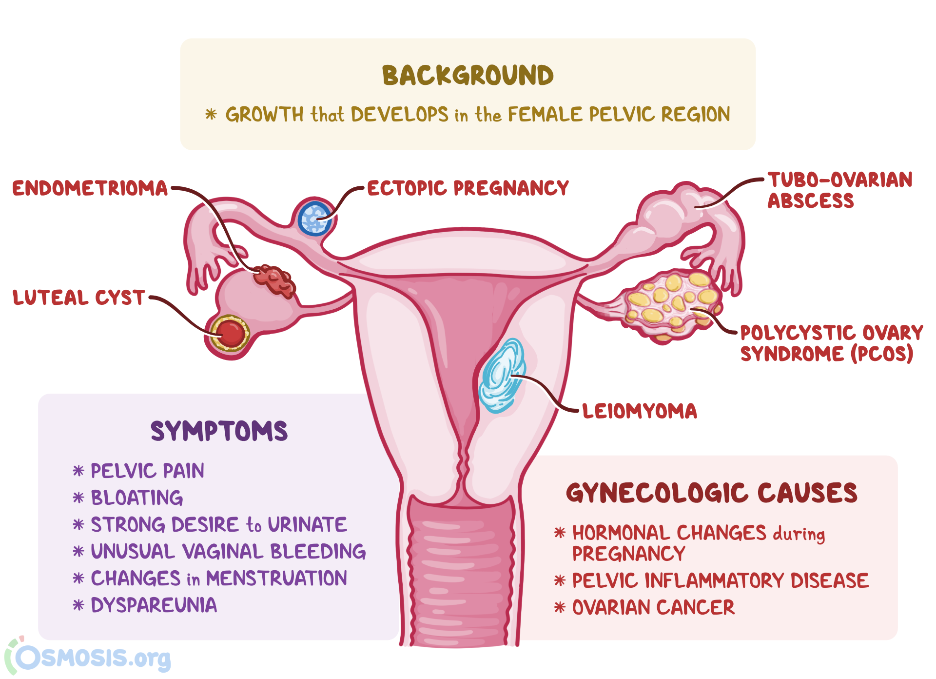
6. Adnexal masses are growths that develop around the uterus, commonly in the ovaries, fallopian tubes, and neighboring tissues.
7. Adnexal tumors can be either fluid-filled or solid.
8. They can occur at any age and often resolve within a few months.
9. Adnexal tumors are more common during the childbearing years.
10. Around 5% to 10% of women in the United States will experience an adnexal mass during their lifetime.
11. The majority of adnexal tumors are benign, but approximately 15% to 20% are malignant.
12. Postmenopausal individuals have a higher risk of malignant adnexal tumors compared to premenopausal individuals.

Continue Reading

Unlocking the Mysteries of Uterus Subseptus: A Comprehensive Guide

– A subseptate uterus is a mild form of congenital uterine anomaly.
– It is often considered a normal variant.
– The condition involves the presence of a partial septum within the uterus.
– The septum does not extend to the cervix.
– The angle of the central point of the septum is acute (<90°). - The external uterine contour is uniformly convex or has an indentation <10 mm. - The prevalence of a septate uterus is approximately 55% among uterine anomalies. - It is classified as a class V Müllerian duct anomaly. - A septate uterus is associated with subfertility, preterm labor, and reproductive failure in approximately 67% of cases. - 15% of women with recurrent pregnancy loss have a septate uterus. - Concurrent renal anomalies may be associated with a septate uterus. - A septate uterus is considered a type of uterine duplication anomaly that results from the partial or complete failure of resorption of the uterovaginal septum after fusion of the paramesonephric ducts. - There are different subtypes of a septate uterus, including a partial septum (subseptate uterus) and a complete septum that extends to either the internal or external cervical os. - Hysterosalpingogram alone has an accuracy of only 55% in differentiating a septate uterus from a bicornuate uterus. - Ultrasound can show that the echogenic endometrial stripe is separated at the fundus by the septum, which is isoechoic to the myometrium. The external uterine contour should be convex, flat, or mildly concave. - MRI is considered the preferred imaging modality for diagnosing a septate uterus. On MR images, the septate uterus appears normal in size, and each endometrial cavity appears smaller than a normal cavity. The septum may be composed of fibrous tissue, myometrial tissue, or both. - The treatment for a septate uterus involves shaving off the septum during hysteroscopy (metroplasty) to form a single uterine cavity without perforating the uterus. Resection of the septum in the uterus has been shown to improve outcomes, with a reported decrease in the spontaneous abortion rate from 88% to 6% after hysteroscopic metroplasty. Differential diagnosis considerations for a septate uterus include a bicornuate uterus, and it is important to differentiate between the two due to different clinical and interventional approaches. Ultrasound or MRI may also be used for diagnosis.

Continue Reading

Leukorrhea: Causes, Symptoms, and Effective Treatment Options

Vulvovaginal candidiasis, yeast infection, candida albicans infection, other types of infection, fungal infection, diabetes, antibiotic medication, estrogen levels, contraceptive pill, pregnancy, weakened immune system, HIV infection, steroid medication, thick and lumpy discharge, vaginal itching, burning sensation, stinging sensation during urination, pain during intercourse, redness, antifungal medication, creams, vaginal suppositories, oral medication, clotrimazole, miconazole, tioconazole, fluconazole.

Continue Reading