Key Considerations for Abdominal SalpingoOophorectomy: Benefits, Risks, Recovery, and Alternatives

– Abdominal salpingo-oophorectomy is a surgical procedure to remove the uterus, cervix, ovaries, and fallopian tubes.
– It is performed through an incision in the abdomen.
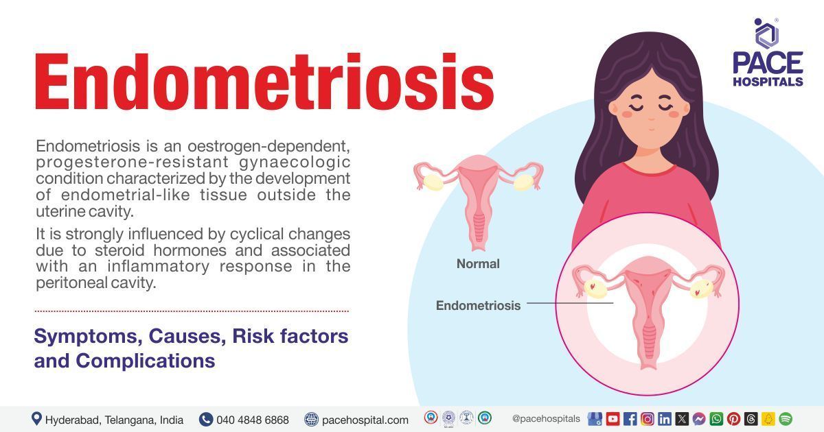
– Reasons for this surgery may include heavy periods, endometriosis, uterine fibroids, and cancer.
– The incision can be either horizontal or vertical.
– Risks of the procedure include bleeding, infection, damage to surrounding organs, and the possibility of further surgery.
– Preparation for the surgery includes fasting before the procedure and arranging for transportation.
– Recovery usually requires a hospital stay of about 2 nights and a full recovery time of 6 weeks.
– Vaginal bleeding and discharge are normal after surgery and should gradually decrease.
– Strenuous exercise, heavy lifting, and sexual activity should be avoided for 6 weeks after surgery.
– Medical attention should be sought if there is fever, severe nausea/vomiting, or abdominal pain, heavy bleeding, or redness/swelling/discharge from incisions.

Continue Reading

Anovarian Syndrome: Understanding Symptoms, Causes, and Treatment Options

List:

1. Conjugating verbs
2. Declining nouns
3. Declining adjectives
4. Declining pronouns
5. Declining numerals
6. Gender of nouns
7. Declension of nouns
8. Degrees of comparison for adjectives
9. Conjugation tables for English
10. Conjugation tables for German
11. Conjugation tables for Russian
12. Conjugation tables for French
13. Conjugation tables for Italian
14. Conjugation tables for Portuguese
15. Conjugation tables for Spanish
16. Learning rules of conjugation and declension
17. Viewing translations in various contexts
18. Filtering search results
19. Displaying exact hits
20. Editing or changing records using a pen tool
21. Accessing further information indicated by an arrow
22. Searching on external websites
23. Closing additional information
24. Using Google Translate to pronounce search results
25. Showing translation information.

Continue Reading

Exploring Borderline Ovarian Tumors: Causes, Symptoms, Treatment

– Borderline ovarian tumors, also known as “low malignant potential” tumors, are not completely benign but also not invasive.
– Diagnosis of a borderline ovarian tumor can be suspected through imaging such as ultrasound or MRI, but the diagnosis is confirmed through microscopic assessment after surgery.
– Surgery is the primary treatment for borderline tumors, even if they have spread to other areas. Endocrine therapy may be considered in some cases.
– Fertility-sparing surgery or preservation of an unaffected ovary is often possible in young patients to avoid surgical menopause.
– Minimally invasive (laparoscopic) surgery is preferred for faster recovery and reduced risk of complications.
– Long-term follow-up care is necessary for patients with borderline ovarian tumors, with periodic imaging recommended for those with one ovary remaining.
– Approximately 10% of borderline tumors may recur, and surgical treatment is often used due to the limited response to systemic treatments like chemotherapy.
– Minimally invasive techniques are used for recurrent tumors whenever possible, and complex debulking surgeries may be performed to remove as much tumor as possible.

Continue Reading

Understanding Atresia of Cervix: Causes, Symptoms, and Treatment

– Cervical agenesis
– Congenital disorder
– Absence of cervix
– Deformation of cervix
– Connecting structure between uterus and vagina
– Early adolescence
– Amenorrhea
– Cyclic pelvic pain
– Hematocolpos
– Endometriosis
– Pelvic adhesions
– Fetal development
– Paramesonephric duct
– Magnetic resonance imaging (MRI)
– Ultrasound
– Hormonal therapy
– Surgery
– Poor surgery outcomes
– Vaginal deformities
– Obstruction of menstrual flow
– Hematosalpinx
– Endometrioma
– Oral contraceptives
– Hysterectomy
– Neovaginoplasty
– Recanalization of cervix
– Low success rate
– 1 in 80,000 females

Continue Reading

Exploring the Intricate Cavity of the Uterus: Unveiling Mysteries

List of pertinent information about the cavity of the uterus (no duplications):

– The uterine cavity is the inside portion of the uterus.
– It is triangular in shape and formed by the internal surface of the body of the uterus.
– The base of the cavity is located between the openings of the fallopian tubes.
– The apex is the internal opening of the uterus that connects to the cervix.
– The part of the uterine cavity that enters the openings of the fallopian tubes is a narrow, flattened area.
– Abnormalities of the womb or congenital uterine abnormalities refer to women who have a womb that is different in shape or size from the norm.
– These abnormalities can be discovered during an ultrasound scan or if a woman experiences miscarriage, bleeding, or difficulties conceiving.
– Women with womb abnormalities may have an increased risk of miscarriage, preterm birth, or fertility problems depending on the shape of the womb.
– Women with bicornuate wombs have a slightly higher risk of miscarriage and preterm birth.
– Women with a unicornate womb have half the size of a normal womb and an increased risk of ectopic pregnancy, late miscarriage, or preterm birth.
– Women with a didelphic womb, which is split in two, may have a small increased risk of preterm birth.
– Women with a septate/subseptate womb may have an increased risk of miscarriage, preterm birth, or fertility problems.
– Septate wombs may cause difficulties with conception.
– Septate wombs have an increased risk of early miscarriage and preterm birth.
– Babies in later pregnancy with septate wombs may not be in a head-down position, leading to a higher likelihood of needing a C-section.
– Many women with congenital uterine abnormalities, like a septate uterus, are not aware of their condition.
– Surgery to resect the septum before pregnancy is common for women with a septate uterus, but it was not recommended by the consultant in this case.
– Despite having a complete septate uterus, the woman in the case was able to carry her baby to term but had an elective C-section because the baby was breech.
– An arcuate womb has a dip at the top but resembles a normal womb.
– Having an arcuate womb does not increase the risk of preterm birth or early miscarriage.
– An arcuate womb may increase the risk of late miscarriage.
– Babies in later pregnancy with an arcuate womb may not be in a head-down position, increasing the likelihood of needing a C-section.

Continue Reading

Ovarian Tumour: Understanding Symptoms, Diagnosis, Treatment, and Support

– Ovarian tumors can be noncancerous (benign) or cancerous (malignant).
– Symptoms of ovarian tumors include stomach bloating, increased belly size, stomach or pelvic pain, constipation, difficulty urinating or urinating frequently, feeling full more quickly, painful cramps during menstruation, lower back pain, nausea or vomiting, pain during sex, and vaginal bleeding after menopause.
– The causes of ovarian tumors are still being studied.
– There are two broad categories of ovarian tumors: benign and malignant.
– Benign tumors can develop into malignant tumors if left untreated.
– Types of ovarian tumors include surface epithelial tumors, stromal tumors, and germ cell tumors.
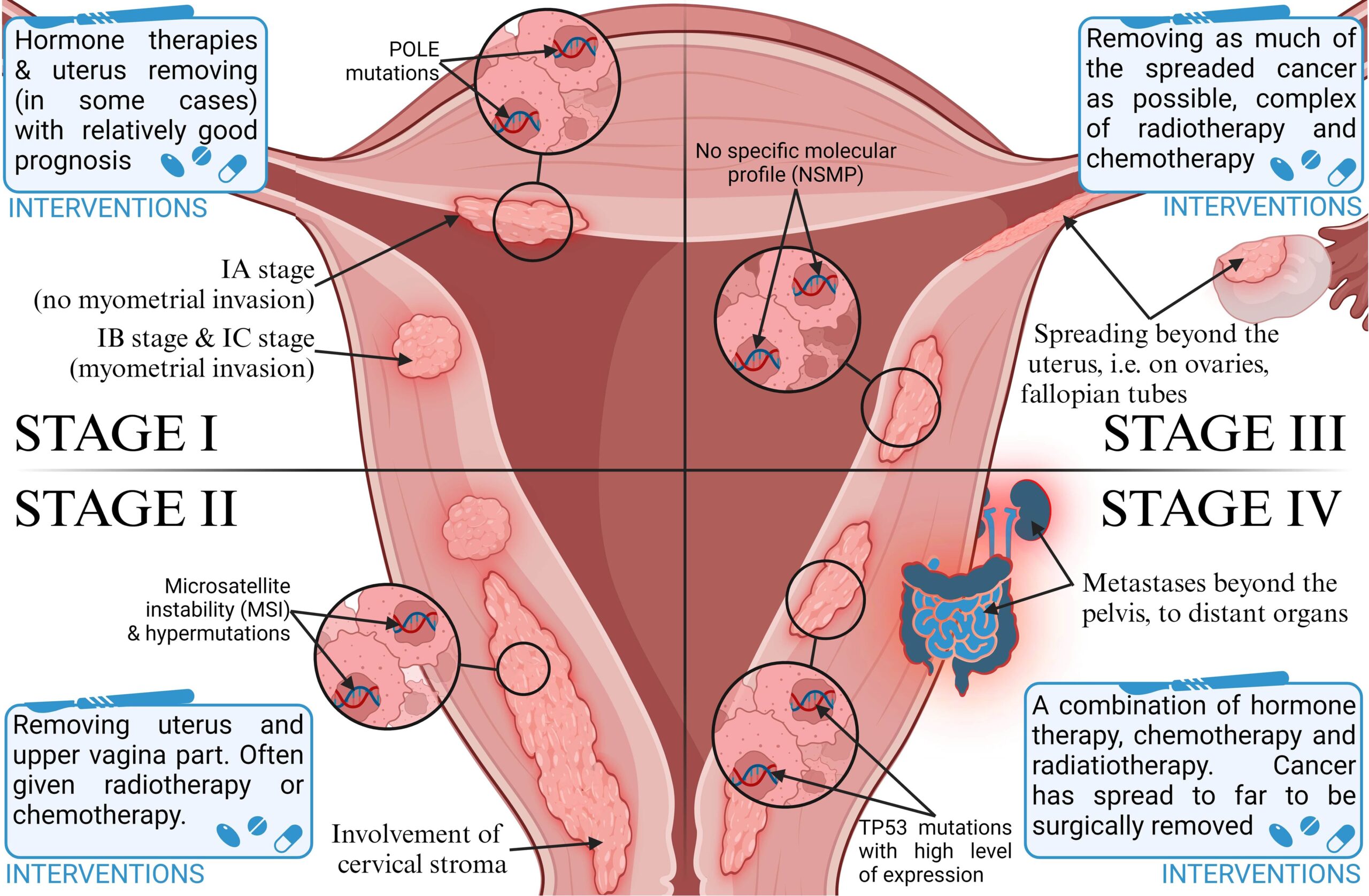
– Ovarian tumors can be classified into four stages if they are malignant: Stage I, Stage II, Stage III, and Stage IV.
– Risk factors for ovarian tumors include age, family history, genetic mutations (BRCA1 or BRCA2), and obesity.
– Women who have never conceived and carried a pregnancy or have done so after age 35 may have an increased risk of ovarian cancer.
– Continuous use of estrogen after menopause can also increase the risk of ovarian cancer.
– The reason for ovarian tumors forming is unknown, so prevention methods are currently unknown.
– To lower the risk of ovarian cancer, steps that can be taken include eating a healthy diet, exercising regularly, maintaining a healthy weight, not smoking, and taking birth control pills.
– The information in the article is for educational purposes only and should not replace advice from a healthcare provider.

Continue Reading

Myoma of Uterus: Understanding Symptoms, Treatment, and Prevention

– A myoma, also known as a uterine fibroid or leiomyoma, is a common noncancerous tumor that grows in or around the uterus.
– Myomas can vary in size and may cause symptoms such as abdominal pain and heavy menstrual bleeding.
– Risk factors for myomas include a family history of myoma, obesity, and age.
– More than half of all people with a uterus will experience a myoma by age 50.
– Myomas can be categorized by location, size, and symptoms they may cause.
– Types of myomas include intramural myomas (located within the wall of the uterus), subserosal myomas (grow on the outside of the uterine wall), pedunculated myomas (develop a stalk or stem attaching them to the uterus), and submucosal myomas (found just under the lining of the uterus).
– A large myoma is considered to be 10 centimeters or more in diameter.
– Emergency room visits for myoma symptoms have increased from 2006 to 2017, including pelvic pain and heavy bleeding.
– Myomas are noncancerous growths that can cause various symptoms depending on their size and location.
– Symptoms of myomas include heavy and painful periods, bleeding between periods, pelvic pain, abdominal pressure, a feeling of fullness in the lower abdomen, constipation, diarrhea, frequent urination, pain during sex, lower back pain, trouble getting pregnant, fatigue, and weakness.
– Myomas are not life-threatening but can cause complications such as heavy blood loss or organ obstruction.
– Myomas are a top cause of hysterectomy surgeries.
– If a myoma bursts, immediate medical care is necessary.
– The exact cause of myomas is unknown but is likely associated with hormone activity, particularly high levels of estrogen and progesterone.
– Risk factors for myomas include a family history of the condition, obesity, high blood pressure, age, and certain dietary factors.
– Myomas are more common among Black people with a uterus.
– Diagnosis of myomas involves a series of steps, including medical history, physical examination, imaging tests such as ultrasound or MRI, and sometimes a biopsy.
– Treatment options for myomas include medication, noninvasive procedures, surgery, or a combination of therapies.
– Medications that may be used include over-the-counter pain medications, iron supplements, and birth control methods.
– Surgical options include laparoscopic myomectomy and uterine fibroid embolization (UFE).
– Lifestyle changes including dietary changes, exercise, stress management, and weight loss may improve symptoms and overall health.
– Complications of untreated myomas include fertility issues, pregnancy complications, and the need for cesarean delivery.
– It is important to discuss myomas with a healthcare provider before pregnancy for potential complications.
– Uterine fibroids, also known as myomas, are non-cancerous tumors that grow in the uterus.
– The most common symptom of myomas is vaginal bleeding.
– Other symptoms of myomas include heavy bleeding, anemia, fatigue, painful intercourse, pain, bleeding, or discharge from the vagina if myomas become infected, a feeling of pressure or lump in the abdomen, difficulties urinating, dribble at the end of urination, or urine retention if a myoma blocks the flow of urine.
– Myomas affect 20 percent of women in their childbearing years.
– The signs and symptoms of uterine fibroids include abnormal bleeding, pelvic masses, pelvic pain, infertility, and pregnancy complications.
– There are five types of uterine fibroids: intramural fibroids, subserosal fibroids, submucosal fibroids, pedunculated fibroids, and intracavitary fibroids.
– Between 70 and 80 percent of women develop a fibroid tumor by the time they reach age 50.
– Estrogen seems to activate the growth of uterine fibroids, and they shrink after menopause, but hormone therapy after menopause may cause their symptoms to continue.
– Factors associated with the development of uterine fibroids include race, age, early menstruation, caffeine and alcohol intake, genetics, obesity, high blood pressure, and diet.
– Nearly one-third of women with uterine fibroids seek treatment because of the severity of their symptoms.
– Treatment options for uterine fibroids include hormonal contraception, intrauterine devices, antifibrinolytic nonsteroidal agents, endometrial ablation, medications called gonadotropin-releasing hormone agonists, myomectomy, MRI-guided ultrasound surgery, and uterine fibroid embolization.
– Uterine fibroid embolization is a minimally invasive option that blocks the blood supply to fibroids, causing them to shrink and die.
– Myoma, leiomyoma, and uterine fibroids all refer to the same thing, a non-cancerous tumor of the uterus.
– The management of uterine fibroids, also known as myomas, requires further research to improve treatment outcomes.

Continue Reading

Infantile Uterus: Understanding the Causes, Symptoms, and Treatments

– Uterine hypoplasia is a condition that affects women’s reproductive health, where the uterus is abnormally small or underdeveloped.
– This condition can cause fertility issues, menstrual irregularities, and difficulties during pregnancy.
– Causes of uterine hypoplasia can include genetic abnormalities, exposure to toxins, hormonal imbalances, infections or inflammations of the uterus, and past surgeries or other conditions.
– Symptoms of uterine hypoplasia may include abnormal menstrual cycles, infertility, pelvic pain, and abnormalities detected through imaging tests.
– The severity of symptoms can vary depending on the degree of uterine hypoplasia.
– There are three degrees of uterine hypoplasia: first degree, second degree, and third degree.
– Treatment options for hypoplastic uterus include hormonal medications, intrauterine insemination, in vitro fertilization, surrogacy, uterine reconstruction surgery, and psychological counseling.
– Diagnostic tests that may be performed include ultrasound imaging, hysterosalpingography, and magnetic resonance imaging.
– It is important to consult with a healthcare provider for personalized recommendations based on individual needs and medical history.
– A small uterus refers to a uterus that is smaller than the average size.
– The causes of a small uterus can include genetic factors, congenital disorders, exposure to certain chemicals or radiation, and health issues such as Asherman’s Syndrome or Turner Syndrome.
– Treatment options for a small uterus can vary and commonly involve hormone therapy.
– Women with a small uterus who experience amenorrhea may receive hormonal therapy and further investigation into underlying conditions.
– Pregnancy without a uterus is possible through gestational surrogacy.
– A small uterus can present challenges during pregnancy, but with regular prenatal care and personalized care plans, successful pregnancies can still occur.
– With the right care and fertility treatments, dreams of parenthood can be realized.

Continue Reading