Myoma of the uterus: Understanding causes, symptoms, treatment

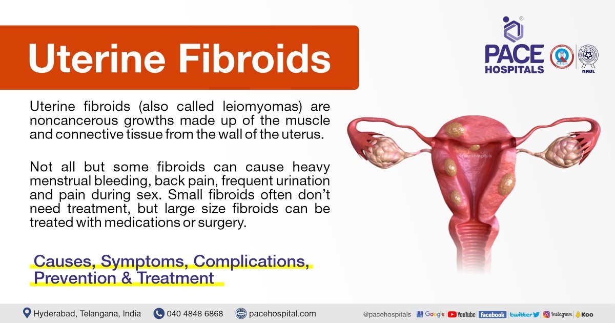
– Uterine fibroids are non-cancerous tumors that grow in the uterus, also known as myomas.
– Myomas are smooth, non-cancerous tumors made partly of muscle tissue that may develop in or around the uterus.
– Myomas in the larger, upper part of the uterus are called fibroids or leiomyomas.
– Most myomas can be seen or felt during a pelvic examination and those causing symptoms can be removed surgically or through less invasive procedures.
– The most common symptom of myomas is vaginal bleeding, which may be irregular or heavy.
– Other symptoms may include heavy bleeding, anemia, fatigue, weakness, painful intercourse, pain, bleeding, or discharge from the vagina if myomas become infected, pressure or lump in the abdomen, difficulties urinating, and urinary tract infections.
– Uterine fibroids, or myomas, affect 20 percent of women in their childbearing years.
– Uterine fibroids can cause abnormal bleeding, pelvic masses, pelvic pain, infertility, and pregnancy complications.
– There are five types of uterine fibroids: intramural, subserosal, submucosal, pedunculated, and intracavitary fibroids.
– Between 70 and 80 percent of women develop a fibroid tumor by the age of 50.
– Estrogen seems to activate the growth of uterine fibroids, and they usually shrink after menopause.
– Hormone therapy after menopause may cause fibroid symptoms to continue.
– African American women are more susceptible to developing fibroids.
– Fibroids tend to grow faster in white women younger than 35 years compared to those older than 45 years.
– Delaying pregnancy until age 30 or older increases the risk of developing fibroids.
– Early menstruation increases the risk of developing fibroids.
– Alcohol and caffeine intake may increase the risk of developing fibroids.
– Specific genetic alterations are linked to fibroid growth.
– Obesity and high blood pressure may play a role in fibroid development and growth.
– A diet rich in red meat may increase the chance of developing fibroids.
– Treatment options for uterine fibroids vary depending on the severity of symptoms and the patient’s desire to have children.
– Hormonal contraception, intrauterine devices, antifibrinolytic drugs, and nonsteroidal agents are options for managing heavy bleeding.
– Endometrial ablation can be performed if the patient does not want to have children.
– Gonadotropin-releasing hormone agonists can shrink fibroids, but they may grow back.
– Myomectomy is a procedure that removes fibroids while preserving the uterus, but fibroids may grow back.
– MRI-guided ultrasound surgery can shrink fibroids and reduce heavy bleeding.
– Uterine fibroid embolization is a minimally invasive procedure that blocks the blood supply to fibroids, causing them to shrink and die.
– Myomas can cause symptoms such as abdominal pain and heavy menstrual bleeding, but some people may remain symptom-free.
– The cause of myomas is unclear, but risk factors include a family history of myoma, obesity, and age.
– More than half of all people with a uterus will experience a myoma by age 50.
– Myomas can vary in size, from as small as a pea to as large as a melon.
– They can be located inside the uterus (intramural myomas), on the outside of the uterine wall (subserosal myomas), have a stalk or stem attaching them to the uterus (pedunculated myomas), or be found just under the lining of the uterus (submucosal myomas).
– Large myomas are considered to be 10 centimeters (cm) or more in diameter.
– Emergency room visits for myoma symptoms, such as pelvic pain and heavy bleeding, have increased from 2006 to 2017, according to a recent study.
– Myomas of the uterus are noncancerous tumors that can cause a variety of symptoms.
– Symptoms may include heavy or prolonged periods, bleeding between periods, pelvic pain, abdominal pressure, a feeling of fullness in the lower abdomen, constipation or diarrhea, frequent urination or trouble urinating, pain during sex, lower back pain, reproductive issues, fatigue, and weakness.
– Myomas are a top cause of hysterectomy surgeries, which come with their own risks and complications.
– The exact cause of myomas is unknown, but they are likely associated with hormone activity.
– High levels of estrogen and progesterone may stimulate their growth, and myomas tend to shrink when hormone levels decrease after menopause.
– Risk factors for developing myomas include a family history of myomas, obesity, high blood pressure, age, and dietary factors such as a diet high in red meat or vitamin D deficiency.
– Myomas are more common among Black individuals with a uterus, and factors such as low vitamin D levels, obesity, stress, genetics, and unequal access to healthcare have been proposed as potential risk factors.
– Medical tests used to diagnose myomas include pelvic examination, ultrasound or transvaginal ultrasound, and magnetic resonance imaging (MRI).
– Treatment for myomas depends on factors such as the severity of symptoms, the size and location of the myomas, the desire for future pregnancy, age, and proximity to menopause.
– Medications can be used to treat myomas, including over-the-counter pain medications, iron supplements for depleted iron levels, and birth control methods to control heavy menstrual bleeding.
– GnRH agonists (hormone-stimulating medications) can be used to temporarily shrink myomas, especially if surgery is planned.
– Surgical options for myomas include laparoscopic myomectomy and uterine fibroid embolization (UFE).
– No specific figures or statistics are mentioned in the article.
– One treatment option is a radiology procedure that uses injections to block blood flow to the myomas, causing them to shrink and sometimes die.
– Another option is MRI-guided ultrasound surgery, which uses ultrasound waves to shrink myomas.
– In more severe cases, other surgical options may be considered.
– Hysterectomy is a surgery to completely remove the uterus, eliminating the fibroids but also making pregnancy impossible in the future.
– Abdominal myomectomy is a surgical procedure that removes the fibroids without removing the uterus, with the possibility of future pregnancy but a risk of the fibroids returning.
– While there are no home remedies that directly treat fibroids, certain complementary therapies like acupuncture, yoga, massage, traditional Chinese medicine, and heating pads may help manage symptoms.
– Lifestyle changes such as dietary changes, exercise, stress management, and weight loss can also be beneficial.
– Myomas can cause complications related to fertility, pregnancy, and childbirth.
– These complications can include fertility issues, pregnancy complications such as miscarriage or early labor, and the need for a cesarean delivery.
– It’s important for those with myomas who want to become pregnant to discuss the condition with their healthcare provider to assess potential risks.
– It is recommended to communicate symptoms affecting one’s life to healthcare providers to determine the most suitable treatments.
Continue Reading

Unlocking the Wonders of Uteroscopy: A Glimpse inside

– Ureteroscopy is a procedure used to address kidney stones.
– It involves the use of a small telescope called a ureteroscope.
– The procedure is typically performed under general anesthesia.
– The duration of the procedure is usually between one to three hours.
– Small stones can be removed whole using a basket device.
– Large stones or narrow ureters may require fragmentation, often done with a laser.
– Once the stone is broken into small pieces, they are removed.
– The use of the ureteroscope may cause swelling in the ureter.
– To ensure proper drainage of urine, a small tube called a ureteral stent may be temporarily left inside the ureter.
– Ureteroscopy is usually an outpatient procedure but may require an overnight hospital stay if it is lengthy or difficult.
– Ureteroscopy is a minimally invasive method to treat kidney stones and stones in the ureter.
– It is performed in the operating room with general or spinal anesthesia.
– Pre-op lab tests and X-rays are done to ensure safety.
– An antibiotic is given before the procedure.
– Monitoring devices are attached to the patient.
– The inside of the bladder is examined with an optical cystoscope.
– X-ray images with contrast may be taken to locate the stone.
– The ureteroscope is passed through the natural urinary channel to reach the stone.
– A wire basket can be used to extract the stone, or it can be fragmented with laser or electrohydraulic energy.
– The procedure takes 1-2 hours.
– The patient is monitored in the post-operative recovery area for about 2 hours before being discharged.
– Some patients may have a temporary stent placed in the ureter to prevent blockage.
– The stent is usually removed 1 to 2 weeks after the procedure.
– Urine may be bloody for several days after the procedure.
– Pain relief medication is provided.
– Activity should be limited after the procedure.
– Most people can resume normal activities without pain several days after the procedure or once the stent is removed.
– A follow-up visit with a Urologist will be scheduled within 1-2 weeks to remove the stent if one was placed without a thread.
– If a stent was not required or was already removed at home, the follow-up visit will be scheduled within 4-6 weeks after surgery.
– During the follow-up visit, an X-ray will be performed to determine the success of the procedure and check for any complications.
– Depending on the individual’s risk of stone recurrence, further testing may be offered to prevent future stones.

Continue Reading

The Joys and Challenges of a Simple Vulvectomy:

– Simple vulvectomy is a surgical procedure for severe vulvar lesions that cannot be treated with local excision or other conservative therapy.
– Conditions that may require simple vulvectomy include extensive carcinoma, Paget’s disease, and severe leukoplakia.
– Unlike radical vulvectomy, simple vulvectomy does not require an incision all the way to the perineal fascia.
– The procedure involves removing the skin and subcutaneous tissues of the vulva.
– Attention must be paid to controlling hemorrhage around the urethra and lateral pudendal vessels to avoid complications.
– The patient is placed in the dorsal lithotomy position during the procedure.
– An elliptical incision is made around the lesion, starting from above the labial folds on the mons pubis and extending down the lateral fold of the labia majora and across the posterior fourchette.
– The pudendal artery and vein should be clamped before incising to prevent major blood loss.
– Additional incisions may be made above the urethra and laterally to avoid damaging the urethral meatus and rectum, respectively.
– The specimen is transected between perforations made in the vaginal mucosa, leaving it attached only to the fat pad in the mons pubis and the vascular plexus surrounding the suspensory ligaments.
– The clitoris is clamped and tied before being transected with scissors.
– Closure of the wound starts with closure of the posterior wall of the vaginal mucosa to avoid contracture of the vaginal introitus. Closure then continues in the mons pubis, levator ani muscles, perineal body, and urethral meatus.
– Closure is done using synthetic absorbable sutures.
– A catheter is inserted into the urethral meatus and removed after 24 hours.
– The patient is ambulated immediately after the procedure.
– Laxatives and stool softeners are administered on the third postoperative day.
– After surgery, drains may be placed to remove fluid build-up.
– Risks and side effects of vulvectomy include bleeding, infection, wound issues, fluid-filled cysts, urinary tract infections, lymphedema, changes in appearance and libido, genital numbness, and discomfort.
– Recovery may involve a hospital stay, catheter placement, Sitz baths, and medication.
– At home, soft, clean towels and a Sitz bath are needed for hygiene.
– Loose clothing and cotton underwear are recommended for comfort.
– Patients may require assistance with daily tasks until they feel better.
– Patients are advised to take prescribed medications as directed to manage pain, prevent infection, and avoid constipation.
– Patients are encouraged to contact their healthcare team if they experience any new or worsening symptoms.
– Recommendations for managing constipation include dietary changes, increased fluid intake, and over-the-counter medications (with consultation with healthcare team).
– Deep breathing and rest are suggested for pain management, lung health after anesthesia, and lymphatic fluid drainage.
– A relaxation exercise is provided as an example.
– It is emphasized that the specific plan and recovery should be discussed with the healthcare team.

Continue Reading

Cervix 101: Understanding the Female Reproductive System Better

Cervicitis
Inflammation of the cervix
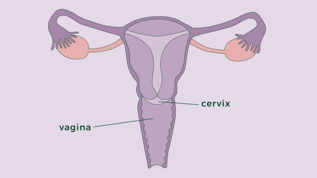
Lower, narrow end of the uterus
Opens into the vagina
Symptoms:
– Bleeding between menstrual periods
– Pain during intercourse or pelvic exam
– Abnormal vaginal discharge
Causes of cervicitis:
– Sexually transmitted infections (STIs) such as chlamydia and gonorrhea
– Noninfectious causes
– Allergic reactions to contraceptives or latex in condoms
– Allergic reactions to feminine hygiene products
Risk factors:
– High-risk sexual behavior
– Early age of sexual intercourse
– History of STIs
Complications:
– Pelvic inflammatory disease
– Fertility problems if left untreated
– Increased risk of getting HIV
Prevention:
– Consistent and correct use of condoms
– Being in a committed, monogamous relationship
Location and structure of the cervix:
– Located inside the pelvic cavity, 3 to 6 inches inside the vaginal canal
– Begins at the base of the uterus and extends downward onto the top part of the vagina
– Wider in the middle and narrows at both ends (opens into the uterus and vagina)
– Consists of the internal OS, endocervical canal, ectocervix, and external OS
– Transformation zone (TZ) is the most common site for abnormal cell growth
– About an inch long and varies in size
– Texture and location change during the menstrual cycle
– Made of fibromuscular tissue, lined with glandular cells and squamous cells
– Contains different cell types, including those covering the outermost part of the cervix and vagina
– Transitional zone (TZ) is the focus of screenings for cervical cancer

Continue Reading

Salpingectomy: A Guide to Tubal Removal Surgery

– Salpingectomy is the surgical removal of one or both of a woman’s fallopian tubes.
– It is performed to treat conditions such as fallopian tube cancer, ovarian cancer, ectopic pregnancy, endometriosis, blocked fallopian tubes, and infected fallopian tubes.
– Before the surgery, patients meet with the surgeon and anesthesiologist to discuss the operation.
– Patients may need to avoid eating and drinking for a few hours before the surgery.
– The procedure can be performed under general anesthesia, with a cut in the abdomen to remove the fallopian tubes, or using laparoscopy.
– After the procedure, patients may need to stay in the hospital for a few days to recover.
– The recovery may require a few days of hospital stay.
– Salpingectomy may be part of fertility treatment for blocked fallopian tubes with fluid build-up.
– It can also be performed as a form of permanent contraception or to reduce the risk of ovarian cancer.
– The article mentions that recovery is usually faster with keyhole surgery compared to laparotomy.
– No specific facts, figures, or statistics are provided in the article.

Continue Reading

Hysteroscopy: A Window into the Female Reproductive System

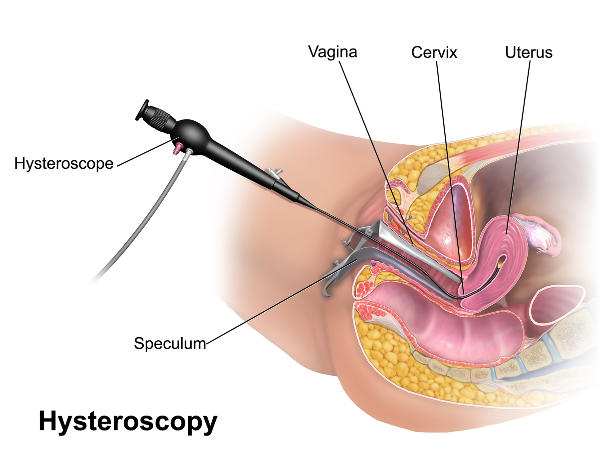
– Hysteroscopy is a procedure used to examine the inside of the womb
– It can be used to investigate symptoms or problems such as heavy periods, unusual vaginal bleeding, and pelvic pain
– It can also be used to diagnose conditions such as fibroids and polyps
– Hysteroscopy can be used to treat conditions and problems such as removing fibroids, polyps, and intrauterine devices
– The procedure is usually done on an outpatient or day-case basis
– Anesthesia may or may not be required
– A speculum may be inserted into the vagina during the procedure
– Fluid is pumped inside to facilitate visualization, and pictures are sent to a monitor for observation and treatment of abnormalities
– The procedure can take up to 30 minutes, or shorter if done only for diagnosis
– Recovery may include discomfort similar to period cramps, spotting or bleeding for a few days, and avoiding sex for a week
– Most women can return to their normal activities the following day
– There is a small risk of complications, such as accidental damage to the womb or cervix
– Excessive bleeding can occur during or after surgery, but can be treated with medication or another procedure
– Infection of the womb can be treated with antibiotics
– Feeling faint after a hysteroscopy affects 1 in every 200 women
– Hysteroscopy will only be performed if the benefits outweigh the risks.

Continue Reading