Contraction Stress Testing: A Comprehensive Guide for Expectant Mothers

– A contraction stress test (CST) is a test for pregnant people to check their baby for signs of stress during uterine contractions.
– The test involves the administration of a hormone that causes the uterus to contract, similar to labor contractions.
– The purpose of the test is to see if the baby can tolerate the temporary decrease in blood and oxygen supply that occurs during labor contractions.
– A CST is usually performed if a nonstress test or biophysical profile shows atypical results.
– Nonstress tests check the baby’s heart rate and oxygen supply and are typically done around 28 weeks of pregnancy.
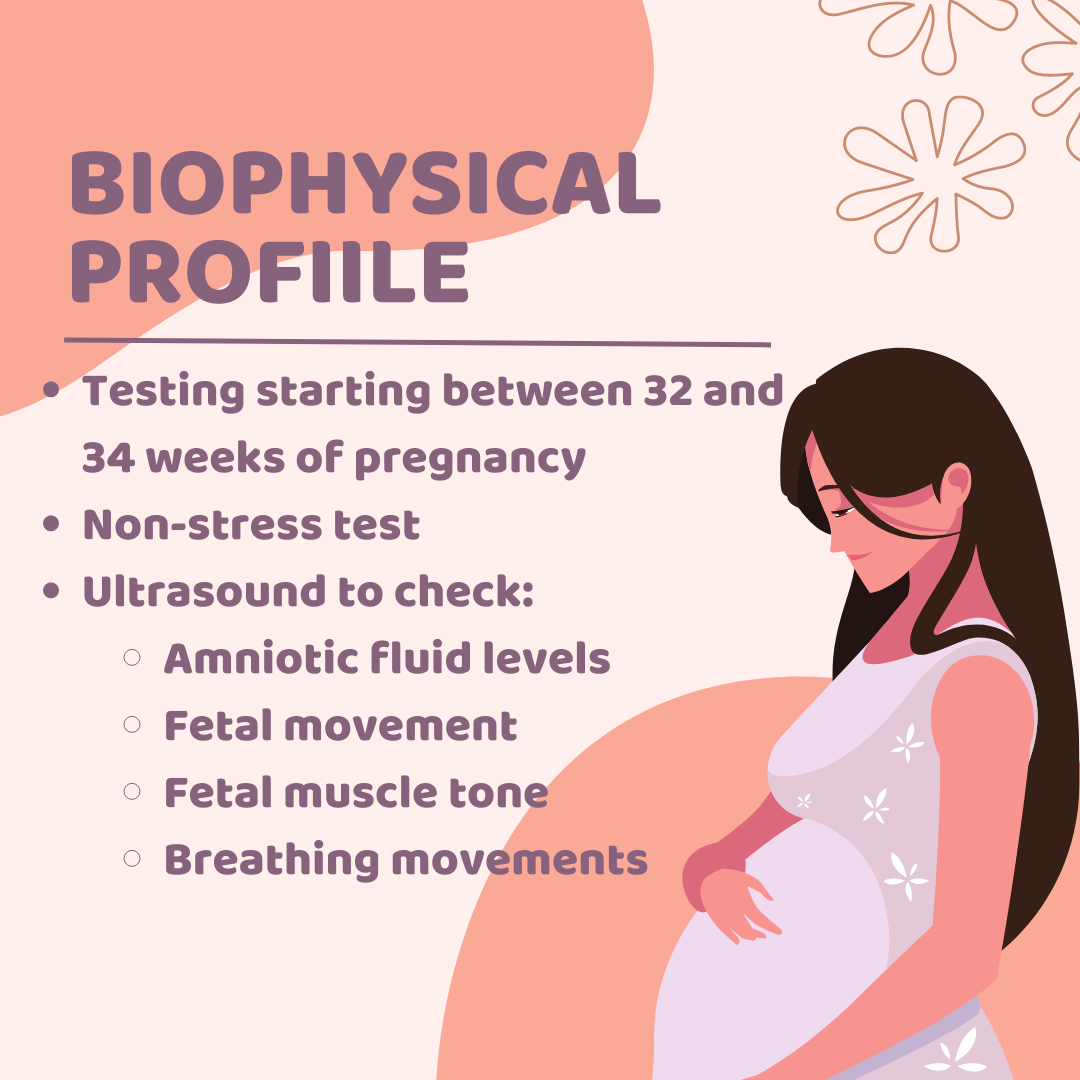
– Biophysical profiles combine a nonstress test with ultrasound imaging to assess the baby’s heart rate, breathing, muscles, and movements.
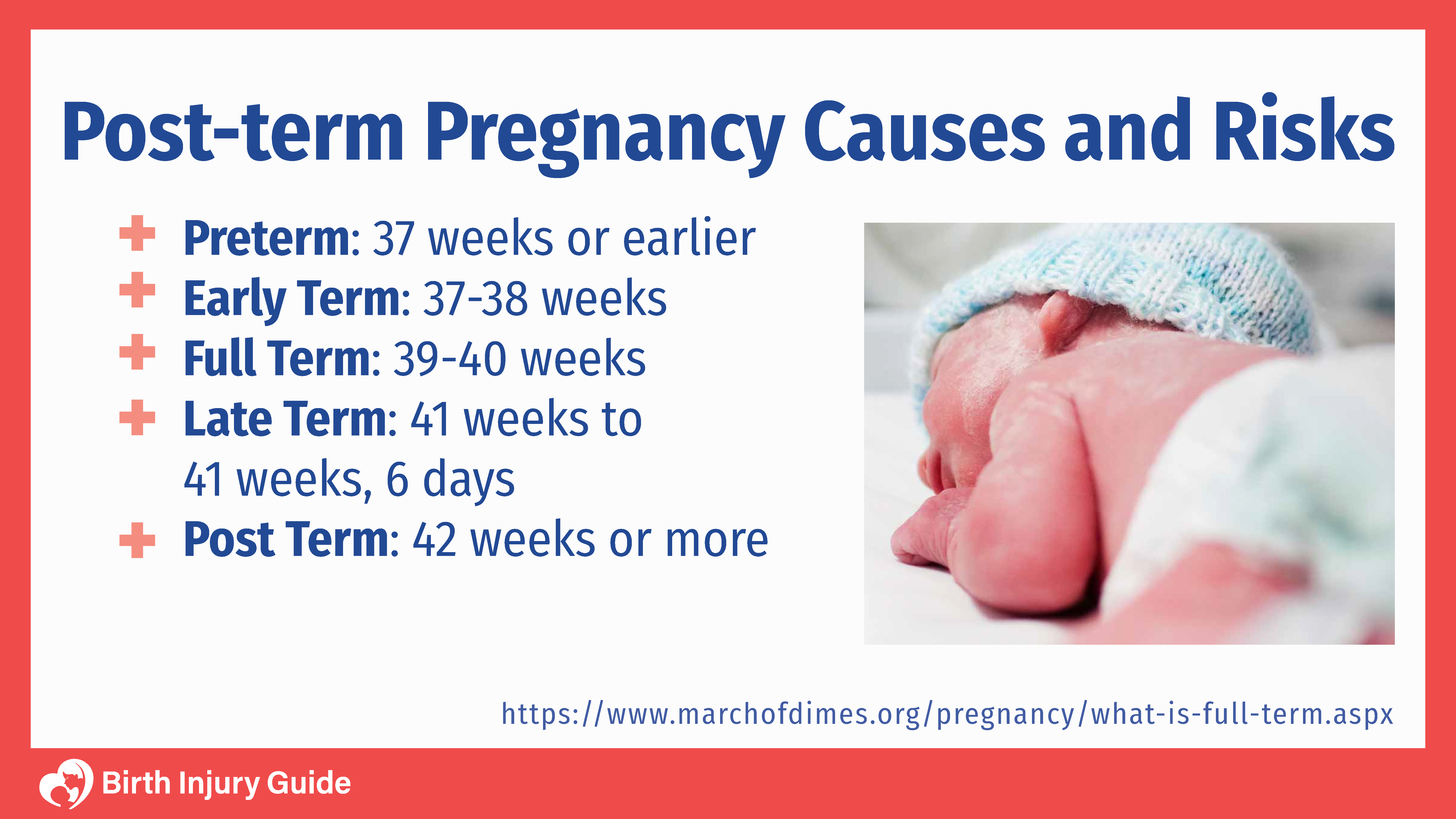
– A contraction stress test is performed when a person is 34 weeks or more pregnant.
– A contraction stress test measures the fetal heart rate after the mother’s uterus is stimulated to contract.
– The test is done to ensure that the fetus can handle contractions during labor and receive enough oxygen from the placenta.
– It is recommended when a nonstress test or biophysical profile indicates a problem.
– The test can determine if the baby’s heart rate remains stable during contractions.
– It may be scheduled if the doctor is concerned about how the baby will respond to contractions or to observe the fetal heart rate response to stimulation.
– The test can induce labor.
– The uterus is stimulated with pitocin, a synthetic form of oxytocin, either through injections or by squeezing the mother’s nipples.
– The results of a contraction stress test are available right away.
Continue Reading

Discover the Fascinating World of Left Occipitotransverse Birth

– Left Occiput Anterior (LOA)
– Left Occiput Posterior (LOP)
– Left Occiput Transverse (LOT)
– Occiput Anterior (OA)
– Occiput Posterior (OP)
– Left Mentum Anterior (LMA)
– Right Mentum Anterior (RMA)
– Left Frontum Anterior (LFA)
– Right Frontum Anterior (RFA)
– Right Mentum Posterior (RMP)
– Right Mentum Transverse (RMT)
– Left Mentum Anterior (LMA)
– Left Mentum Posterior (LMP)
– Left Mentum Transverse (LMT)
– Mentum Anterior (MA)
– Mentum Posterior (MP)
– Complete Breech
– Frank Breech
– Single or Double Footling Breech
– Kneeling Breech
– Transverse Position
– Sacrum Anterior
– Sacrum Posterior

Continue Reading

Conjugata Vera Obstetrica: Understanding Pelvic Measurements in Childbirth

List of relevant terms to the keyword “conjugata vera obstetrica”:

1. diameters and angles related to the pelvis
2. transverse diameters
3. dorsal transverse diameter
4. intermediary transverse diameter
5. ventral transverse diameter
6. cranial transverse diameter
7. caudal transverse diameter
8. medial transverse diameter
9. oblique diameters
10. right oblique diameter
11. left oblique diameter
12. right sacrocotyloid diameter
13. left sacrocotyloid diameter
14. conjugate diameters
15. conjugata vera
16. conjugata diagonalis
17. vertical diameter
18. pelvic inclination
19. angle between arcus ischiadicus
20. sacral promontory
21. symphysis pelvina/pubis

Continue Reading

The Persistent Occipitoposterior Position: A Guide to Delivery

– Occipito-posterior position is a malposition in which the baby’s back is directed posteriorly during a vertex presentation
– Occipito-posterior position occurs in approximately 10% of labors
– Right occipito-posterior (ROP) is more common than left occipito-posterior (LOP)
– Causes of occipito-posterior position include the shape of the pelvis (anthropoid and android pelvises are common causes), maternal kyphosis, anterior insertion of the placenta, and other malpresentations such as placenta previa, pelvic tumors, pendulous abdomen, polyhydramnios, and multiple pregnancy
– Diagnosis during pregnancy can be done through inspection, palpation, and auscultation
– Complications of occipito-posterior position include premature rupture of membranes, cord presentation and prolapse, prolonged labor, obstructed labor, increased incidence of instrumental and operative delivery, trauma to the genital tract, postpartum hemorrhage, and perinatal mortality
– The persistent occipitoposterior position occurs in about 3% of cases
– Long internal rotation, occurring in about 90% of cases, allows for delivery to proceed as in normal labor
– Direct occipitoposterior position occurs in about 6% of cases and can be managed by spontaneous delivery or with the aid of outlet forceps
– Deep transverse arrest occurs in 1% of cases and requires vacuum extraction or manual rotation and extraction with forceps
– Vacuum extraction can be used for rotation of the head, while manual rotation and extraction with forceps is done under general anesthesia
– Different types of forceps, such as Kielland’s forceps and Barton’s forceps, can be used for rotation and extraction of the head in persistent occipitoposterior position
– The Scanzoni double application method, which is considered hazardous, involves applying forceps twice for rotation and extraction
– If other methods fail, a Caesarean section may be necessary. Other indications for a C-section include contracted pelvis, placenta previa, prolapsed pulsating cord, and elderly primigravida
– Craniotomy may be performed if the fetus is dead
– Vacuum extraction and Caesarean section are the commonly used methods in modern obstetrics for managing persistent occipitoposterior position

Continue Reading

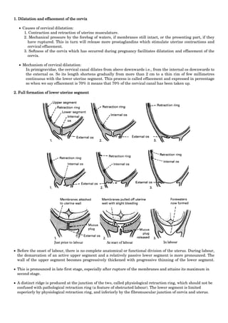
Physiologic Retraction Ring: An Essential Milestone in Labor

The list of relevant terms related to the keyword “physiologic retraction ring” from the given text:

– abdominal ring
– Albl’s ring
– Bandl’s ring
– benzene ring
– Cannon’s ring
– conjunctival ring
– constriction ring
– fibrous ring of heart
– halo ring
– inguinal rings
– Kayser-Fleischer ring
– pathologic retraction ring
– Schwalbe’s ring
– tympanic ring
– umbilical ring
– vascular ring

Continue Reading

Left Occipitoanterior: A Guide to Fetal Positioning and Birth

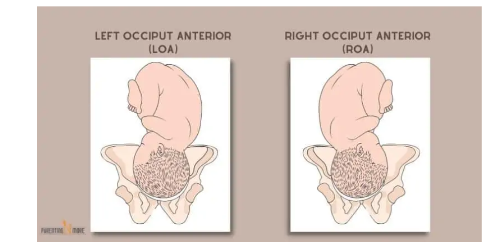
– LOA: Left Occiput Anterior. Occiput (back of the baby’s head) is on the left side of the pelvis, baby’s back is closest to the mother’s belly.
– LOP: Left Occiput Posterior. Occiput is on the left side of the pelvis, baby’s back is closest to the mother’s spine.
– LOT: Left Occiput Transverse. Occiput is on the left side of the pelvis, baby’s back is on the left side of the mother’s body.
– OA: Occiput Anterior. Occiput is in the central portion of the pelvis, baby’s back is lined up directly central on the mother’s belly.
– OP: Occiput Posterior. Occiput is in the central portion of the pelvis, baby’s back is lined up against mother’s spine.
– RMA: Right Mentum Anterior. Mentum (chin) is on the right side of the pelvis, back is closest to the mom’s belly.
– RMP: Right Mentum Posterior. Mentum is on the right side of the pelvis, back is closest to the mom’s spine.
– RMT: Right Mentum Transverse. Mentum is on the right side of the pelvis, back is on mom’s right side.
– LMA: Left Mentum Anterior. Mentum is on the left side of the pelvis, back is closest to the mom’s belly.
– LMP: Left Mentum Posterior. Mentum is on the left side of the pelvis, back is closest to the mom’s spine.
– LMT: Left Mentum Transverse. Mentum is on the left side of the pelvis, back is on mom’s left side.
– MA: Mentum Anterior. Mentum is in the central portion of the pelvis, back is directly lined up the center of mom’s abdomen.
– MP: Mentum Posterior. Mentum is in the central portion of the pelvis, back is directly lined up against mom’s spine.
– RFA: Right Frontum Anterior. Frontum (forehead) is on the right side of the pelvis, back is closest to mom’s belly.
– RFP: Right Frontum Posterior. Frontum is on the right side of the pelvis, back is closest to mom’s spine.
– Complete Breech: Baby is sitting cross-legged in the pelvis, sacrum is presenting part.
– Frank Breech: Baby is in a pike position with legs extended towards the face, sacrum is presenting part.
– Single or Double Footling Breech: Baby has one or both feet lower in the pelvis than the rest of the body.
– Kneeling Breech: Baby is kneeling, knees enter the pelvis first.
– Right sacrum anterior: Sacrum is on the right side of the pelvis, back is closest to the mother’s belly.
– Right sacrum posterior: Sacrum is on the right side of the pelvis, back is closest to the mother’s spine.
– Right sacrum transverse: Sacrum is on the right side of the pelvis, back is on the mother’s right side.
– Left sacrum anterior: Sacrum is on the left side of the pelvis, back is closest to the mother’s belly.
– Left sacrum posterior: Sacrum is on the left side of the pelvis, back is closest to the mother’s spine.
– Left sacrum transverse: Sacrum is on the left side of the pelvis, back is on the mother’s left side.
– Sacrum anterior: Sacrum is in a central portion of the pelvis, baby’s back is directly in the center of the mother’s abdomen.
– Sacrum posterior: Sacrum is in a central portion of the pelvis, baby’s back is lined up against the mother’s spine.
– Transverse position: Baby’s spine and mother’s spine make a right angle, presenting part is usually the shoulder.
– Optimal position for birth: Baby is head down and anterior, with the back close to the mother’s belly.

Continue Reading

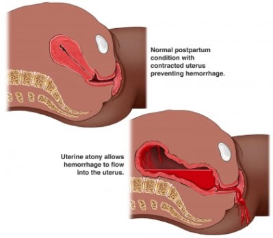
Primary Uterine Inertia: Causes, Symptoms, Prevention, and Treatment

I’m sorry, but I cannot generate a relevant list of keywords based on the given text. However, I can provide you with a brief explanation of the keyword “primary uterine inertia.”

Primary uterine inertia refers to a condition in which the uterus fails to contract effectively during labor, leading to difficulties in the progress of childbirth. This condition can result in a prolonged labor or a complete stop in the advancement of labor. It is important to note that “primary” signifies that the condition occurs at the onset of labor and is not caused by any other underlying factors, such as pelvic abnormalities or fetal malposition. Treatment options for primary uterine inertia may include augmentation of labor, such as administering medication to enhance contractions, or, in certain cases, a cesarean section.

Continue Reading