New Research Revealed: The Shocking Truth About Abruption

– Placental abruption is when a part or all of the placenta separates from the uterus prematurely, causing vaginal bleeding.
– There are two main types of placental abruption: revealed and concealed. Revealed abruption results in visible vaginal bleeding, while concealed abruption involves bleeding that remains within the uterus and may not be visible.
– Major risk factors for placental abruption include previous placental abruption, pre-eclampsia, abnormal lie of the baby, abdominal trauma, smoking or drug use, bleeding in the first trimester, and underlying thrombophilias.
– Clinical features of placental abruption include painful vaginal bleeding, woody and painful uterus on palpation, and the need for systematic assessment and resuscitation.
– General examination involves assessing pallor, distress, peripheral circulation, abdominal tenderness, the feel of the uterus, and the lie and presentation of the fetus/fetuses. The article provides guidance on how to assess bleeding during pregnancy and discusses differential diagnoses for antenatal hemorrhage. It suggests using a cardiotocograph (CTG) at 26 weeks gestation or above to check fetal wellbeing. It advises checking hand-held pregnancy notes for scan reports and looking for signs of placenta praevia. The article also recommends assessing the bleeding externally by looking at pads, avoiding speculum examination until placenta praevia is excluded, and taking triple genital swabs to exclude infection. It warns against performing a digital vaginal examination with known placenta praevia as it could cause massive bleeding. The article mentions placental abruption, placenta praevia, marginal placental bleed, vasa praevia, uterine rupture, and local genital causes as differential diagnoses for antenatal hemorrhage.
– Placental abruption is a common cause of antepartum hemorrhage.
– Investigations that should be performed include hematology (full blood count, Kleihauer test, group and save, cross-match), biochemistry (urea and electrolytes, liver function tests), and fetal wellbeing assessment (cardiotocograph).
– Ultrasound scan should be performed to assess placental abruption, but ultrasound should not be used to exclude abruption.
– Management of placental abruption depends on the health of the fetus: emergency delivery is indicated in the presence of maternal and/or fetal compromise, induction of labor is recommended for hemorrhage at term without compromise, and conservative management is an option for some partial or marginal abruptions without compromise.
– Anti-D should be given within 72 hours of bleeding onset if the woman is rhesus D negative.
– Placental abruption complicates approximately 1% of pregnancies and increases the risk of maternal, fetal, and neonatal morbidity and mortality.
– Risk factors for placental abruption include smoking, alcohol or cocaine use during pregnancy, advanced maternal age, history of maternal hypertension, and preeclampsia.
– Previous placental abruption and multiple gestational pregnancies also increase the risk.
– Trauma from a motor vehicle accident, fall, or blow to the abdomen can cause placental abruption.
– Radiologic imaging, such as ultrasonography, may assist in diagnosing placental abruption.
– Ultrasonography is usually the preferred study due to its benefits of avoiding ionizing radiation, dynamic nature, and availability.
– In severe trauma cases, CT scanning may be required to evaluate for abdominopelvic injuries.
– American College of Radiology guidelines recommend ultrasound FAST scan as a limited bedside adjunct for triage in a pregnant patient with major blunt trauma. The article discusses the use of imaging techniques for diagnosing major blunt trauma in pregnant patients. The American College of Radiology (ACR) states that there are two options for these procedures, but only one should be used to gather clinical information. No other facts, stats, or figures are mentioned in the article.

Continue Reading

The Shocking Truth: Premature Separation of Normally Implanted Placenta

– Placental abruption is a condition in which the placenta separates from the wall of the uterus before birth.
– It can happen partially or completely, leading to a lack of oxygen and nutrients for the baby.
– Symptoms include vaginal bleeding, pain, contractions, discomfort, and tenderness.
– It occurs in about 1 in 100 pregnant individuals.
– Placental abruption is more likely to occur in the third trimester, but can happen after 20 weeks of pregnancy.
– Mild cases can lead to complete separation and may require close monitoring.
– Placental abruption is related to about 1 in 10 premature births.
– Premature babies are at higher risk for health problems, disabilities, and death.
– Placental abruption can cause anemia and life-threatening complications for the pregnant individual and the baby.
– Immediate diagnosis and treatment are necessary to prevent hemorrhage and blood clotting complications.
– Delivery by cesarean birth may be required.
– The causes of placental abruption may include previous abruption, high blood pressure, smoking, cocaine use, physical trauma, age 35 or older, infection in the uterus, preterm labor, early water breaking, issues with the uterus or umbilical cord, excess fluid around the baby, carrying multiples, asthma, family history of abruption, previous c-section, and exposure to air pollution.
– If a person has previously experienced placental abruption, they have a 10% chance of it occurring again in a later pregnancy.
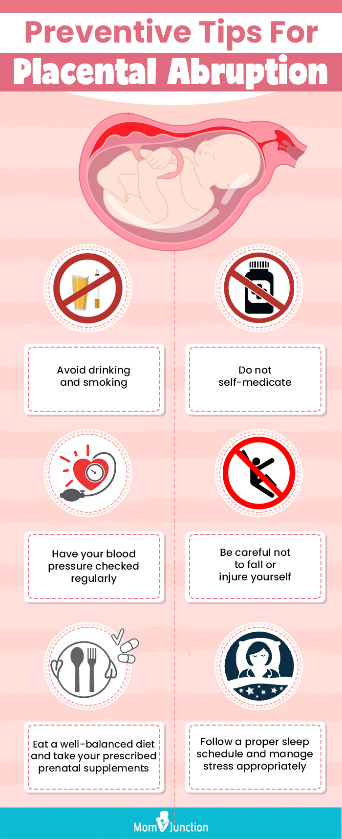
– Measures to reduce the risk of placental abruption include closely monitoring and treating high blood pressure, avoiding smoking or using street drugs, always wearing a seatbelt when in a car, and taking prenatal vitamins with folic acid.

Continue Reading

Ablatio Placentae: Causes, Symptoms, Treatments, and Prevention Methods

– Placental abruption is most likely to occur in the last trimester of pregnancy, particularly in the last few weeks before birth.
– The signs and symptoms of placental abruption include vaginal bleeding, although it is possible to have no bleeding, abdominal pain, back pain, uterine tenderness or rigidity, uterine contractions that often come one after another.
– The onset of abdominal and back pain is sudden.
– The amount of vaginal bleeding can vary greatly and does not indicate how much of the placenta has separated from the uterus.
– Even with severe placental abruption, there might be no visible bleeding as the blood can become trapped inside the uterus.
– In some cases, placental abruption develops slowly (chronic abruption) and can cause light, intermittent vaginal bleeding.
– This can result in the baby not growing as expected and complications such as low amniotic fluid.
– Complications may include slow fetal growth and low amniotic fluid.

Continue Reading

Puerperant Health: Essential Tips for Postpartum Recovery

List of pertinent terms for the keyword ‘puerperant’:

– Puerperant
– Lactation
– Medical criteria
– Exclusions
– Control group
– Non-pregnant
– Patients
– 18 years or older
– Medical conditions
– Auricular point sticking
– Practices
– Rituals
– 40th day of delivery
– Covering belly
– Thyroid hormone
– Oral contraceptive pills
– GH (growth hormone)
– Pregnant
– HIV
– Hepatitis
– Alcoholics
– Diabetics
– Comorbidities
– Kirkcikarma
– Bath
– Sufficient and balanced diet
– Mother and baby
– Albasmasi
– Harmless
– Psychologically beneficial
– Unsafe places
– Women with periods
– Prohibiting visits
– Red ribbon

Continue Reading

Puerperal Endometritis: Causes, Symptoms, Treatment, and Prevention Strategies

List of pertinent information to the keyword ‘puerperal endometritis’:

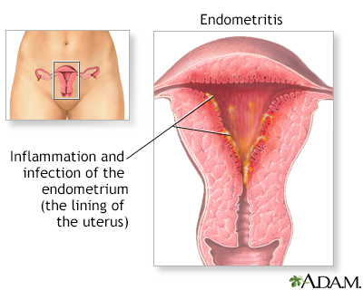
1. Postpartum endometritis is an infection of the lining of the womb.
2. It can occur up to six weeks after childbirth.
3. Risk is higher for caesarean section births.
4. Common between the second and tenth day after delivery.
5. Occurs in 1-3 out of every 100 women who have had a normal delivery.
6. More common in women who have had a caesarean section.
7. Risk factors include long labors, membranes breaking before birth, meconium-stained amniotic fluid, difficulty removing the placenta, multiple internal examinations during labor, infection in the genital area, past history of pelvic inflammatory disease (PID), bacterial vaginosis (BV), Group B streptococcus infection, HIV infection, obesity, diabetes, and delivery in circumstances of poor hygiene.
8. Symptoms include fever, lower tummy pain, smelly discharge from the vagina, increased bleeding from the vagina, pain during sex, pain during urination, and general feeling of being unwell.
9. Diagnosis is usually based on typical symptoms and signs, and tests are usually not necessary.
10. Treatment involves the use of antibiotics, commonly clindamycin and gentamicin.
11. If left untreated, the infection can spread to other parts of the body and potentially lead to sepsis.
12. Complications are rare when treated with antibiotics.
13. Most women recover quickly with antibiotics, typically within 2-3 days.
14. Women undergoing caesarean section are offered antibiotics beforehand to reduce the likelihood of infection.
15. Antibiotics are also given during labor if the woman has Group B streptococcus in her vagina to protect both her and the newborn baby from infections caused by this germ.

Continue Reading

Perinatal Cardiomyopathy: Causes, Symptoms, Treatment, and Prevention Guidelines

Perinatal cardiomyopathy is a rare condition where weakness in the heart muscle occurs sometime during the final month of pregnancy through about five months after delivery. It can have mild or severe symptoms. The seriousness of the condition can be measured by the ejection fraction, which is the percentage of blood the heart pumps out with each beat. A normal ejection fraction is about 60%. The degree of severity does not affect the rate of recovery. Some patients with a low ejection fraction can fully recover from perinatal cardiomyopathy. Recovery can occur over a period of six months or longer, but in some cases, the heart can return to full strength in as little as two weeks. Perinatal cardiomyopathy has a relatively high recovery rate compared to other forms of cardiomyopathy. Symptoms of perinatal cardiomyopathy, such as swelling in the feet and legs and shortness of breath, can be similar to the symptoms of the third trimester of a normal pregnancy. Mild cases may go undiagnosed and recover without medical attention. Severe cases can cause shortness of breath and swollen feet. Cardiomyopathy can be detected through an echocardiogram which shows the diminished functioning of the heart.

Continue Reading

Discover the Fascinating Functions of the Accessory Placenta

– Accessory Lobe of Placenta is a variation in the normal shape of the placenta
– It is a small lobe of the placenta attached to the main disc through blood vessels
– There can be one or more accessory lobes attached to the main placenta
– A routine abdominal ultrasound scan during pregnancy can help establish the presence of an Accessory Lobe of Placenta
– The condition is not associated with an increased risk of fetal anomalies
– There may be an increased risk of bleeding after delivery
– Accessory Lobe of Placenta occurs in approximately 2 per 1000 pregnancies
– There are no distinct racial, ethnic or geographical predilections
– Currently, no risk factors have been identified for Accessory Lobe of Placenta
– Accessory Lobe of Placenta is formed by non-involution of the chorionic villi
– The disc shape of the placenta is due to remodeling of placental tissue
– There are no specific signs and symptoms associated with Accessory Lobe of Placenta
– It is diagnosed by an ultrasound scan of the abdomen
– Possible complications include increased chances of postpartum hemorrhage, increased incidence of Vasa Previa, and rupture of vessels connecting the main and accessory lobe of placenta causing fetal compromise
– It does not require specific treatment but careful monitoring is needed for increased risk of bleeding after delivery
– Currently, there are no definitive methods to prevent it
– The prognosis is excellent with suitable care and management during delivery
– The incidence of Accessory Lobe of Placenta is higher in pregnancies using in-vitro fertilization
– A succenturiate (accessory) lobe is a smaller placental lobe that is in addition to the largest lobe
– The smaller succenturiate lobe often has areas of infarction or atrophy
– Risk factors for a succenturiate placenta include advanced maternal age, primigravida (first-time pregnancy), proteinuria in the first trimester, and major malformations in the fetus
– The membranes between the lobes of a succenturiate placenta can tear during delivery
– The extra lobe can be retained after the rest of the placenta is delivered, leading to postpartum bleeding
– Succenturiate lobes are generally not a major concern unless they are large and have a weak blood supply
– Vasa previa can occur if the fetal blood vessels connecting the two lobes of the placenta are located between the baby’s presenting part and the cervix or if the cord insertion is located between the two lobes.

Continue Reading

Placenta: Unraveling Its Role in Prenatal Development

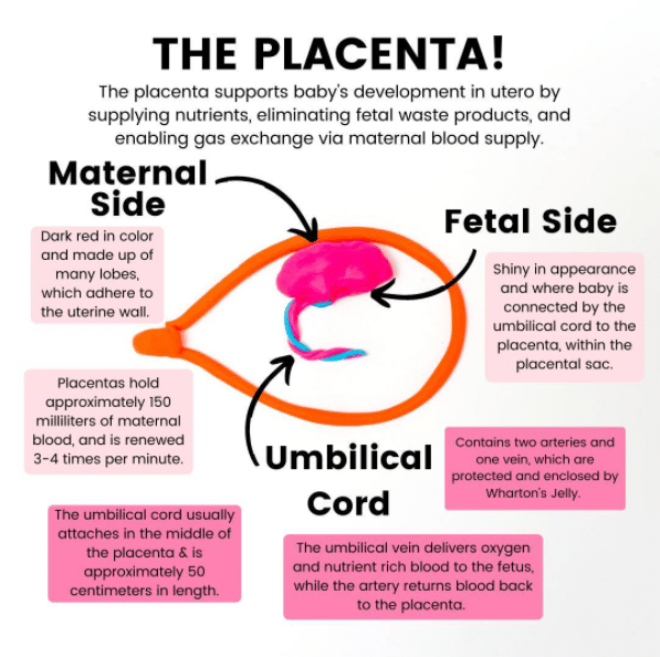
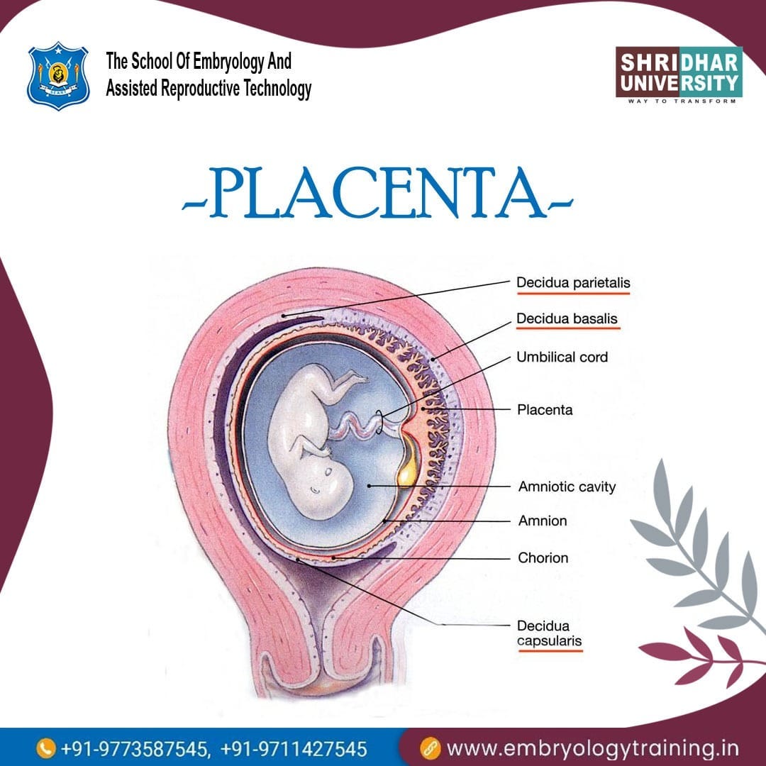
– The placenta is a temporary organ that develops during pregnancy and attaches to the lining of the uterus.
– It delivers oxygen and nutrients to the growing baby through the umbilical cord.
– Complications with the placenta can be serious and life-threatening to both the mother and baby.
– The placenta passes oxygen, nutrients, and antibodies from the mother’s blood to the baby and carries waste products back to the mother’s blood.
– It produces hormones like estrogen and progesterone that are needed during pregnancy.
– The normal position of the placenta is usually at the top, side, front, or back of the uterus, but it can sometimes develop low and move higher as the uterus stretches.
– Fraternal twins have separate placentas, while identical twins can share a placenta or have their own.
– Alcohol, nicotine, medicines, and other drugs can cross the placenta and affect the baby’s health.
– Regular visits to a healthcare provider during pregnancy are important to monitor the placenta and identify any complications.
– Prior problems with the placenta in a previous pregnancy or surgery to the uterus should be disclosed to the doctor.
– Smoking, drinking alcohol, and taking certain drugs increase the likelihood of problems with the placenta.
– Consult a doctor before taking any medicines, including over-the-counter medicines, natural therapies, and supplements during pregnancy.
– Seek medical attention if experiencing severe abdominal or back pain, vaginal bleeding, contractions, or trauma to the abdomen.
– After the baby is born, the placenta needs to be birthed during the third stage of labor.
– Placental abruption is when the placenta detaches from the uterus wall before the baby is born.
– Placenta previa is when the placenta partially or fully covers the cervix.
– Placental insufficiency occurs when the placenta does not function properly during pregnancy, resulting in a lack of oxygen and nutrients for the baby.
– Placenta accreta is when the placenta grows too deeply into the uterine wall, potentially causing severe bleeding during or after delivery.
– Retained placenta occurs when the placenta does not fully detach or come out after birth. This can be due to it being stopped by the cervix or still attached to the uterus.

Continue Reading