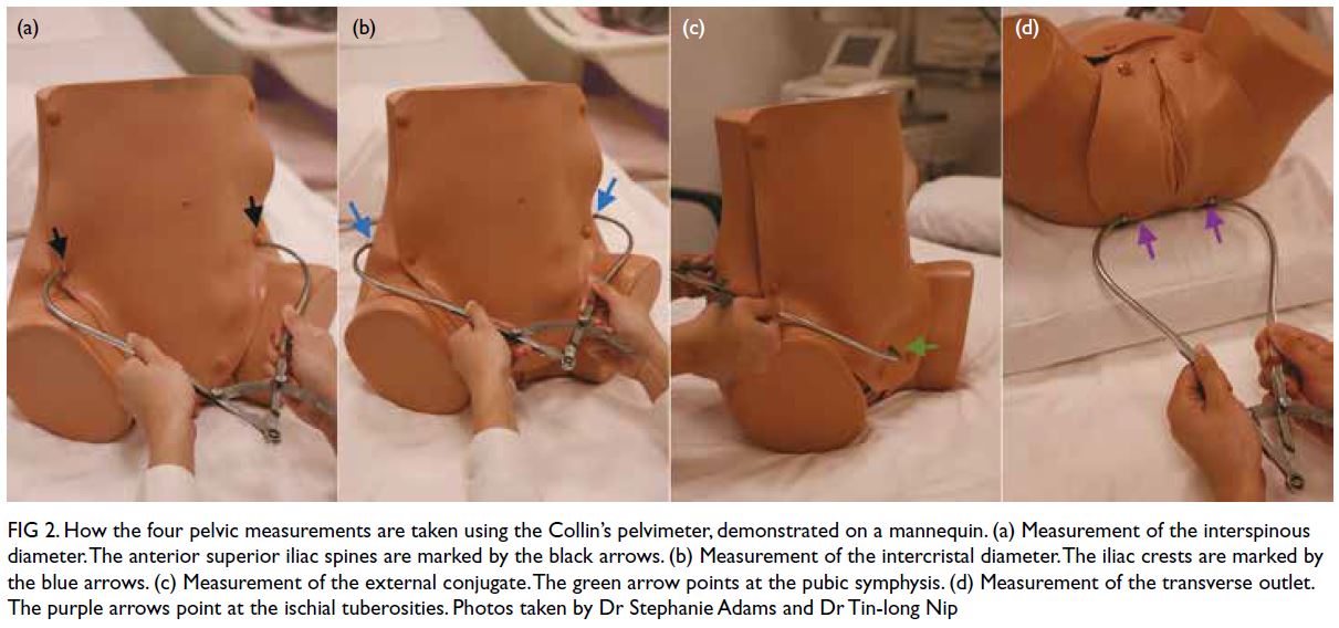
External Pelvimetry: A Comprehensive Guide to Evaluating Childbirth Potential

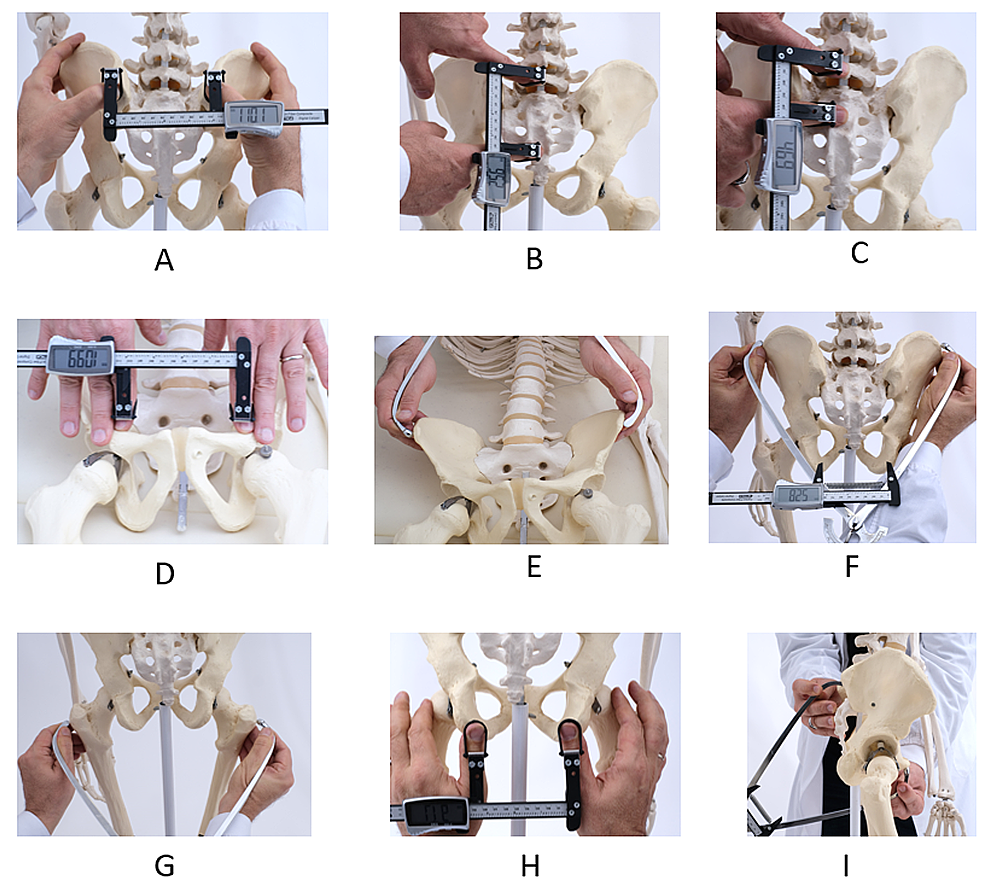
– external pelvimetry
– measuring pelvis size and shape
– predicting success of vaginal delivery
– third trimester of pregnancy
– calipers
– dimensions of the pelvis
– inlet, mid-pelvis, outlet
– limitations of external pelvimetry
– low sensitivity and specificity
– identifying cephalopelvic disproportion (CPD)
– false positive rate
– ultrasound pelvimetry
– clinical assessment
– more reliable methods
– non-invasive method
– evaluating maternal pelvic dimensions
– measuring pelvic landmarks
– calipers or tape measures
– adjunct to traditional methods
– internal pelvimetry
– determining suitability for vaginal delivery
– cesarean section
– limitations of external pelvimetry
– obesity
– fetal position
– pelvic soft tissue
– valuable information
– obstetric care

Continue Reading

Contraction Stress Testing: A Comprehensive Guide for Expectant Mothers

– A contraction stress test (CST) is a test for pregnant people to check their baby for signs of stress during uterine contractions.
– The test involves the administration of a hormone that causes the uterus to contract, similar to labor contractions.
– The purpose of the test is to see if the baby can tolerate the temporary decrease in blood and oxygen supply that occurs during labor contractions.
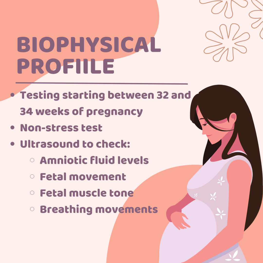
– A CST is usually performed if a nonstress test or biophysical profile shows atypical results.
– Nonstress tests check the baby’s heart rate and oxygen supply and are typically done around 28 weeks of pregnancy.
– Biophysical profiles combine a nonstress test with ultrasound imaging to assess the baby’s heart rate, breathing, muscles, and movements.
– A contraction stress test is performed when a person is 34 weeks or more pregnant.
– A contraction stress test measures the fetal heart rate after the mother’s uterus is stimulated to contract.
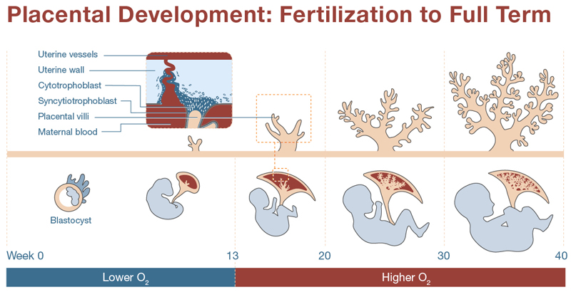
– The test is done to ensure that the fetus can handle contractions during labor and receive enough oxygen from the placenta.
– It is recommended when a nonstress test or biophysical profile indicates a problem.
– The test can determine if the baby’s heart rate remains stable during contractions.
– It may be scheduled if the doctor is concerned about how the baby will respond to contractions or to observe the fetal heart rate response to stimulation.
– The test can induce labor.
– The uterus is stimulated with pitocin, a synthetic form of oxytocin, either through injections or by squeezing the mother’s nipples.
– The results of a contraction stress test are available right away.

Continue Reading

The Left Mentoanterior Position: Understanding Fetal Presentation during Labor

– Face presentation is a cephalic presentation in which the head is completely extended.
– The incidence of face presentation is about 1 in 300 labours.
– Primary face presentation occurs during pregnancy and may be caused by factors such as anencephaly, loops of the cord around the neck, foetal neck tumors, hypertonicity of neck muscles, dolicocephaly (long antero-posterior diameter of the head), dead or premature foetus, or idiopathic causes.
– Secondary face presentation occurs during labour and may be due to factors such as contracted pelvis, pendulous abdomen, marked lateral obliquity of the uterus, further deflexion of brow or occipito-posterior positions, or other malpresentations such as polyhydramnios and placenta praevia.
– Left mento-anterior (LMA) and right mento-anterior (RMA) are more common positions of face presentation.
– Diagnosis during pregnancy is difficult, but the back is difficult to feel and the limbs may be felt more prominently in mento-anterior position. Ultrasound or X-ray can confirm the diagnosis.
– Diagnosis during labour is done through vaginal examination, which shows identifying features such as supra-orbital ridges, malar processes, nose, mouth, and chin.
– Late in labour, the face may become oedematous (tumefaction), which can be misdiagnosed as a buttock (breech presentation). Differentiating factors include the formation of a triangle with foetal mouth and malar processes as apexes, anus on the same line as ischial tuberosities, feeling of a hard gum through the mouth, and no hard object through the anus.
– The mechanism of labour in mento-anterior position involves descent.
– Engagement by submento-bregmatic diameter: 9.5 cm
– Submental region hinges below the symphysis in flexion position
– Submento-vertical diameter: 11.5 cm
– Biparietal diameter does not pass the plane of pelvic inlet until the chin is below the level of the ischial spines and the face begins to distend the perineum
– In about 2/3 of cases, long anterior rotation of 3/8 circle occurs during mento-posterior position
– In about 1/3 of cases, deep transverse arrest of the face, persistent mento-posterior, or direct mento-posterior occur during mento-posterior position
– Direct mento-posterior cannot be delivered due to obstruction caused by the length of the sacrum and neck
– Management of labour includes excluding foetal anomalies and contracted pelvis
– Spontaneous delivery usually occurs in mento-anterior position during second stage of labour
– Forceps delivery and episiotomy may be indicated in prolonged second stage of labour in mento-anterior position
– Wait for long anterior rotation of 3/8 circle in mento-posterior position during second stage of labour
– Oxytocin is used to compete inertia during this period if there are no contraindications
– Caesarean section is the safest option if long anterior rotation fails or there is foetal or maternal distress
– Manual rotation and forceps extraction or rotation and extraction by Kielland forceps are alternative methods, but are hazardous and not commonly used
– Craniotomy may be performed if the foetus is dead
– Complications may occur, refer to complications of malpresentations and malposition for more information.
– There is an increased risk of trauma to the baby in face presentation, so internal manipulation, vacuum extractors, and manual extraction should be avoided.
– Abnormalities in fetal heart rate are more common in face presentation. Monitoring is crucial during labor.
– Complications of face presentation include prolonged labor, facial trauma, facial edema, skull molding, respiratory distress, spinal cord injury, abnormal fetal heart rate patterns, and low Apgar scores.
– Informed consent should be obtained from the mother, and failure to do so is considered negligence.
– Forceps and oxytocin used during labor can put a baby at risk of complications. Forceps can cause head injuries and oxytocin can deprive the baby of oxygen due to strong contractions.
– Mothers should be given the option of a C-section if facing complications.
– Face presentation babies should be closely monitored and delivered by an experienced physician.
– If negligent practices cause injury to the baby, it can be considered medical malpractice. ABC Law Centers specialize in birth injury cases and offer free legal consultations.

Continue Reading
The Shocking Truth: Premature Separation of Normally Implanted Placenta

– Placental abruption is a condition in which the placenta separates from the wall of the uterus before birth.
– It can happen partially or completely, leading to a lack of oxygen and nutrients for the baby.
– Symptoms include vaginal bleeding, pain, contractions, discomfort, and tenderness.
– It occurs in about 1 in 100 pregnant individuals.
– Placental abruption is more likely to occur in the third trimester, but can happen after 20 weeks of pregnancy.
– Mild cases can lead to complete separation and may require close monitoring.
– Placental abruption is related to about 1 in 10 premature births.
– Premature babies are at higher risk for health problems, disabilities, and death.
– Placental abruption can cause anemia and life-threatening complications for the pregnant individual and the baby.
– Immediate diagnosis and treatment are necessary to prevent hemorrhage and blood clotting complications.
– Delivery by cesarean birth may be required.
– The causes of placental abruption may include previous abruption, high blood pressure, smoking, cocaine use, physical trauma, age 35 or older, infection in the uterus, preterm labor, early water breaking, issues with the uterus or umbilical cord, excess fluid around the baby, carrying multiples, asthma, family history of abruption, previous c-section, and exposure to air pollution.
– If a person has previously experienced placental abruption, they have a 10% chance of it occurring again in a later pregnancy.
– Measures to reduce the risk of placental abruption include closely monitoring and treating high blood pressure, avoiding smoking or using street drugs, always wearing a seatbelt when in a car, and taking prenatal vitamins with folic acid.

Continue Reading

Understanding Hypertonic Uterine Inertia: Causes, Symptoms, and Solutions

List of pertinent details about ‘hypertonic uterine inertia’:

1. Hypertonic uterine inertia is defined as infrequent, weak, and short-duration uterine contractions.
2. The causes of hypertonic uterine inertia are unknown.
3. Factors that may be involved in hypertonic uterine inertia include primigravida (particularly elderly), anemia, nervousness, hormonal deficiencies, improper use of analgesics, uterine overdistension, developmental anomalies, myomas, malpresentations, malpositions, cephalopelvic disproportion, and a full bladder or rectum.
4. Hypertonic uterine inertia can be categorized as primary or secondary inertia.
5. Primary inertia occurs when weak contractions occur from the start of labor.
6. Secondary inertia occurs when contractions may initially be strong but become weak and inadequate to overcome an obstruction.
7. Clinical symptoms of hypertonic uterine inertia include prolonged labor, infrequent and weak contractions, slow cervical dilatation, intact membranes, and little impact on the fetus and mother apart from maternal anxiety.
8. Complications from hypertonic uterine inertia can include retained placenta and postpartum hemorrhage.
9. Diagnosis of hypertonic uterine inertia is done through tocography to measure uterine contractions and examination to detect any abnormalities.
10. Management of hypertonic uterine inertia may include proper management of the first stage of labor, prophylactic antibiotics in prolonged labor, amniotomy, and the use of oxytocin to stimulate contractions.
11. Operative delivery, such as vaginal delivery using forceps or vacuum, or caesarean section, may be necessary in cases of failure of other methods or complications.
12. Hypertonic uterine inertia is more common in primigravidae.
13. Management includes general measures such as analgesics and antispasmodics.
14. Constriction (contraction) ring is a persistent localized spasm of the uterine muscles that typically occurs at the junction of the upper and lower uterine segments.
15. The cause of constriction ring is unknown but may be associated with factors such as malpresentations, improper use of oxytocin, and intrauterine manipulations.
16. Complications of constriction ring can include prolonged first or second stage of labor and retained placenta and postpartum hemorrhage.
17. Two conditions discussed in the article are hypertonic uterine inertia and cervical dystocia.
18. Delivery of the fetus is the only way to relieve hypertonic uterine inertia.
19. Treatment for hypertonic uterine inertia includes excluding malpresentations, malposition, and disproportion, as well as the use of medication such as pethidine or deep general anesthesia and amyl nitrite inhalation.
20. Cervical dystocia is the failure of the cervix to dilate within a reasonable time despite regular uterine contractions.
21. There are two varieties of cervical dystocia: organic and functional dystocia.
22. Complications of cervical dystocia include annular detachment of the cervix, rupture of the uterus, and postpartum hemorrhage.
23. Management of organic dystocia is cesarean section.
24. For functional dystocia, pethidine and antispasmodics may be effective. If medical treatment fails or fetal distress develops, a cesarean section is performed.

Continue Reading

Conjugata Vera Obstetrica: Understanding Pelvic Measurements in Childbirth

List of relevant terms to the keyword “conjugata vera obstetrica”:

1. diameters and angles related to the pelvis
2. transverse diameters
3. dorsal transverse diameter
4. intermediary transverse diameter
5. ventral transverse diameter
6. cranial transverse diameter
7. caudal transverse diameter
8. medial transverse diameter
9. oblique diameters
10. right oblique diameter
11. left oblique diameter
12. right sacrocotyloid diameter
13. left sacrocotyloid diameter
14. conjugate diameters
15. conjugata vera
16. conjugata diagonalis
17. vertical diameter
18. pelvic inclination
19. angle between arcus ischiadicus
20. sacral promontory
21. symphysis pelvina/pubis

Continue Reading

The Intricate Process: Retention of Placenta Explained

– Physical examination
– Ultrasound
– Blood tests
– Emptying the bladder
– Gentle pulling on the umbilical cord
– Surgical procedure under anesthesia
– Symptoms of retained placenta
– Ultrasound scan
– Treatment for retained placenta
– Close monitoring
– Heavy bleeding
– Imaging tests
– Surgery
– Complications of retained placenta
– Life-threatening infections
– Postpartum hemorrhage
– Early diagnosis and management

Continue Reading

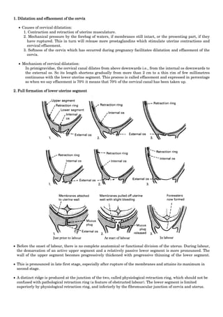
Physiologic Retraction Ring: An Essential Milestone in Labor

The list of relevant terms related to the keyword “physiologic retraction ring” from the given text:

– abdominal ring
– Albl’s ring
– Bandl’s ring
– benzene ring
– Cannon’s ring
– conjunctival ring
– constriction ring
– fibrous ring of heart
– halo ring
– inguinal rings
– Kayser-Fleischer ring
– pathologic retraction ring
– Schwalbe’s ring
– tympanic ring
– umbilical ring
– vascular ring

Continue Reading

Left Occipitoanterior: A Guide to Fetal Positioning and Birth

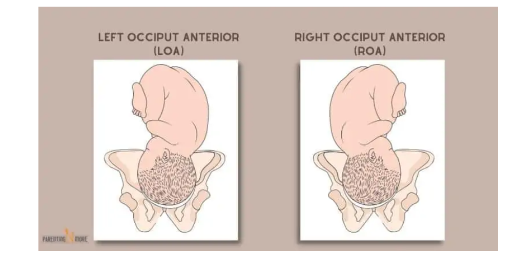
– LOA: Left Occiput Anterior. Occiput (back of the baby’s head) is on the left side of the pelvis, baby’s back is closest to the mother’s belly.
– LOP: Left Occiput Posterior. Occiput is on the left side of the pelvis, baby’s back is closest to the mother’s spine.
– LOT: Left Occiput Transverse. Occiput is on the left side of the pelvis, baby’s back is on the left side of the mother’s body.
– OA: Occiput Anterior. Occiput is in the central portion of the pelvis, baby’s back is lined up directly central on the mother’s belly.
– OP: Occiput Posterior. Occiput is in the central portion of the pelvis, baby’s back is lined up against mother’s spine.
– RMA: Right Mentum Anterior. Mentum (chin) is on the right side of the pelvis, back is closest to the mom’s belly.
– RMP: Right Mentum Posterior. Mentum is on the right side of the pelvis, back is closest to the mom’s spine.
– RMT: Right Mentum Transverse. Mentum is on the right side of the pelvis, back is on mom’s right side.
– LMA: Left Mentum Anterior. Mentum is on the left side of the pelvis, back is closest to the mom’s belly.
– LMP: Left Mentum Posterior. Mentum is on the left side of the pelvis, back is closest to the mom’s spine.
– LMT: Left Mentum Transverse. Mentum is on the left side of the pelvis, back is on mom’s left side.
– MA: Mentum Anterior. Mentum is in the central portion of the pelvis, back is directly lined up the center of mom’s abdomen.
– MP: Mentum Posterior. Mentum is in the central portion of the pelvis, back is directly lined up against mom’s spine.
– RFA: Right Frontum Anterior. Frontum (forehead) is on the right side of the pelvis, back is closest to mom’s belly.
– RFP: Right Frontum Posterior. Frontum is on the right side of the pelvis, back is closest to mom’s spine.
– Complete Breech: Baby is sitting cross-legged in the pelvis, sacrum is presenting part.
– Frank Breech: Baby is in a pike position with legs extended towards the face, sacrum is presenting part.
– Single or Double Footling Breech: Baby has one or both feet lower in the pelvis than the rest of the body.
– Kneeling Breech: Baby is kneeling, knees enter the pelvis first.
– Right sacrum anterior: Sacrum is on the right side of the pelvis, back is closest to the mother’s belly.
– Right sacrum posterior: Sacrum is on the right side of the pelvis, back is closest to the mother’s spine.
– Right sacrum transverse: Sacrum is on the right side of the pelvis, back is on the mother’s right side.
– Left sacrum anterior: Sacrum is on the left side of the pelvis, back is closest to the mother’s belly.
– Left sacrum posterior: Sacrum is on the left side of the pelvis, back is closest to the mother’s spine.
– Left sacrum transverse: Sacrum is on the left side of the pelvis, back is on the mother’s left side.
– Sacrum anterior: Sacrum is in a central portion of the pelvis, baby’s back is directly in the center of the mother’s abdomen.
– Sacrum posterior: Sacrum is in a central portion of the pelvis, baby’s back is lined up against the mother’s spine.
– Transverse position: Baby’s spine and mother’s spine make a right angle, presenting part is usually the shoulder.
– Optimal position for birth: Baby is head down and anterior, with the back close to the mother’s belly.

Continue Reading