Repair of Vesicovaginal Fistula: A LifeChanging Procedure

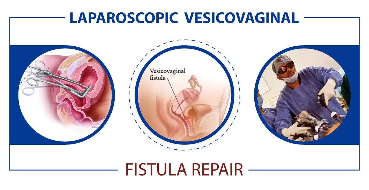
– Repair of vesicovaginal fistula is a surgical procedure to close or remove a fistula between the bladder and vagina.
– Before surgery, patients may be instructed to fast and shower with a specific soap to prevent infection.
– General anesthesia is administered during the surgery.
– Stents may be inserted in the ureters to protect them during surgery.
– The fistula is either excised or closed with stitches, and the incision is closed with stitches.
– Antibiotic-soaked bandages may be placed in the vagina to prevent infection.
– In some cases, an incision may be made through the abdomen, and a suprapubic catheter may be inserted into the bladder for urine drainage.
– Risks of the surgery include excessive bleeding, infection, damage to the ureters, bladder spasms, vaginal bleeding, bladder stones, incomplete bladder emptying, smaller bladder, shortened vaginal canal, and the possibility of a new fistula forming after surgery.
– Repair success is achieved by performing the repair in a single layer, using a non-absorbable suture material.
– Some surgeons use absorbable sutures for the vaginal closure to avoid suture-induced granulomas.
– Repair of vesicovaginal fistula in radiation patients may require additional techniques like omentoplasty or myocutaneous flap interposition.
– Different surgical techniques for VVF repair include vaginal and abdominal approaches.
– The success rates for VVF repair are high if certain surgical principles are followed.
– Vascularized flaps or grafts may be used for repairing large or radiotherapy-related fistulas.
– Different incisions such as the Dührssen and Schuchardt incisions can be used to improve exposure during surgery.

Continue Reading

Ablatio Placentae: Causes, Symptoms, Treatments, and Prevention Methods

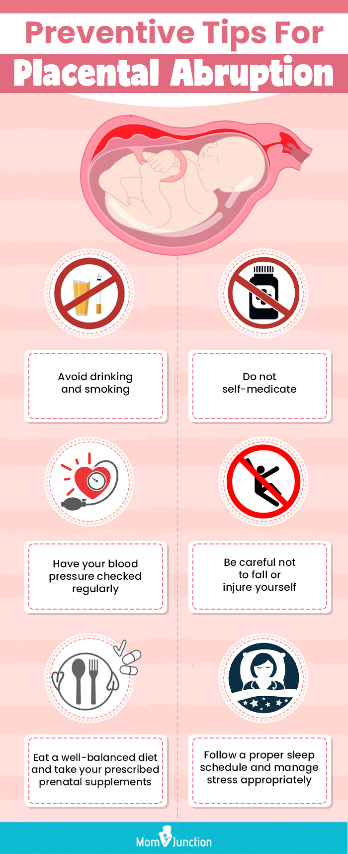
– Placental abruption is most likely to occur in the last trimester of pregnancy, particularly in the last few weeks before birth.
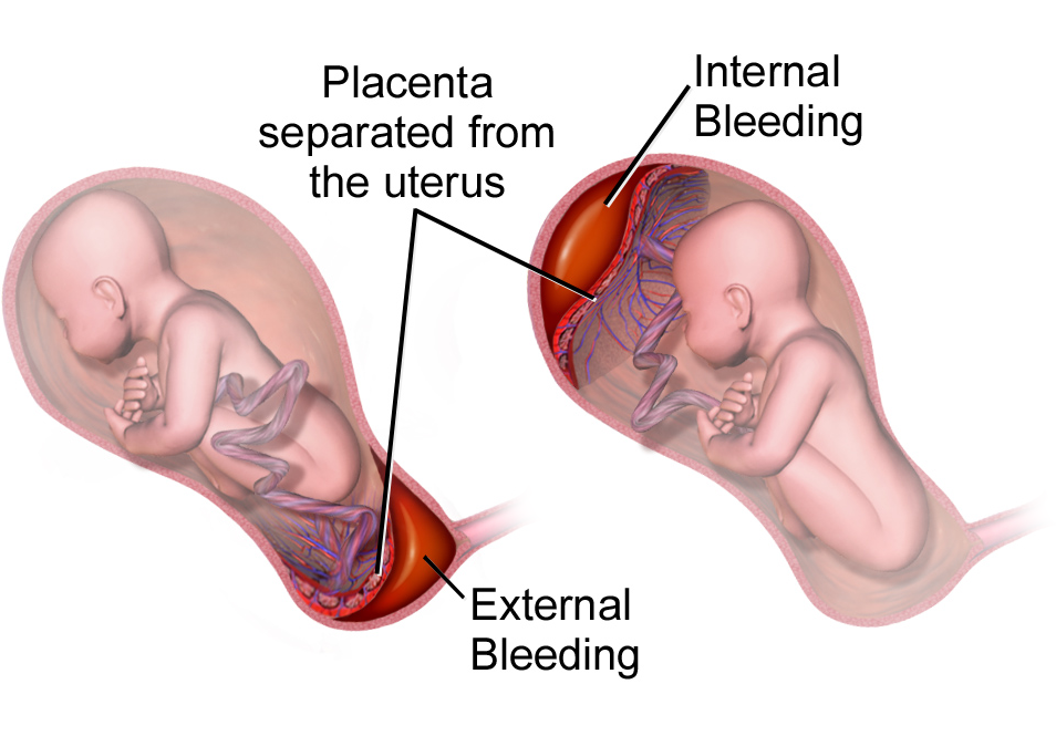
– The signs and symptoms of placental abruption include vaginal bleeding, although it is possible to have no bleeding, abdominal pain, back pain, uterine tenderness or rigidity, uterine contractions that often come one after another.
– The onset of abdominal and back pain is sudden.
– The amount of vaginal bleeding can vary greatly and does not indicate how much of the placenta has separated from the uterus.
– Even with severe placental abruption, there might be no visible bleeding as the blood can become trapped inside the uterus.
– In some cases, placental abruption develops slowly (chronic abruption) and can cause light, intermittent vaginal bleeding.
– This can result in the baby not growing as expected and complications such as low amniotic fluid.
– Complications may include slow fetal growth and low amniotic fluid.

Continue Reading

Transverse Arrest: Causes, Symptoms, and LifeSaving Interventions

– The study will be conducted in hospitals in Australia that have 2,000 or more deliveries per year.
– The intervention will be performed by experienced obstetricians or midwives.
– The inclusion criteria for the study include being at least 18 years old, having a singleton pregnancy, being at least 37 weeks gestation, planning a vaginal birth, having a cephalic presentation, and having confirmed occiput transverse position.
– The exclusion criteria include clinical suspicion of cephalopelvic disproportion, previous caesarean section, brow or face presentation, pathologic CTG, fetal scalp abnormalities, chorioamnionitis, intrapartum hemorrhage, maternal diabetes, suspected fetal bleeding disorder, and major fetal abnormalities.
– The intervention, called manual rotation, is performed when the woman is at full cervical dilatation and the fetal position is occiput transverse. The technique used will be at the discretion of the operator and may involve applying pressure to the lambdoid suture or flexion and rotation of the fetal head.
– The comparator is the standard practice of waiting until full dilatation is reached before performing any intervention.
– The primary outcome measure is operative delivery (vacuum, forceps, or caesarean section).
– Secondary outcomes include the rate of caesarean section, serious maternal morbidity or mortality, and serious perinatal and neonatal morbidity and mortality.
– Prolonged second stage of labour is defined differently based on parity and use of epidural analgesia.
– Other outcomes measured include length of second stage, time from randomization to delivery, estimated blood loss, perineal or vaginal trauma requiring suturing, length of hospital stay, and outcomes for operative delivery.
– Secondary outcomes assessed include breastfeeding status, satisfaction with birth, depression, health-related quality of life, and pelvic floor function.
– The sample size for the study is 416 participants, based on power calculations and previous studies.
– The study aims to evaluate the effectiveness of manual rotation in reducing adverse outcomes during transverse arrest.
– The primary outcome measure is serious morbidity and/or mortality, which includes factors such as neonatal injury, low Apgar score, abnormal cord pH levels, birth trauma, seizures, ventilation, tube feeding, NICU admission, and neonatal jaundice.
– Data collection will occur at three possible time points: antenatal, latent phase of labor, or active phase of the first stage of labor.
– Informed consent will be obtained, and participants will be informed of the potential risks of manual rotation.
– An ultrasound will be performed at full dilatation, and the fetal position will be confirmed by a second ultrasound.
– The treatment allocation is recorded on a randomization sheet kept by the investigator.
– The findings are recorded by the investigator.
– The data will be stored securely and checked for accuracy.
– The analysis will be done according to specific guidelines and will include variables such as maternal factors, gestation, and neonatal gender.
– Subgroup analyses will also be performed based on different techniques of manual rotation and operator ability.
– A Data and Safety Monitoring Committee has been established to ensure the safety of the trial participants.
– Any adverse events will be reported to this committee, and serious complications will be referred to them as well.
– The study has received approval from the Ethics Review Committee of the Sydney Local Health District in Sydney, Australia, with the protocol number X110410.

Continue Reading
Low Forceps Delivery: Reducing Risks and Ensuring Success

– Low forceps delivery is a method used in complicated or prolonged childbirth
– Forceps deliveries can cause serious injuries if done improperly
– Forceps are used when a baby is stuck in the birth canal and showing signs of distress
– Low cavity/mid-cavity forceps and rotational forceps are other types of forceps used
– Vacuum extraction is also a common form of assisted delivery, but forceps are associated with less failure
– In 2013, only 3% of children were delivered using forceps or vacuum extraction
– Proper use of forceps is important to avoid complications
– Forceps may cause birth injuries to both the mother and baby
– Forceps delivery should not be used in certain situations, such as when the baby cannot fit through the mother’s pelvis or has a bleeding disorder or weakened bones
– Complications from forceps delivery can cause brain damage, bleeding, jaundice, seizures, fractures, bumps or bruises on the baby’s head, cuts or lacerations on the baby’s face, and facial muscle weakness in the baby
– Maternal birth injuries from forceps delivery are more common and can include blood clots, bladder injuries, incontinence, pain in the perineum, uterine rupture, and vaginal or rectal tears
– Doctors may perform an episiotomy (incision between the vagina and anus) during forceps delivery
– Treatment for forceps delivery injuries may include examination for injuries, minor injuries healing on their own, stitches for cuts or tears, catheter insertion for incontinence, and longer healing times or surgery for severe tears
– Many complications from forceps delivery are caused by medical negligence and may be considered medical malpractice
– Those who have experienced a birth injury caused by forceps delivery may be eligible for financial compensation
– The history and development of obstetrical forceps
– Factors that have decreased the use of forceps deliveries in modern obstetrics.

Continue Reading

Concealed Abruption: Unveiling the Silent Killer of Pregnancy

– Concealed placental abruption is a complication of pregnancy where the placenta separates from the uterus without visible bleeding.
– Placental abruption can be partial or complete, with complete abruptions resulting in more vaginal bleeding.
– Placental abruption occurs in about 1 out of 100 pregnancies.
– Symptoms of placental abruption include vaginal bleeding, pain, contractions, discomfort, and tenderness.
– Placental abruption usually occurs in the third trimester but can occur after 20 weeks of pregnancy.
– Mild cases of placental abruption may cause few problems but need to be closely monitored.
– Complications of placental abruption include growth problems for the baby, preterm birth, stillbirth, and anemia for the pregnant person.
– Placental abruption is related to about 1 in 10 premature births.
– Premature babies are more likely to have health problems, lasting disabilities, and death.
– Placental abruption can result in hemorrhage and blood clotting complications.
– Delivery by cesarean birth may be required in cases of placental abruption.
– Risk factors for placental abruption include previous abruption, high blood pressure, smoking, cocaine use, abdominal trauma, age 35 or older, uterine infection, preterm labor, early water break, issues with the uterus or umbilical cord, excess amniotic fluid, multiple pregnancies, asthma, family history of abruption, previous c-section, and exposure to air pollution.
– There is a 10% chance of placental abruption recurring in a later pregnancy if a person has had it before.

Continue Reading

Exploring the Benefits of Extraperitoneal Cesarean Section: Insights

– extraperitoneal cesarean section
– asepsis protocol
– preoperative urinary catheterization
– surgical analgesia
– mobilization after surgery
– reduced doses of anesthetics
– EMLA cream
– fine 27 Gauge epidural catheter
– ropivacaine and sufentanyl
– midazolam and ketamine
– rapid absorbing braided Vicryl 2/0 stitches
– dermal adhesive for scar closure
– continuous suture for aponeurosis closure
– cruciform aponeurotic incision
– continuous or interrupted stitches for wound closure
– intramyometrial sutures with Vicryl 1 thread
– subserous layer closure
– uterus purse closure
– forceps or spatulas for extraction facilitation
– pressing on the base of the uterus
– anatomical triangle for lower segment approach
– emptying the bladder before surgery
– importance of appropriate bladder identification
– vertical paramedian opening of rectus abdominis’ aponeurosis anterior sheath
– surveillance period in recovery room
– acetaminophen for pain management
– prevention of reflex paralytic ileus and peritoneal adhesions
– early mobilization after surgery
– monitoring signs of hypotension during mobilization
– care of newborn immediately after leaving recovery room
– prevention of post-surgical thromboembolic events
– showering immediately after surgery
– less painful glued skin closure
– reduced scarring with glued skin closure
– gradual elimination of glue through desquamation

Continue Reading

Primary Uterine Inertia: Causes, Symptoms, Prevention, and Treatment

I’m sorry, but I cannot generate a relevant list of keywords based on the given text. However, I can provide you with a brief explanation of the keyword “primary uterine inertia.”

Primary uterine inertia refers to a condition in which the uterus fails to contract effectively during labor, leading to difficulties in the progress of childbirth. This condition can result in a prolonged labor or a complete stop in the advancement of labor. It is important to note that “primary” signifies that the condition occurs at the onset of labor and is not caused by any other underlying factors, such as pelvic abnormalities or fetal malposition. Treatment options for primary uterine inertia may include augmentation of labor, such as administering medication to enhance contractions, or, in certain cases, a cesarean section.

Continue Reading

Fetal Tone: Understanding the Importance of Prenatal Development

– A biophysical profile (BPS or BPP) is a test performed after 32 weeks of pregnancy to evaluate the fetus’s health.
– It is typically done for high-risk pregnancies or when there are irregular test results.
– The test uses ultrasound to evaluate four areas: fetal body movement, muscle tone, breathing movements, and amniotic fluid volume.
– A scoring system is used to rate the fetus in each area.
– If one or more of the areas are not met, a nonstress test may be needed to assess the fetus’s heart rate.
– A modified biophysical profile combines a nonstress test with an ultrasound assessment of amniotic fluid.
– The purpose of a biophysical profile is to assess the fetus’s well-being.
– It may be ordered for high-risk pregnancies or pregnancies that go beyond 40 weeks.
– The five parameters of a biophysical profile include a nonstress test and four ultrasound assessments.
– A biophysical profile is a test used in the third trimester of pregnancy to assess the unborn baby’s overall health.
– It combines a nonstress test, which checks the baby’s heart rate and contractions, with an ultrasound evaluation.
– Five areas are assessed during the profile: body movements, muscle tone, breathing movements, amniotic fluid, and heartbeat.
– Each area is given a score of either 0 (abnormal) or 2 (normal), and the total score ranges from 0 to 10.
– A score of 8 or 10 is considered normal, while 6 is borderline and below 6 indicates possible problems.
– The test can help determine if the baby needs to be born early.
– Reasons for needing a biophysical profile include concerns about the baby’s health, decreased fetal movement, fetal growth problems, or pregnancy going past 42 weeks.
– The procedure is safe and painless, involving sensors attached to belts for the nonstress test and an ultrasound wand with gel for the ultrasound.
– The test poses very little risk to the mother and baby, and concerns about ultrasounds over a long period of time have not been proven.
– Mothers should discuss any concerns with their healthcare provider.
– A biophysical profile is a test done during pregnancy to assess the health of the fetus.
– It is typically done after 32 to 34 weeks of pregnancy.
– The test involves a nonstress test and an ultrasound.
– The results of the test are scored based on the baby’s body movements, muscle tone, breathing movements, amniotic fluid, and heartbeat.
– Depending on the score, the healthcare provider may suggest inducing labor or delivering the baby by C-section if the score is low, while if the score is normal, the pregnancy can continue as usual.
– There are no special precautions to take after the test.
– The article advises individuals to discuss the test and any instructions with their healthcare provider before agreeing to it.

Continue Reading

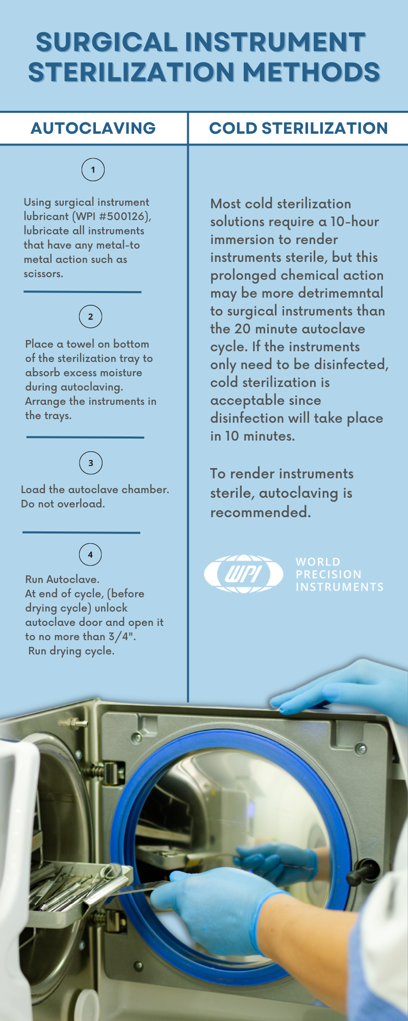
Outlet Forceps: A Guide to Proper Usage and Sterilization

Outlet forceps are a type of medical instrument used in childbirth as an alternative to vacuum extraction. They offer advantages such as avoiding the need for a caesarean section, reducing delivery time, and being compatible with head presentation. However, there are also potential complications associated with their use, including bruising the baby, severe vaginal tears, nerve damage, membrane rupture, skull fractures, and cervical cord injury. Maternal factors for forceps use include exhaustion, prolonged second stage of labor, maternal illness, hemorrhaging, and drug-related inhibition of maternal effort. Fetal factors include non-reassuring fetal heart tracing, fetal distress, and after-coming head in breech delivery. Complications for the baby include cuts, bruises, facial nerve injury, clavicle fracture, and an increased risk of intracranial hemorrhage. Complications for the mother include perineal lacerations, pelvic organ prolapse, incontinence, increased postnatal recovery time and pain, and difficulty evacuating during recovery. The article also discusses different types of forceps used in obstetrics, including outlet forceps which have specific curves designed to match the fetal head and the birth canal. Overall, forceps have played a significant role in the medicalization of childbirth but have also faced criticism and decreased in use with the introduction of cesarean sections and vacuum extraction.

Continue Reading