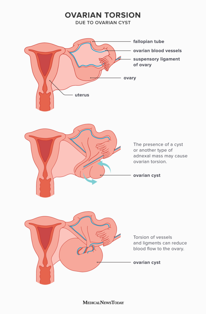
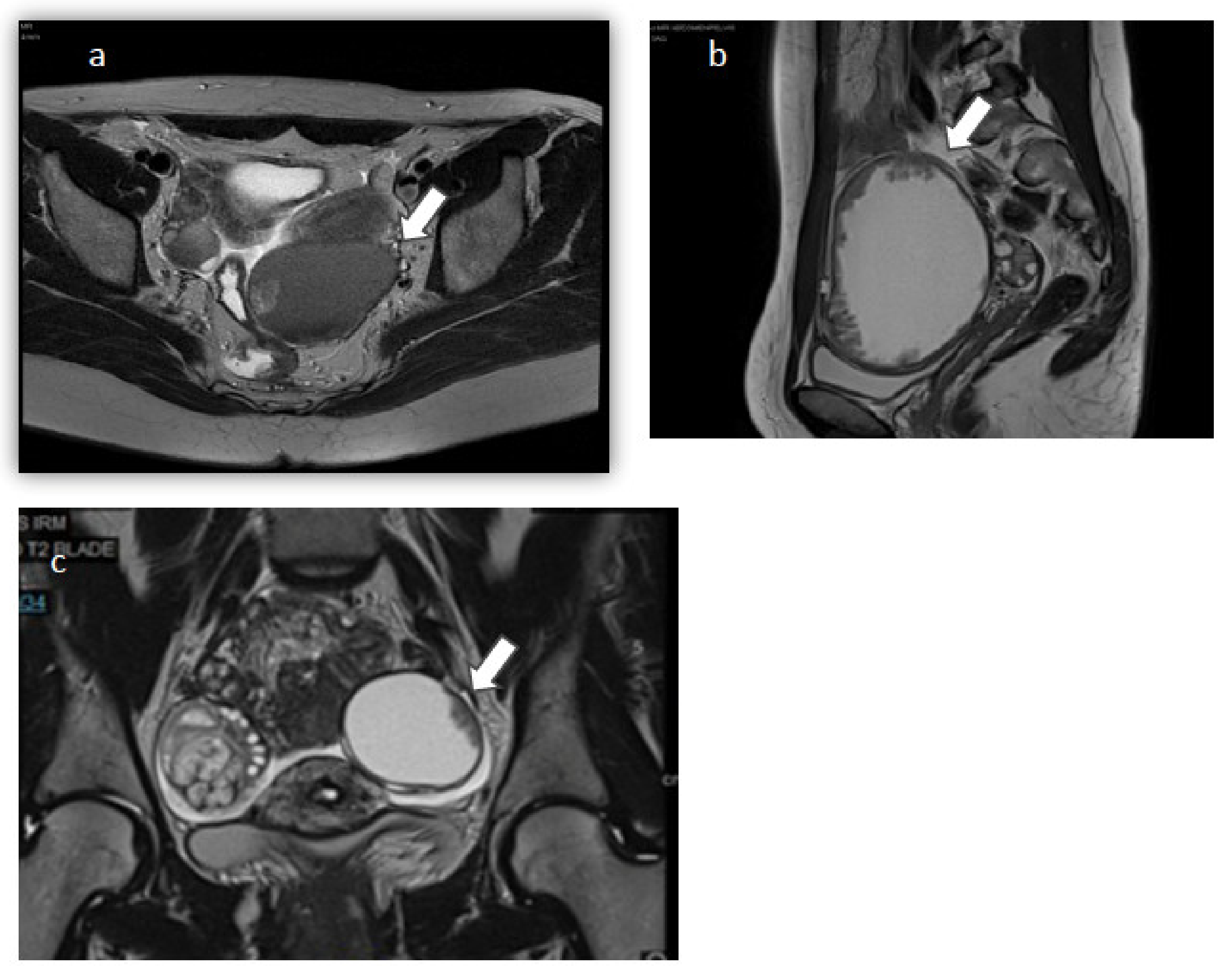
Understanding the Risks and Treatment of Torsion: Ovarian Tumor

– Ovarian torsion
– Twisting of ovarian tumor
– Cutting off blood supply to the ovary
– Organ death due to ovarian torsion
– Intense pain caused by ovarian torsion
– Vomiting as a symptom of ovarian torsion
– Peritonitis caused by ovarian torsion
– Ovarian torsion in women of reproductive age
– Ovarian torsion in girls
– Surgery for untwisting or removal of the ovary
– Full recovery with prompt treatment of ovarian torsion
– Impact of untreated ovarian torsion on fertility
– State-of-the-art ultrasound technology for diagnosis of ovarian torsion
– Minimally invasive surgery for ovarian torsion
– Adnexal torsion as another name for ovarian torsion
– Twisting of the fallopian tube in ovarian torsion
– Tissue death due to lack of blood supply in ovarian torsion
– Abdominal infection as a complication of ovarian torsion
– Diagnosis and treatment of ovarian torsion at Yale Medicine.

Continue Reading

SertoliLeydig Cell Tumour: A Comprehensive Understanding of Diagnosis, Treatment, and Prognosis

List for the keyword ‘sertoli-leydig cell tumour’:

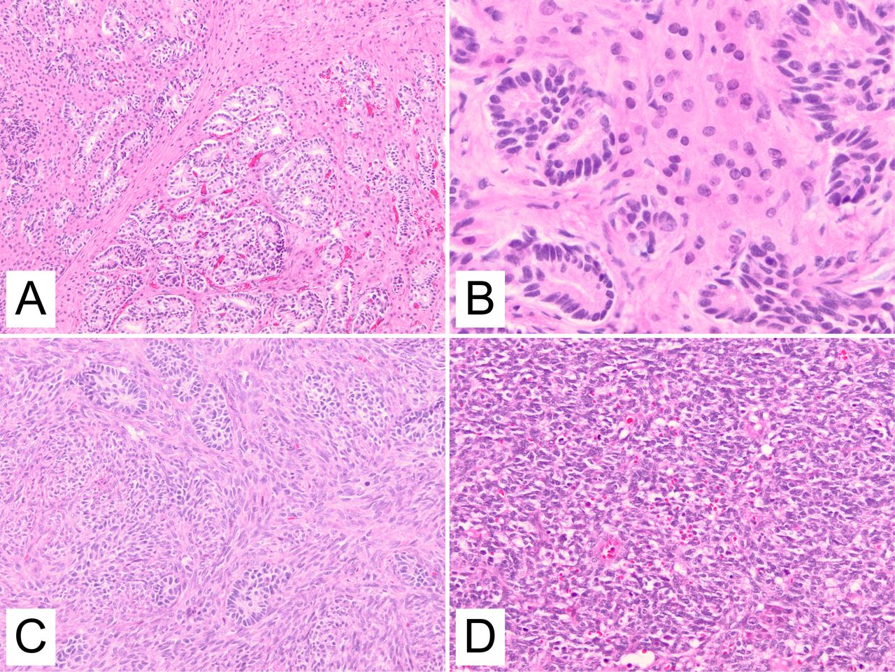
– The Sertoli-Leydig cell tumour (SLCT) originates in the ovaries of females.
– It is a rare form of cancer that primarily affects one ovary.
– SLCT cells release a male sex hormone, which can cause symptoms such as a deep voice, enlarged clitoris, facial hair, loss in breast size, and cessation of menstrual periods.
– Pain in the lower belly may be experienced due to the tumor pressing on nearby structures.
– SLCT typically begins in one ovary of a woman.
– The cancer cells release a male sex hormone, leading to symptoms such as a deep voice, enlarged clitoris, facial hair, loss in breast size, and stopped menstrual periods.
– Pain in the lower belly (pelvic area) can also occur due to the tumor pressing on nearby structures.

Continue Reading

Exploring the Origins, Symptoms, and Treatment of Endometrioid Adenoma

List:

– Endometrioid adenoma
– Adenoma-malignum-like
– Rare variant
– Endometrial endometrioid adenocarcinoma
– 58 reported cases
– Microscopic examination
– Deep invasion of glandular cells
– Myometrium
– pT2 stage
– Cervical stromal involvement
– History of endometrial adenocarcinoma
– Pelvic mass
– Malignant peripheral nerve sheath tumor
– Banal glands invading the myometrium
– Prognosis of well-differentiated adenocarcinomas
– “Adenoma malignum-like” pattern of invasion
– Recurrence-free survival
– Small sample size
– Adenocarcinoma with similar pattern of invasion
– Endometrioid type minimal deviation adenocarcinoma
– Diagnostic significance
– Benign-looking endometrial glands in the myometrium
– Consider as a differential diagnosis

Continue Reading

Exploring the Diagnostic Criteria and Management of Borderline Serous Cystadenoma: A Comprehensive Guide

– Ovarian borderline serous cystadenomas represent approximately 15% of all serous tumors.
– They are more common in younger age groups, with a peak age of presentation of around 45 years.
– Clinical presentation is often silent until the tumor reaches an advanced size or stage.
– The most frequent initial symptoms are abdominal pain, increasing abdominal girth or distension, or an abdominal mass.
– Borderline tumors develop on the surface of the ovary without invading the underlying tissue and have characteristic papillary projections.
– They can develop extra-ovarian tumor implants in advanced stages, but these implants behave in a benign manner and remain on the surface of underlying tissues.
– Serum CA-125 level is typically mildly elevated.
– Radiographic features of borderline tumors include bilateral adnexal masses with profuse papillary projections.
– They can display aggressive behavior and occasionally present with peritoneal or nodal metastases.
– Doppler ultrasound can detect intratumoral blood flow, similar to more malignant neoplasms.
– Post-surgical prognosis is better than for ovarian cystadenocarcinoma.
– They are staged using the same ovarian cancer staging as malignant ovarian neoplasms.
– Borderline serous cystadenomas were first described in 1929 and received separate classification in the early 1970s by the World Health Organization.

Continue Reading

Theca Cell Tumor: Understanding the Rare Ovarian Condition

– Prognosis for theca cell tumors (GCTs) is generally very favorable and considered to be tumors of low malignant potential
– Approximately 90% of GCTs are at stage I at the time of diagnosis
– 10-year survival rate for stage I tumors in adults is 90-96%
– GCTs of more advanced stages have 5- and 10-year survival rates of 33-44%
– Overall 5-year survival rates for patients with adult-type GCTs (AGCTs) or juvenile-type GCTs (JGCTs) are 90% and 95-97% respectively
– 10-year survival rate for AGCTs is approximately 76%
– Recurrence rate for AGCTs is 43%
– Average recurrence for AGCTs is approximately 5 years after treatment, with more than half occurring more than 5 years after primary treatment
– Mean survival after recurrence is diagnosed is 5 years for AGCTs
– 10-year overall survival after AGCT recurrence is 50-60%
– JGCTs tend to recur much sooner, with more than 90% of recurrences occurring in the first 2 years
– Tumor stage at initial surgery is the most important prognostic variable
– Other factors predicting survival include early stage disease, age younger than 50 years, high mitotic rates, moderate-to-severe atypia, preoperative spontaneous rupture of the capsule, and tumors larger than 15 cm
– True thecomas have a 5-year survival rate of nearly 100%, but may cause increased morbidity due to estrogen-producing capabilities
– More than 90% of AGCTs and JGCTs are diagnosed before spread occurs outside the ovary
– Five-year survival rates for stage I tumors are usually 90-95%
– AGCTs have a 5-year survival rate of 25-50% for patients with advanced-stage disease
– Late recurrence can occur up to 37 years after diagnosis
– Approximately 20% of GCT patients die from the disease in their lifetime
– Morbidity is primarily due to endocrine manifestations of the tumor
– Physical changes caused by high estrogen levels usually regress after tumor removal, but some patients may present with symptoms of androgen excess
– Estrogen production can stimulate the endometrium, leading to endometrial hyperplasia in 30-50% of patients and endometrial adenocarcinoma in 8-33% of patients
– There may also be an increased risk of breast cancer, although it’s difficult to prove a direct correlation
– Acute abdominal symptoms can occur in 10-15% of cases due to rupture, hemorrhage, or ovarian torsion
– Adverse effects from chemotherapy vary depending on the type given
– The standard of care for initial management of GCTs is surgical
– Preoperative evaluation, including imaging and laboratory studies, helps measure the extent of the disease
– Complete surgical staging is important and involves examination of the pelvic and intra-abdominal structures
– Optimal tumor debulking improves overall survival and decreases recurrences
– In younger patients who desire future fertility, unilateral salpingo-oophorectomy is usually sufficient treatment
– Staging typically involves pelvic washings, lymph node sampling, biopsies, and examination of the contralateral ovary
– The need for lymphadenectomy is being questioned due to the low risk of lymph node metastasis even in advanced stage disease
– Dilatation and curettage should be considered to rule out a neoplastic process of the endometrium in younger patients
– Surgical staging/biopsy based on incidence of microscopic extraovarian disease is important
– For patients who do not require future fertility, surgical therapy should consist of bilateral salpingo-oophorectomy and total abdominal hysterectomy, in addition to staging procedures
– The treatment of recurrent GCTs is not standardized, and surgical debulking may be beneficial if the tumor appears to be focal on imaging studies
– Chemotherapy, radiotherapy, and hormonal treatments have been used with varying success
– The mean survival after a recurrence has been diagnosed is approximately 5 years for adult GCTs.

Continue Reading