Partial Placenta Praevia: Causes, Symptoms, and Management Strategies

List:

– Placenta previa
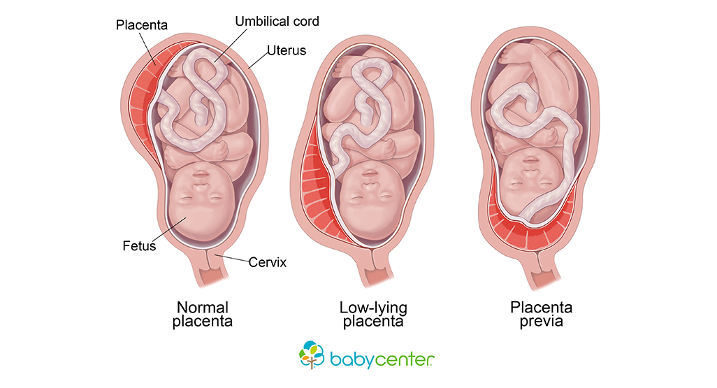
– Condition where the placenta implants at the bottom of the uterus, covering the cervix
– Painless vaginal bleeding after 20 weeks of pregnancy
– 1 in every 200 pregnancies affected by placenta previa
– Other causes of vaginal bleeding during pregnancy
– Thinning and spreading of the bottom part of the uterus can cause bleeding if the placenta is anchored to the bottom
– Sexual intercourse can cause bleeding
– Complications of placenta previa: major bleeding, shock, fetal distress, premature labor or delivery, health risks to the baby, emergency cesarean delivery, hysterectomy, blood loss for the baby, and death
– Causes and risk factors for placenta previa: low implantation of the fertilized egg, abnormalities of the uterine lining, scarring of the uterine lining, abnormalities of the placenta, and multiple pregnancies
– Tests used to diagnose placenta previa: ultrasound scans, feeling the mother’s belly
– Differentiating between placenta previa and placental abruption
– Life-threatening condition for both the mother and baby
– Diagnosis involves ultrasound scan and gentle speculum vaginal examination
– Treatment options depend on factors: type and location of the placenta, amount of blood loss, gestational age of the baby, and the health of both the baby and mother
– Medical treatment during pregnancy: bed rest, hospitalization, close monitoring, blood transfusion, avoiding activities that trigger contractions
– Delivery usually done through a caesarean section
– Postpartum monitoring for complications such as postpartum bleeding
– Baby closely monitored for health issues related to prematurity or lack of oxygen during delivery
– Prompt medical attention needed if experiencing vaginal bleeding during pregnancy
– Treatment options depend on factors such as the type and location of the placenta, amount of blood lost, gestational age of the baby, and the health of both the baby and mother
– Treatment during pregnancy may include bed rest, hospitalization, close monitoring, blood transfusion, and avoiding activities that trigger contractions
– Caesarean section usually performed once the baby is old enough
– Tests to check the mother’s blood cell counts and clotting ability needed.
Continue Reading

Discover the Fascinating Functions of the Accessory Placenta

– Accessory Lobe of Placenta is a variation in the normal shape of the placenta
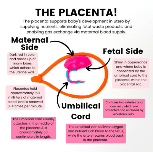
– It is a small lobe of the placenta attached to the main disc through blood vessels
– There can be one or more accessory lobes attached to the main placenta
– A routine abdominal ultrasound scan during pregnancy can help establish the presence of an Accessory Lobe of Placenta
– The condition is not associated with an increased risk of fetal anomalies
– There may be an increased risk of bleeding after delivery
– Accessory Lobe of Placenta occurs in approximately 2 per 1000 pregnancies
– There are no distinct racial, ethnic or geographical predilections
– Currently, no risk factors have been identified for Accessory Lobe of Placenta
– Accessory Lobe of Placenta is formed by non-involution of the chorionic villi
– The disc shape of the placenta is due to remodeling of placental tissue
– There are no specific signs and symptoms associated with Accessory Lobe of Placenta
– It is diagnosed by an ultrasound scan of the abdomen
– Possible complications include increased chances of postpartum hemorrhage, increased incidence of Vasa Previa, and rupture of vessels connecting the main and accessory lobe of placenta causing fetal compromise
– It does not require specific treatment but careful monitoring is needed for increased risk of bleeding after delivery
– Currently, there are no definitive methods to prevent it
– The prognosis is excellent with suitable care and management during delivery
– The incidence of Accessory Lobe of Placenta is higher in pregnancies using in-vitro fertilization
– A succenturiate (accessory) lobe is a smaller placental lobe that is in addition to the largest lobe
– The smaller succenturiate lobe often has areas of infarction or atrophy
– Risk factors for a succenturiate placenta include advanced maternal age, primigravida (first-time pregnancy), proteinuria in the first trimester, and major malformations in the fetus
– The membranes between the lobes of a succenturiate placenta can tear during delivery
– The extra lobe can be retained after the rest of the placenta is delivered, leading to postpartum bleeding
– Succenturiate lobes are generally not a major concern unless they are large and have a weak blood supply
– Vasa previa can occur if the fetal blood vessels connecting the two lobes of the placenta are located between the baby’s presenting part and the cervix or if the cord insertion is located between the two lobes.

Continue Reading

Ectopic Pregnancy: Causes, Symptoms, Diagnosis, Treatment, and Prevention

– Symptoms of an ectopic pregnancy usually develop between the 4th and 12th weeks of pregnancy
– Main symptoms include missed period, positive pregnancy test, and other signs of pregnancy
– Vaginal bleeding during ectopic pregnancy may be watery and dark brown in color
– Tummy pain, typically low down on one side, can develop suddenly or gradually
– Shoulder tip pain, felt where the shoulder ends and the arm begins, can be a sign of internal bleeding in ectopic pregnancy
– Discomfort when going to the toilet, including pain or pressure in the bottom, pain when going for a pee, and diarrhea, may occur
– These symptoms may also be caused by urinary tract infections and stomach bugs, but medical advice should be sought if pregnant and experiencing these symptoms
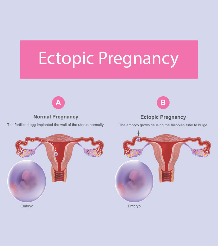
– An ectopic pregnancy occurs when a fertilized egg implants and grows outside the main cavity of the uterus.
– It most often occurs in a fallopian tube, but can also occur in other areas of the body such as the ovary, abdominal cavity, or the lower part of the uterus (cervix).
– If left untreated, it can cause life-threatening bleeding.

Continue Reading

The Hidden Dangers of Placenta Accreta: A Complicated Pregnancy Complication Explained

– Placenta accreta is a serious pregnancy condition.
– It occurs when the placenta grows too deeply into the uterine wall.
– Typically, the placenta detaches from the uterine wall after childbirth.
– With placenta accreta, part or all of the placenta remains attached.
– Placenta accreta can cause severe blood loss after delivery.
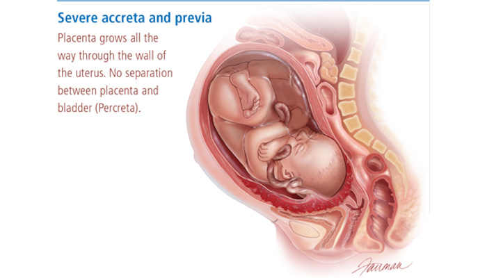
– In some cases, the placenta can invade the muscles of the uterus (placenta increta) or grow through the uterine wall (placenta percreta).

Continue Reading