Understanding Pregnancy Induced Hypertension Syndrome: Risks and Management

– pregnancy-induced hypertension syndrome
– hypertension during pregnancy
– gestational hypertension
– high blood pressure during pregnancy
– preeclampsia
– eclampsia
– complications of pregnancy-induced hypertension
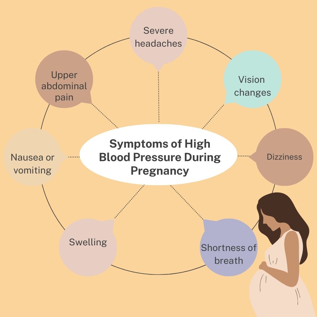
– symptoms of pregnancy-induced hypertension
– causes of pregnancy-induced hypertension
– treatment for pregnancy-induced hypertension
– management of gestational hypertension
– prevention of pregnancy-induced hypertension
– long-term effects of pregnancy-induced hypertension

Continue Reading

New Research Revealed: The Shocking Truth About Abruption

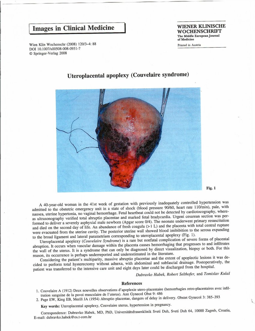
– Placental abruption is when a part or all of the placenta separates from the uterus prematurely, causing vaginal bleeding.
– There are two main types of placental abruption: revealed and concealed. Revealed abruption results in visible vaginal bleeding, while concealed abruption involves bleeding that remains within the uterus and may not be visible.
– Major risk factors for placental abruption include previous placental abruption, pre-eclampsia, abnormal lie of the baby, abdominal trauma, smoking or drug use, bleeding in the first trimester, and underlying thrombophilias.
– Clinical features of placental abruption include painful vaginal bleeding, woody and painful uterus on palpation, and the need for systematic assessment and resuscitation.
– General examination involves assessing pallor, distress, peripheral circulation, abdominal tenderness, the feel of the uterus, and the lie and presentation of the fetus/fetuses. The article provides guidance on how to assess bleeding during pregnancy and discusses differential diagnoses for antenatal hemorrhage. It suggests using a cardiotocograph (CTG) at 26 weeks gestation or above to check fetal wellbeing. It advises checking hand-held pregnancy notes for scan reports and looking for signs of placenta praevia. The article also recommends assessing the bleeding externally by looking at pads, avoiding speculum examination until placenta praevia is excluded, and taking triple genital swabs to exclude infection. It warns against performing a digital vaginal examination with known placenta praevia as it could cause massive bleeding. The article mentions placental abruption, placenta praevia, marginal placental bleed, vasa praevia, uterine rupture, and local genital causes as differential diagnoses for antenatal hemorrhage.
– Placental abruption is a common cause of antepartum hemorrhage.
– Investigations that should be performed include hematology (full blood count, Kleihauer test, group and save, cross-match), biochemistry (urea and electrolytes, liver function tests), and fetal wellbeing assessment (cardiotocograph).
– Ultrasound scan should be performed to assess placental abruption, but ultrasound should not be used to exclude abruption.
– Management of placental abruption depends on the health of the fetus: emergency delivery is indicated in the presence of maternal and/or fetal compromise, induction of labor is recommended for hemorrhage at term without compromise, and conservative management is an option for some partial or marginal abruptions without compromise.
– Anti-D should be given within 72 hours of bleeding onset if the woman is rhesus D negative.
– Placental abruption complicates approximately 1% of pregnancies and increases the risk of maternal, fetal, and neonatal morbidity and mortality.
– Risk factors for placental abruption include smoking, alcohol or cocaine use during pregnancy, advanced maternal age, history of maternal hypertension, and preeclampsia.
– Previous placental abruption and multiple gestational pregnancies also increase the risk.
– Trauma from a motor vehicle accident, fall, or blow to the abdomen can cause placental abruption.
– Radiologic imaging, such as ultrasonography, may assist in diagnosing placental abruption.
– Ultrasonography is usually the preferred study due to its benefits of avoiding ionizing radiation, dynamic nature, and availability.
– In severe trauma cases, CT scanning may be required to evaluate for abdominopelvic injuries.
– American College of Radiology guidelines recommend ultrasound FAST scan as a limited bedside adjunct for triage in a pregnant patient with major blunt trauma. The article discusses the use of imaging techniques for diagnosing major blunt trauma in pregnant patients. The American College of Radiology (ACR) states that there are two options for these procedures, but only one should be used to gather clinical information. No other facts, stats, or figures are mentioned in the article.

Continue Reading
Hypertonic Uterine Contraction: Understanding Causes, Risks, and Management

List of Pertinent Information:

1. Uterine hyperstimulation, also known as hypertonic uterine dysfunction, can occur as a complication of labor induction.
2. It is characterized by frequent contractions (more than five in 10 minutes) or contractions lasting more than two minutes.
3. Uterine hyperstimulation can result in fetal heart rate abnormalities, uterine rupture, or placental abruption.
4. The drug Misoprostol, used for peptic ulcers, can cause uterine hyperstimulation when used to induce labor.
5. Terbutaline is commonly used to treat uterine hyperstimulation.
6. Prostaglandin E2 can be administered before labor to minimize the risk of hyperstimulation and its effects on the fetal heart rate.
7. Tocolytic treatment with β2-adrenergic drugs has been used to stabilize uterine contractions and lower fetal heart rate.
8. Using a balloon catheter for labor induction instead of Prostaglandin E2 can reduce the risk of uterine hyperstimulation and its impact on the fetal heart rate.

Continue Reading

The Shocking Truth: Premature Separation of Normally Implanted Placenta

– Placental abruption is a condition in which the placenta separates from the wall of the uterus before birth.
– It can happen partially or completely, leading to a lack of oxygen and nutrients for the baby.
– Symptoms include vaginal bleeding, pain, contractions, discomfort, and tenderness.
– It occurs in about 1 in 100 pregnant individuals.
– Placental abruption is more likely to occur in the third trimester, but can happen after 20 weeks of pregnancy.
– Mild cases can lead to complete separation and may require close monitoring.
– Placental abruption is related to about 1 in 10 premature births.
– Premature babies are at higher risk for health problems, disabilities, and death.
– Placental abruption can cause anemia and life-threatening complications for the pregnant individual and the baby.
– Immediate diagnosis and treatment are necessary to prevent hemorrhage and blood clotting complications.
– Delivery by cesarean birth may be required.
– The causes of placental abruption may include previous abruption, high blood pressure, smoking, cocaine use, physical trauma, age 35 or older, infection in the uterus, preterm labor, early water breaking, issues with the uterus or umbilical cord, excess fluid around the baby, carrying multiples, asthma, family history of abruption, previous c-section, and exposure to air pollution.
– If a person has previously experienced placental abruption, they have a 10% chance of it occurring again in a later pregnancy.
– Measures to reduce the risk of placental abruption include closely monitoring and treating high blood pressure, avoiding smoking or using street drugs, always wearing a seatbelt when in a car, and taking prenatal vitamins with folic acid.

Continue Reading

Antepartum fetal death: Understanding causes, prevention, and healing

– Stillbirth is when a baby dies in the womb after 20 weeks of pregnancy.
– Stillbirth affects about 1 in 160 pregnancies each year in the United States.
– Risk factors for stillbirth include medical conditions such as obesity, diabetes, high blood pressure, and substance abuse.
– Pregnancy conditions and history can also be risk factors, such as being pregnant with multiples, having intrahepatic cholestasis of pregnancy (ICP), having complications in a previous pregnancy (preterm birth, preeclampsia, fetal growth restriction), never having given birth before, having had a miscarriage or stillbirth in a previous pregnancy, and being pregnant after the age of 35.
– Unmarried people may have a higher risk of stillbirth due to a lack of social support.
– Health disparities and racism play a role in stillbirth rates.
– Social determinants of health can affect stillbirth rates, including living conditions, access to healthcare, and exposure to chronic stress caused by racism.
– Black people have the highest stillbirth rate, more than double the rate of other groups, except for American Indian/Alaskan Native people.
– Signs and symptoms of stillbirth include the cessation of fetal movement, cramps, pain, or bleeding from the vagina.
– Tests can be conducted to determine the cause of stillbirth, including amniocentesis, autopsy, genetic tests, and tests for infections.
– After experiencing a stillbirth, it is recommended to give oneself time to heal physically and emotionally before having another baby.
– Lowering the risk of stillbirth in a subsequent pregnancy involves getting a preconception checkup, treating existing medical conditions, achieving a healthy weight, avoiding harmful substances, reporting any pain or bleeding during pregnancy, and following recommended tests and monitoring.
– Placental problems cause about 24% of stillbirths, while problems with the umbilical cord may lead to about 10% of stillbirths.
– Complications during pregnancy, health conditions like lupus and thyroid disorders, preterm labor, and fetal growth restriction can contribute to stillbirth.
– Rh disease, trauma or injuries, birth defects, and genetic conditions are also factors in stillbirth.
– Grieving after a stillbirth is a painful process, and families have various options for remembering their baby, such as spending time alone with the baby, naming the baby, holding the baby, and partaking in cultural or religious traditions.

Continue Reading

Unraveling the Secrets of the Malignant Hydatidiform Mole

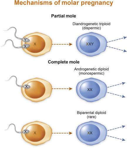
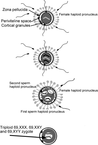
– Molar pregnancy, also known as HM or malignant hydatidiform mole, occurs due to abnormal fertilization of the egg.
– It results in an abnormal fetus and normal growth of the placenta with little or no fetal tissue growth.
– The placental tissue forms a mass in the uterus with a grape-like appearance on ultrasound, containing small cysts.
– Older women have a higher chance of developing a molar pregnancy.
– A history of molar pregnancy in earlier years is also a risk factor.
– There are two types of molar pregnancy: partial molar pregnancy, where there is an abnormal placenta and some fetal development, and complete molar pregnancy, where there is an abnormal placenta and no fetus.
– There is no known prevention for the formation of these masses.
– Mortality rate from hydatidiform mole is essentially zero due to early diagnosis and appropriate treatment.
– Approximately 20% of women with a complete mole develop a trophoblastic malignancy, which is almost 100% curable.
– Risk factors for malignant disease include advanced maternal age, high levels of hCG (>100,000 mIU/mL), eclampsia, hyperthyroidism, and bilateral theca lutein cysts.
– Predicting who will develop gestational trophoblastic neoplasia is difficult.
– Study suggests that outcomes of subsequent pregnancies in women who have had molar pregnancies are similar to those in the general population.
– Incidence of another molar pregnancy in women with a molar pregnancy is about 1.7%.
– Incidence of stillbirth in subsequent pregnancies in women with gestational trophoblastic neoplasia is 1.3%.
– Women with gestational trophoblastic neoplasia who conceive after chemotherapy have similar obstetric outcomes to those of the general population.
– Following a molar pregnancy, the risk of preterm birth is increased.
– Likelihood of large-for-gestational-age birth and stillbirth is greater if at least one birth occurs between the molar pregnancy and the index birth.
– Risk of adverse maternal outcomes is not increased following molar pregnancy.
– Malignancy is diagnosed in 15-20% of patients with a complete hydatidiform mole and 2-3% of partial moles.
– Lung metastases are found in 4-5% of patients.
– Perforation of the uterus during suction curettage is a potential complication, which may require laparoscopic guidance to complete the procedure.
– Hemorrhage is a common complication during the evacuation of a molar pregnancy, and intravenous oxytocin should be started at the beginning of suctioning.
– Other medications such as Methergine and Hemabate should also be available, and blood for possible transfusion should be readily available.
– Malignant trophoblastic disease develops in 20% of molar pregnancies, so quantitative hCG should be monitored regularly.
– Factors released by the molar tissue could trigger the coagulation cascade, leading to disseminated intravascular coagulopathy (DIC).
– Acute respiratory insufficiency can also occur due to trophoblastic embolism.
– The greatest risk factor for this complication is a larger uterus size compared to the expected gestational age.

Continue Reading

Unraveling the Mystery: Foetus Papyraceus, an Extraordinary Phenomenon

– Fetus papyraceus
– Rare condition
– Compressed in-utero
– Resorption or paper thin
– Increased incidence with assisted reproductive techniques (ART)
– High morbidity and mortality for mother and fetus
– Incidence: 1 in 12,000 pregnancies
– Incidence in twin pregnancies: 1 in 190
– Can occur in monozygotic or dizygotic twins
– Associated with ovulation induction or in vitro fertilization
– Associated complications: pre-eclampsia, disseminated intravascular coagulation, thrombotic thrombocytopenic purpura
– Intrauterine complications: premature birth, low birth weight, hypoxic ischemic encephalopathy
– Case presentation of a 43-year-old primigravida with severe oligohydramnios and anemia
– Conceived through ART
– Three fetus papyraceus identified
– Emergency caesarean section performed
– Two live births
– Diagnosis through sonographic examinations
– Monitoring the effect on surviving fetus and mother

Continue Reading