Concealed Abruption: Unveiling the Silent Killer of Pregnancy

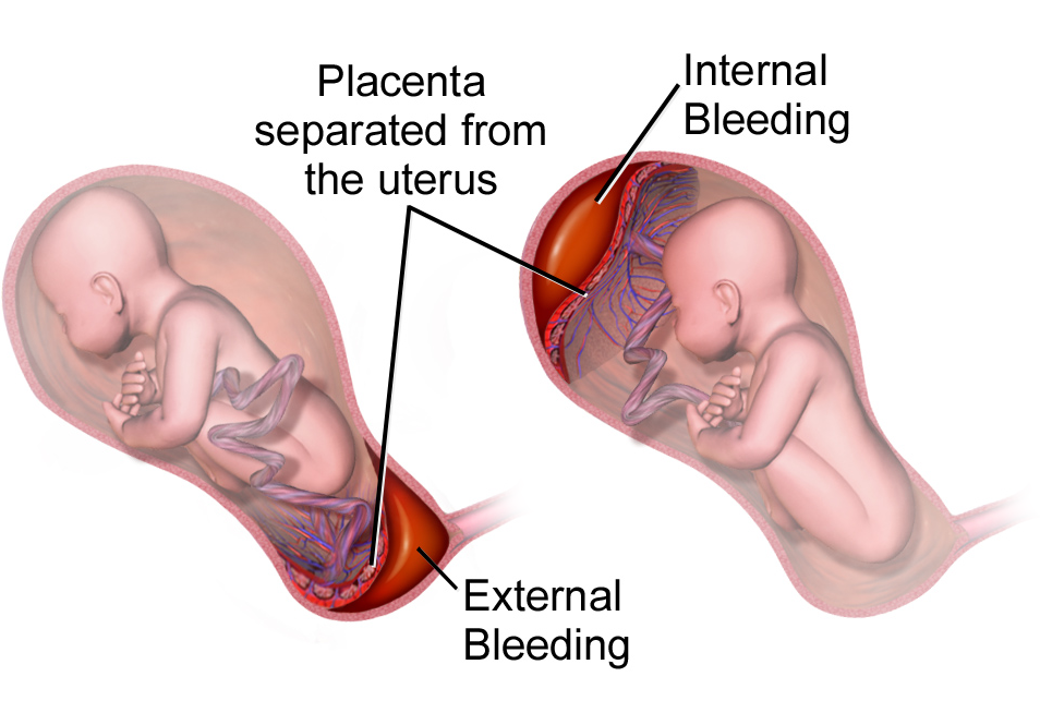
– Concealed placental abruption is a complication of pregnancy where the placenta separates from the uterus without visible bleeding.
– Placental abruption can be partial or complete, with complete abruptions resulting in more vaginal bleeding.
– Placental abruption occurs in about 1 out of 100 pregnancies.
– Symptoms of placental abruption include vaginal bleeding, pain, contractions, discomfort, and tenderness.
– Placental abruption usually occurs in the third trimester but can occur after 20 weeks of pregnancy.
– Mild cases of placental abruption may cause few problems but need to be closely monitored.
– Complications of placental abruption include growth problems for the baby, preterm birth, stillbirth, and anemia for the pregnant person.
– Placental abruption is related to about 1 in 10 premature births.
– Premature babies are more likely to have health problems, lasting disabilities, and death.
– Placental abruption can result in hemorrhage and blood clotting complications.
– Delivery by cesarean birth may be required in cases of placental abruption.
– Risk factors for placental abruption include previous abruption, high blood pressure, smoking, cocaine use, abdominal trauma, age 35 or older, uterine infection, preterm labor, early water break, issues with the uterus or umbilical cord, excess amniotic fluid, multiple pregnancies, asthma, family history of abruption, previous c-section, and exposure to air pollution.
– There is a 10% chance of placental abruption recurring in a later pregnancy if a person has had it before.

Continue Reading

Fetal Tone: Understanding the Importance of Prenatal Development

– A biophysical profile (BPS or BPP) is a test performed after 32 weeks of pregnancy to evaluate the fetus’s health.
– It is typically done for high-risk pregnancies or when there are irregular test results.
– The test uses ultrasound to evaluate four areas: fetal body movement, muscle tone, breathing movements, and amniotic fluid volume.
– A scoring system is used to rate the fetus in each area.
– If one or more of the areas are not met, a nonstress test may be needed to assess the fetus’s heart rate.
– A modified biophysical profile combines a nonstress test with an ultrasound assessment of amniotic fluid.
– The purpose of a biophysical profile is to assess the fetus’s well-being.
– It may be ordered for high-risk pregnancies or pregnancies that go beyond 40 weeks.
– The five parameters of a biophysical profile include a nonstress test and four ultrasound assessments.
– A biophysical profile is a test used in the third trimester of pregnancy to assess the unborn baby’s overall health.
– It combines a nonstress test, which checks the baby’s heart rate and contractions, with an ultrasound evaluation.
– Five areas are assessed during the profile: body movements, muscle tone, breathing movements, amniotic fluid, and heartbeat.
– Each area is given a score of either 0 (abnormal) or 2 (normal), and the total score ranges from 0 to 10.
– A score of 8 or 10 is considered normal, while 6 is borderline and below 6 indicates possible problems.
– The test can help determine if the baby needs to be born early.
– Reasons for needing a biophysical profile include concerns about the baby’s health, decreased fetal movement, fetal growth problems, or pregnancy going past 42 weeks.
– The procedure is safe and painless, involving sensors attached to belts for the nonstress test and an ultrasound wand with gel for the ultrasound.
– The test poses very little risk to the mother and baby, and concerns about ultrasounds over a long period of time have not been proven.
– Mothers should discuss any concerns with their healthcare provider.
– A biophysical profile is a test done during pregnancy to assess the health of the fetus.
– It is typically done after 32 to 34 weeks of pregnancy.
– The test involves a nonstress test and an ultrasound.
– The results of the test are scored based on the baby’s body movements, muscle tone, breathing movements, amniotic fluid, and heartbeat.
– Depending on the score, the healthcare provider may suggest inducing labor or delivering the baby by C-section if the score is low, while if the score is normal, the pregnancy can continue as usual.
– There are no special precautions to take after the test.
– The article advises individuals to discuss the test and any instructions with their healthcare provider before agreeing to it.

Continue Reading

Unintended Consequences: Understanding Accidental Abortion and Prevention

– A missed abortion is also known as a missed miscarriage or spontaneous abortion.
– It’s a miscarriage in which the fetus didn’t form or is no longer developing, but the placenta and embryonic tissues are still in the uterus.
– A missed abortion does not cause symptoms of bleeding and cramps like other types of miscarriages.
– Common symptoms of a missed abortion include brownish discharge, lessening or disappearing early pregnancy symptoms like nausea and breast soreness.
– Typical miscarriages can cause vaginal bleeding, abdominal cramps or pain, discharge of fluid or tissue, and lack of pregnancy symptoms.
– About 50% of miscarriages happen because the embryo has the wrong number of chromosomes.
– Uterine problems like scarring can also cause missed abortion. Endocrine or autoimmune disorders and heavy smoking can increase the risk.
– Physical trauma can cause missed miscarriage as well.
– Stress, exercise, sex, and travel do not cause miscarriage.
– It’s important to see a doctor if any miscarriage symptoms occur.
– A lack of pregnancy symptoms may be the only sign of a missed miscarriage.
– A missed miscarriage is usually diagnosed through ultrasound before 20 weeks of gestation.
– Doctors typically diagnose it when they can’t detect a heartbeat during a prenatal checkup.
– If the pregnancy hormone hCG doesn’t rise at a typical rate, it indicates that the pregnancy has ended.
– A follow-up ultrasound may be ordered a week later to check for the heartbeat.
– There are different treatment options for a missed miscarriage.
– Expectant management, where the patient waits for the tissue to pass naturally, is successful in more than 65% of cases.
– Medical management involves taking a medication called misoprostol to trigger the body to pass the tissue.
– Surgical management may be necessary if the tissue doesn’t pass on its own or with medication. Dilation and curettage (D&C) surgery is a common option.
– Physical recovery time after a miscarriage can range from a few weeks to a month or longer.
– Emotional recovery can take longer, and people may choose to perform religious or cultural traditions or seek counseling support.
– It is important to be understanding and supportive of someone who has experienced a miscarriage, giving them time and space to grieve in their own way.
– A miscarriage is the loss of a pregnancy before 20 weeks gestation.
– Most spontaneous miscarriages occur in the first 12 weeks of pregnancy.
– It is estimated that 1 in 4 pregnancies end in miscarriage.
– Miscarriages usually occur because the pregnancy is not developing properly.
– Miscarriages are more common in older women than younger women.
– Another cause of miscarriage may be improper embedding of the developing pregnancy in the uterus lining.
– Symptoms of a miscarriage can include pain and bleeding in early pregnancy, but not always.
– Treatment for a miscarriage is aimed at avoiding heavy bleeding and infection and providing emotional support.
– Once a miscarriage has begun, nothing can be done to stop it.
– If heavy bleeding, severe abdominal pain, fever, dizziness, or other concerning symptoms occur, medical attention should be sought.
– Types of miscarriage include missed miscarriage, threatened miscarriage, incomplete miscarriage, and complete miscarriage. There are different types of miscarriages, including blighted ovum and ectopic pregnancy. Blighted ovum occurs when a pregnancy sac is formed, but there is no developing baby within the sac. Ectopic pregnancy happens when the developing pregnancy implants in the fallopian tubes instead of the uterus. 1-2% of all pregnancies are ectopic.
– Reactions to miscarriage can include feelings of emptiness, anger, disbelief, disappointment, sadness, and isolation. Grief is common after a miscarriage, and partners may react differently. Hormonal changes may cause emotional distress. It is important not to blame yourself for a miscarriage as it is rarely caused by anything the mother did.
– After a miscarriage, it is necessary to remove any remaining pregnancy tissue to avoid complications such as prolonged bleeding or infection. This can be done with a curette under general anesthesia. Women may experience bleeding for 5-10 days after a curette and should contact a doctor if they experience prolonged or heavy bleeding, blood clots, abdominal pain, changes in vaginal discharge, or fever/flu-like symptoms.
– After a miscarriage, the first period should occur within 4 to 6 weeks.
– A check-up with a doctor is recommended 6 weeks after a miscarriage to ensure there are no problems and to check the size of the uterus.
– Most miscarriages happen by chance and are not likely to happen again in future pregnancies.
– Testing is not usually offered to women who have miscarried once or twice.
– Women who have had 3 consecutive miscarriages are at risk of miscarrying again and can seek further investigations and counseling.
– There is no right time to try for another pregnancy after a miscarriage, it varies for each individual.
– It is suggested to wait until after the next period before trying for another pregnancy.
– If a person has an Rh negative blood group, they will require an injection of anti-D immunoglobulin following a miscarriage to prevent problems with the Rh factor in future pregnancies.
– Preparing for another pregnancy after a miscarriage includes stopping smoking, exercising, having a balanced diet, reducing stress, and maintaining a healthy weight.
– Taking folic acid is recommended for all women planning a pregnancy as it helps promote normal development of a baby’s nervous system.

Continue Reading

Decidua vera: Understanding the Biology and Significance Explained

List of pertinent information about ‘decidua vera’:

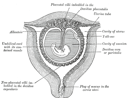
1. Decidua vera, also known as parietal decidua or true decidua, is the decidua that does not include the area occupied by the implanted ovum and chorion.
2. Decidua vera is the exclusive name for the decidua that is not occupied by the implanted ovum and chorion.
3. The decidua is the mucosal lining of the uterus that forms every month in preparation for pregnancy.
4. Decidua vera is shed after childbirth, except for the deepest layer.
5. The decidua forms the maternal part of the placenta during pregnancy and is shed together with the placenta after birth.
6. The decidua has distinct histological features, including large decidual cells.
7. The decidua plays a role in nutrient exchange, gas exchange, and waste removal during pregnancy.
8. In invasive placental disorders like placenta accreta, the decidua is found to be deficient.
9. The decidua secretes hormones, growth factors, and cytokines.
10. The role and interplay of these hormones and factors is not well understood.
11. Chronic deciduitis, a long-lasting infection of the decidua, is associated with pre-term labor.

Continue Reading

The Hidden Dangers of Red Degeneration: Untold Symptoms, Risks

– Red degeneration, also known as carneous degeneration
– Type of degeneration that can occur in uterine leiomyomas
– Most common form of degeneration during pregnancy
– Symptoms of red degeneration include abdominal pain, fever, and leukocytosis
– Red appearance of the leiomyoma
– Caused by venous thrombosis or rupture of intratumoral arteries
– Unusual signal intensity patterns on MRI, such as high signal intensity on T1-weighted images and variable signal intensity on T2-weighted images
– Contrast enhancement can vary

Continue Reading

Fetal Heart Sound: Understanding the Rhythmic Melodies

– Fetal heart monitoring measures the heart rate and rhythm of the baby during late pregnancy and labor.
– The average fetal heart rate is between 110 and 160 beats per minute, with a variation of 5 to 25 beats per minute.
– Abnormal fetal heart rate may indicate oxygen deprivation or other problems.
– There are two methods of fetal heart monitoring: external and internal.
– External monitoring uses a Doppler ultrasound device to listen to and record the baby’s heartbeat through the mother’s abdomen.
– Internal monitoring involves placing a thin wire electrode on the baby’s scalp, connected to a monitor.
– Fetal heart monitoring is especially helpful in high-risk pregnancies or when monitoring the effects of preterm labor medicines.
– Fetal heart rate can be affected by factors such as uterine contractions, pain medicines or anesthesia, tests done during labor, and pushing during the second stage of labor.
– Risks of fetal heart rate monitoring include discomfort caused by the transducer belts and slight discomfort during internal monitoring.
– The accuracy of fetal heart rate monitoring may be affected by factors such as the mother’s obesity, position of the baby or mother, and certain conditions like polyhydramnios.
– The article advises patients to discuss any concerns with their healthcare provider.
– Before the procedure, patients may be asked to sign a consent form and may need to follow specific instructions.
– The amniotic sac must be broken and the cervix dilated for internal monitoring.
– The test can be done at a healthcare provider’s office or as part of a hospital stay.
– Fetal heartbeat can be detected by a vaginal ultrasound as early as 5 1/2 to 6 weeks gestation.
– Embryonic cardiac activity begins approximately 22 days after conception.
– The earliest the baby’s cardiac activity can be detected is between five and six weeks gestation.
– At this stage, the heartbeat is seen through an ultrasound image rather than heard through a Doppler.
– It may still be too early to detect a heartbeat if it is not seen on the ultrasound screen at the first visit.
– Transvaginal ultrasounds are commonly used early on to detect the heartbeat.
– Fetal Dopplers can detect the heartbeat as early as 8 weeks, but other factors may make it difficult.
– Most fetal heart tones can be heard by 10-12 weeks.
– A baby’s heartbeat can be heard with a stethoscope starting at 18 to 20 weeks.

Continue Reading

Placenta: Unraveling Its Role in Prenatal Development

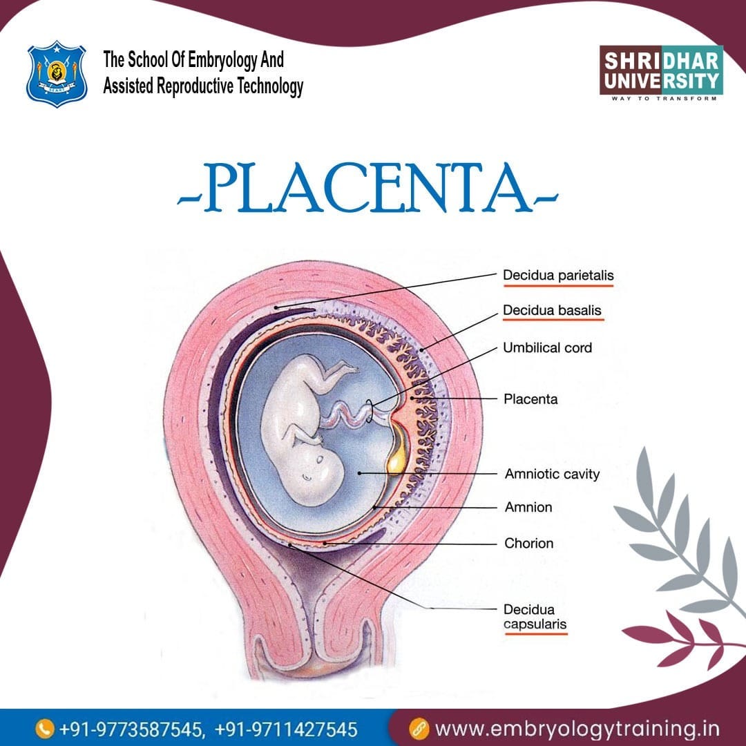
– The placenta is a temporary organ that develops during pregnancy and attaches to the lining of the uterus.
– It delivers oxygen and nutrients to the growing baby through the umbilical cord.
– Complications with the placenta can be serious and life-threatening to both the mother and baby.
– The placenta passes oxygen, nutrients, and antibodies from the mother’s blood to the baby and carries waste products back to the mother’s blood.
– It produces hormones like estrogen and progesterone that are needed during pregnancy.
– The normal position of the placenta is usually at the top, side, front, or back of the uterus, but it can sometimes develop low and move higher as the uterus stretches.
– Fraternal twins have separate placentas, while identical twins can share a placenta or have their own.
– Alcohol, nicotine, medicines, and other drugs can cross the placenta and affect the baby’s health.
– Regular visits to a healthcare provider during pregnancy are important to monitor the placenta and identify any complications.
– Prior problems with the placenta in a previous pregnancy or surgery to the uterus should be disclosed to the doctor.
– Smoking, drinking alcohol, and taking certain drugs increase the likelihood of problems with the placenta.
– Consult a doctor before taking any medicines, including over-the-counter medicines, natural therapies, and supplements during pregnancy.
– Seek medical attention if experiencing severe abdominal or back pain, vaginal bleeding, contractions, or trauma to the abdomen.
– After the baby is born, the placenta needs to be birthed during the third stage of labor.
– Placental abruption is when the placenta detaches from the uterus wall before the baby is born.
– Placenta previa is when the placenta partially or fully covers the cervix.
– Placental insufficiency occurs when the placenta does not function properly during pregnancy, resulting in a lack of oxygen and nutrients for the baby.
– Placenta accreta is when the placenta grows too deeply into the uterine wall, potentially causing severe bleeding during or after delivery.
– Retained placenta occurs when the placenta does not fully detach or come out after birth. This can be due to it being stopped by the cervix or still attached to the uterus.

Continue Reading

Preeclampsia: Understanding, Prevention, and Optimal Management Approaches Revealed

– Preeclampsia is a complication of pregnancy.
– Symptoms include high blood pressure, proteinuria, and organ damage.
– It usually occurs after 20 weeks of pregnancy in women with previously normal blood pressure.
– If left untreated, it can be fatal for both mother and baby.
– Early delivery of the baby is often recommended, based on the severity of the condition and gestational age.
– Treatment includes monitoring and medications to lower blood pressure and manage complications.
– Preeclampsia can also develop after delivery, known as postpartum preeclampsia.
– Preeclampsia is a serious condition that can occur after the 20th week of pregnancy or after giving birth.
– It can cause high blood pressure and affect the functioning of organs such as the kidneys and liver.
– Preeclampsia is responsible for 10 to 15 percent of maternal deaths worldwide.
– In the United States, it affects 5 to 8 percent of pregnancies and often leads to preterm birth.
– Signs and symptoms of preeclampsia include high blood pressure, changes in vision, headaches, nausea, pain in the upper right belly area, sudden weight gain, swelling, and trouble breathing.
– Taking low-dose aspirin may help reduce the risk of preeclampsia and preterm birth for some women.
– Low-dose aspirin, also known as baby aspirin or 81 mg aspirin, can be bought over-the-counter or prescribed by a healthcare provider.
– It is important to follow the recommended dosage and instructions given by the healthcare provider when taking low-dose aspirin for preeclampsia prevention.
– The American College of Obstetricians and Gynecologists (ACOG) states that taking low-dose aspirin during pregnancy has a low risk of serious complications and is considered safe.
– Risk factors for preeclampsia include a history of preeclampsia in a previous pregnancy, pregnancy with multiples (twins, triplets), high blood pressure, diabetes, kidney disease, or autoimmune diseases like lupus. Other risk factors include being a first-time mother, having obesity, a family history of preeclampsia, complications in previous pregnancies, fertility treatment like in vitro fertilization (IVF), and being older than 35.
– Certain groups, such as African-American women and those with lower income, are at higher risk for complications like preeclampsia due to historical health disparities and unequal access to healthcare.
– Without treatment, preeclampsia can have serious health consequences for both the mother and baby, potentially leading to death.
– Preeclampsia is a condition during pregnancy that can lead to serious complications and even death for both the mother and baby.
– The condition can cause kidney, liver, and brain damage, as well as problems with blood clotting.
– Eclampsia, characterized by seizures or a coma, is a rare and life-threatening complication that can occur after preeclampsia.
– Stroke can occur if the blood supply to the brain is interrupted or reduced.
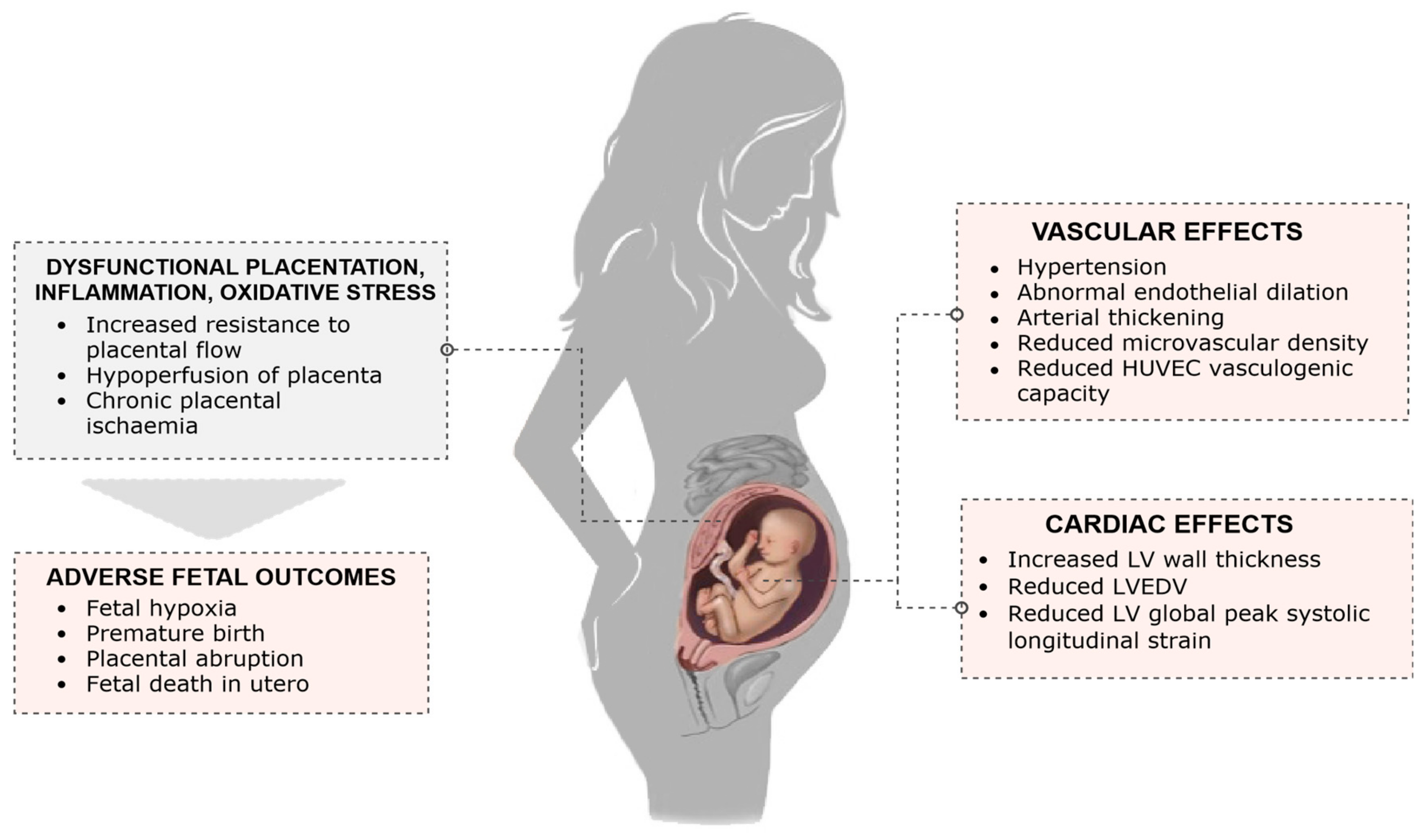
– Pregnancy complications from preeclampsia include preterm birth, placental abruption, and intrauterine growth restriction.
– Preeclampsia narrows blood vessels in the uterus and placenta, resulting in poor growth of the baby in the womb.
– Low birth weight and postpartum hemorrhage are potential complications of preeclampsia.
– Preeclampsia increases the risk of heart disease, diabetes, and kidney disease later in life.
– Diagnosis of preeclampsia involves measuring blood pressure and testing urine for protein at every prenatal visit. Additional lab work and ultrasound tests may be done.
– Treatment for preeclampsia depends on its severity and the stage of pregnancy. Mild cases may require regular monitoring and check-ups, potentially including hospitalization.
– Most women with mild preeclampsia are delivered by 37 weeks of pregnancy.
– Severe preeclampsia combined with HELLP syndrome requires early delivery. Blood transfusions may be necessary.
– Vaginal birth may be safer than a C-section, as long as there are no complications with blood clotting.
– Postpartum preeclampsia is a rare condition that can occur within 48 hours to 6 weeks after giving birth. It requires medical attention.
– Possible risk factors for postpartum preeclampsia include previous gestational hypertension or preeclampsia, obesity, and c-section.
– Complications from postpartum preeclampsia include HELLP syndrome, seizures, pulmonary edema, stroke, and thromboembolism.
– Diagnosis is done through blood and urine tests.
– Treatment may include magnesium sulfate to prevent seizures and medication to lower blood pressure.

Continue Reading
Pregnancy: Navigating the Changes, Challenges, and Delightful Discoveries

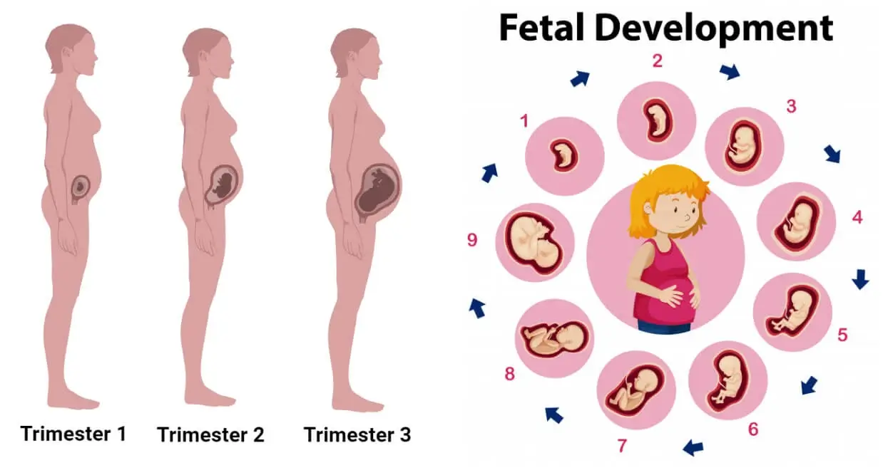
– Pregnancy usually lasts about 40 weeks, or just over 9 months
– Pregnancy is divided into three trimesters
– First Trimester: Conception occurs, fertilized egg implants in uterine wall
– Second Trimester: Sex of baby can often be determined, movement can be felt, footprints and fingerprints form, survival rate for babies born at 28 weeks is 92%
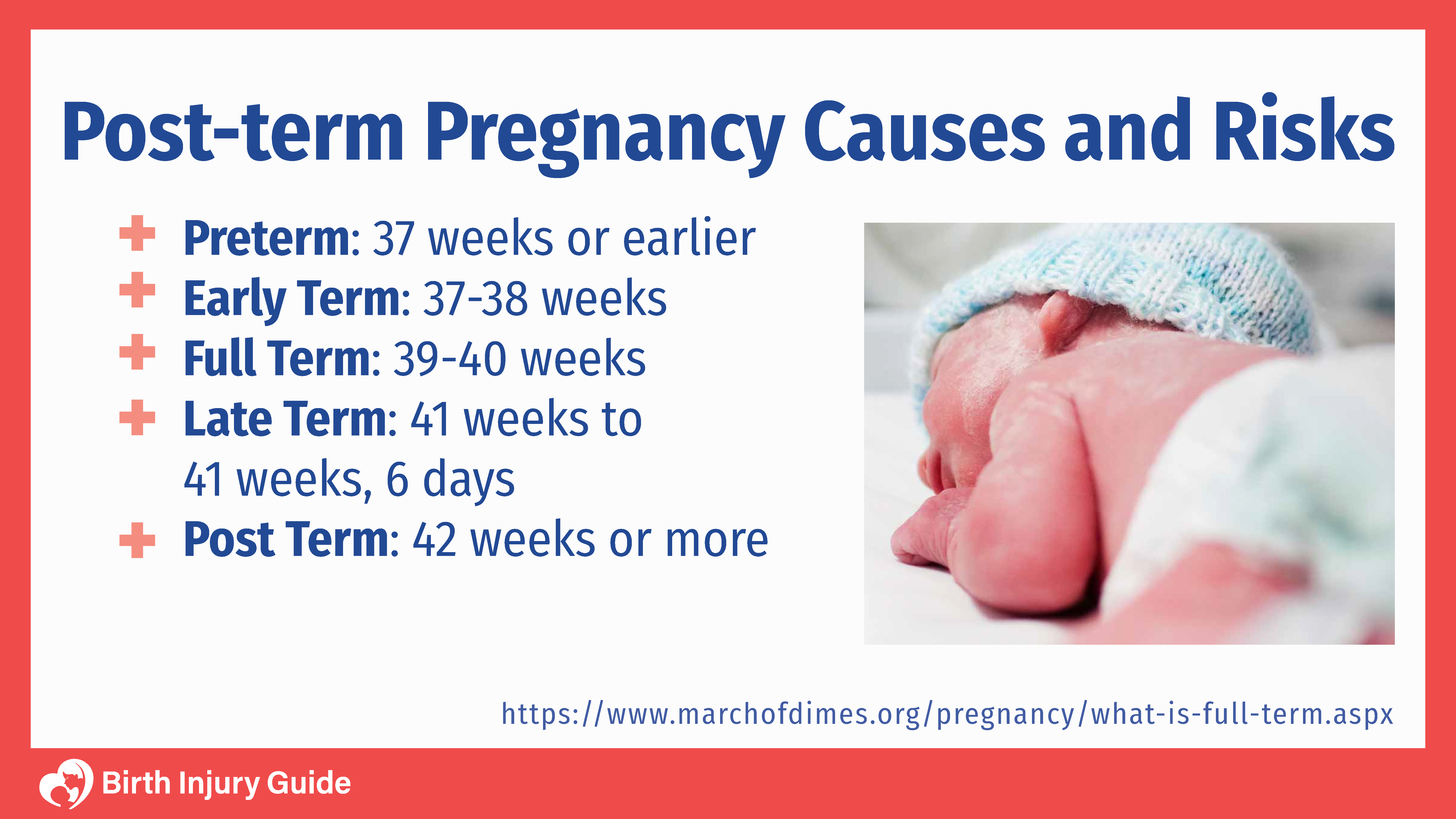
– Third Trimester: Bones become almost fully formed at 32 weeks, infants born before 37 weeks are considered preterm and at risk for complications such as developmental delays, vision and hearing problems, and cerebral palsy, infants born between 34 and 36 weeks are considered “late preterm,” infants born at 37-38 weeks are now considered “early term” and face more health risks than those born at 39 weeks or later, full-term infants born at 39-40 weeks have better health outcomes, infants born at 41-41 weeks and 6 days are considered late term, infants born at 42 weeks and beyond are considered post term.

Continue Reading