Fetal Breath Movement: Understanding Its Significance During Development

Here is the updated list of pertinent information related to the keyword “fetal breath movement” without any duplication:

– Babies don’t breathe in the womb in the traditional sense as they don’t have access to air.
– They practice breathing movements called fetal breathing movements.
– Fetal breathing movements help strengthen the muscles involved in breathing and may support the development of air sacs in the lungs.
– These movements start around week 10 of pregnancy but become more frequent after week 20.
– Fetal breathing movements are not necessary for survival and may not happen constantly.
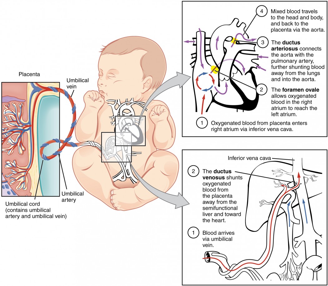
– Oxygen and carbon dioxide exchange in the womb occurs through the umbilical cord and placenta.
– After birth, babies initially rely on the umbilical cord and placenta for oxygen.
– Within about 10 seconds of delivery, babies take their first breath as they respond to the change in environment.
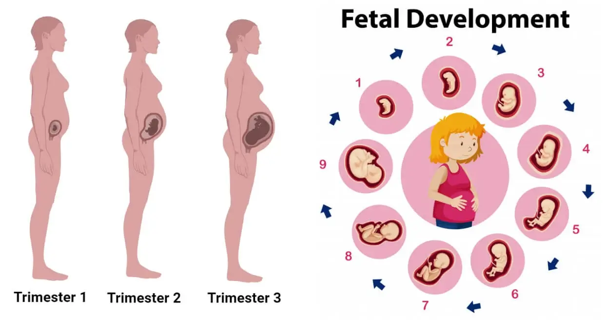
– By week 28 of pregnancy, the lungs of babies are fully developed enough that premature babies can breathe on their own.
– The lungs and circulatory system still need more time to mature.
– By week 37, the lungs have developed more fully and continue to grow and fine-tune until the child is at least 8 years old.
– The lungs begin producing surfactant at around month 6 of pregnancy, which allows them to inflate and deflate.
– Artificial surfactant can be used for babies born extremely preterm, along with breathing assistance from a ventilator, CPAP machine, or small breathing tubes.
– Babies born before week 36 and most born before week 32 may also need breathing support.
– A healthy pregnancy, quitting smoking, following a nutritious diet, avoiding smoke after birth, and encouraging regular exercise can support a baby’s lung development.
– Contractions during delivery squeeze the baby and force amniotic fluid out of the lungs, making it easier for them to breathe.
– As long as the baby is attached to the mother through the placenta and umbilical cord, it is not necessary for them to breathe.
– The baby will take their first breath a few seconds after delivery, without the support of the mother.
– The respiratory system is still growing after birth.
– Alveoli, small air sacs in the lungs, allow the body to transfer oxygen.
– Most newborns have between 20 and 50 million alveoli when born, which can increase to 300 million by the age of eight.
– The ribs surrounding the critical organs will get tougher as the baby develops, making the lungs more secure.
– Newborns may accidentally ingest meconium (first bowel movement) during birth, which can contaminate the lungs if not eliminated promptly.
Continue Reading

Fetal Tone: Understanding the Importance of Prenatal Development

– A biophysical profile (BPS or BPP) is a test performed after 32 weeks of pregnancy to evaluate the fetus’s health.
– It is typically done for high-risk pregnancies or when there are irregular test results.
– The test uses ultrasound to evaluate four areas: fetal body movement, muscle tone, breathing movements, and amniotic fluid volume.
– A scoring system is used to rate the fetus in each area.
– If one or more of the areas are not met, a nonstress test may be needed to assess the fetus’s heart rate.
– A modified biophysical profile combines a nonstress test with an ultrasound assessment of amniotic fluid.
– The purpose of a biophysical profile is to assess the fetus’s well-being.
– It may be ordered for high-risk pregnancies or pregnancies that go beyond 40 weeks.
– The five parameters of a biophysical profile include a nonstress test and four ultrasound assessments.
– A biophysical profile is a test used in the third trimester of pregnancy to assess the unborn baby’s overall health.
– It combines a nonstress test, which checks the baby’s heart rate and contractions, with an ultrasound evaluation.
– Five areas are assessed during the profile: body movements, muscle tone, breathing movements, amniotic fluid, and heartbeat.
– Each area is given a score of either 0 (abnormal) or 2 (normal), and the total score ranges from 0 to 10.
– A score of 8 or 10 is considered normal, while 6 is borderline and below 6 indicates possible problems.
– The test can help determine if the baby needs to be born early.
– Reasons for needing a biophysical profile include concerns about the baby’s health, decreased fetal movement, fetal growth problems, or pregnancy going past 42 weeks.
– The procedure is safe and painless, involving sensors attached to belts for the nonstress test and an ultrasound wand with gel for the ultrasound.
– The test poses very little risk to the mother and baby, and concerns about ultrasounds over a long period of time have not been proven.
– Mothers should discuss any concerns with their healthcare provider.
– A biophysical profile is a test done during pregnancy to assess the health of the fetus.
– It is typically done after 32 to 34 weeks of pregnancy.
– The test involves a nonstress test and an ultrasound.
– The results of the test are scored based on the baby’s body movements, muscle tone, breathing movements, amniotic fluid, and heartbeat.
– Depending on the score, the healthcare provider may suggest inducing labor or delivering the baby by C-section if the score is low, while if the score is normal, the pregnancy can continue as usual.
– There are no special precautions to take after the test.
– The article advises individuals to discuss the test and any instructions with their healthcare provider before agreeing to it.

Continue Reading

Fetal Heart Sound: Understanding the Rhythmic Melodies

– Fetal heart monitoring measures the heart rate and rhythm of the baby during late pregnancy and labor.
– The average fetal heart rate is between 110 and 160 beats per minute, with a variation of 5 to 25 beats per minute.
– Abnormal fetal heart rate may indicate oxygen deprivation or other problems.
– There are two methods of fetal heart monitoring: external and internal.
– External monitoring uses a Doppler ultrasound device to listen to and record the baby’s heartbeat through the mother’s abdomen.
– Internal monitoring involves placing a thin wire electrode on the baby’s scalp, connected to a monitor.
– Fetal heart monitoring is especially helpful in high-risk pregnancies or when monitoring the effects of preterm labor medicines.
– Fetal heart rate can be affected by factors such as uterine contractions, pain medicines or anesthesia, tests done during labor, and pushing during the second stage of labor.
– Risks of fetal heart rate monitoring include discomfort caused by the transducer belts and slight discomfort during internal monitoring.
– The accuracy of fetal heart rate monitoring may be affected by factors such as the mother’s obesity, position of the baby or mother, and certain conditions like polyhydramnios.
– The article advises patients to discuss any concerns with their healthcare provider.
– Before the procedure, patients may be asked to sign a consent form and may need to follow specific instructions.
– The amniotic sac must be broken and the cervix dilated for internal monitoring.
– The test can be done at a healthcare provider’s office or as part of a hospital stay.
– Fetal heartbeat can be detected by a vaginal ultrasound as early as 5 1/2 to 6 weeks gestation.
– Embryonic cardiac activity begins approximately 22 days after conception.
– The earliest the baby’s cardiac activity can be detected is between five and six weeks gestation.
– At this stage, the heartbeat is seen through an ultrasound image rather than heard through a Doppler.
– It may still be too early to detect a heartbeat if it is not seen on the ultrasound screen at the first visit.
– Transvaginal ultrasounds are commonly used early on to detect the heartbeat.
– Fetal Dopplers can detect the heartbeat as early as 8 weeks, but other factors may make it difficult.
– Most fetal heart tones can be heard by 10-12 weeks.
– A baby’s heartbeat can be heard with a stethoscope starting at 18 to 20 weeks.

Continue Reading

Discover the Hidden Secrets: Amniotic Fluid Unveiled

Amniotic fluid is a vital component of pregnancy, surrounding and protecting the baby in the womb. It provides cushioning, helps regulate temperature, promotes lung and digestive system development, aids in muscle and bone growth, and prevents compression of the umbilical cord.

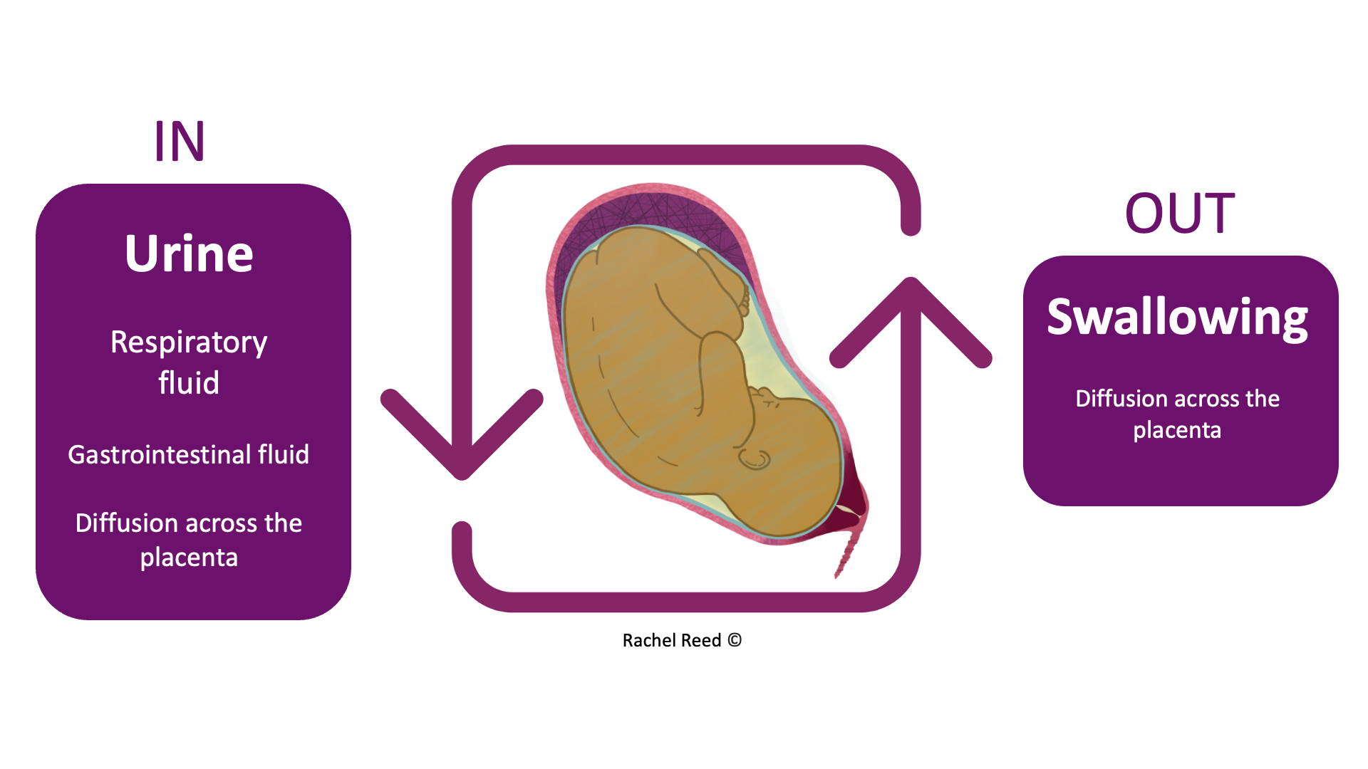
The fluid is primarily water in the early weeks of pregnancy but is later composed mostly of the baby’s urine. It also contains nutrients, hormones, and antibodies.

The amount of amniotic fluid increases until around 36 weeks of pregnancy and then gradually decreases. Too little (oligohydramnios) or too much (polyhydramnios) amniotic fluid can be problematic for both the mother and the baby, although most babies are born healthy even with these conditions.

Normal amniotic fluid is clear or tinted yellow, but green or brown fluid indicates that the baby may have passed their first bowel movement (meconium) in the womb. Meconium in the amniotic fluid can lead to breathing difficulties, known as meconium aspiration syndrome. Some babies may require immediate treatment at birth to prevent complications, while others may be healthy and not require treatment.

Continue Reading

Unveiling the Dangers and Causes of Placental Abruption

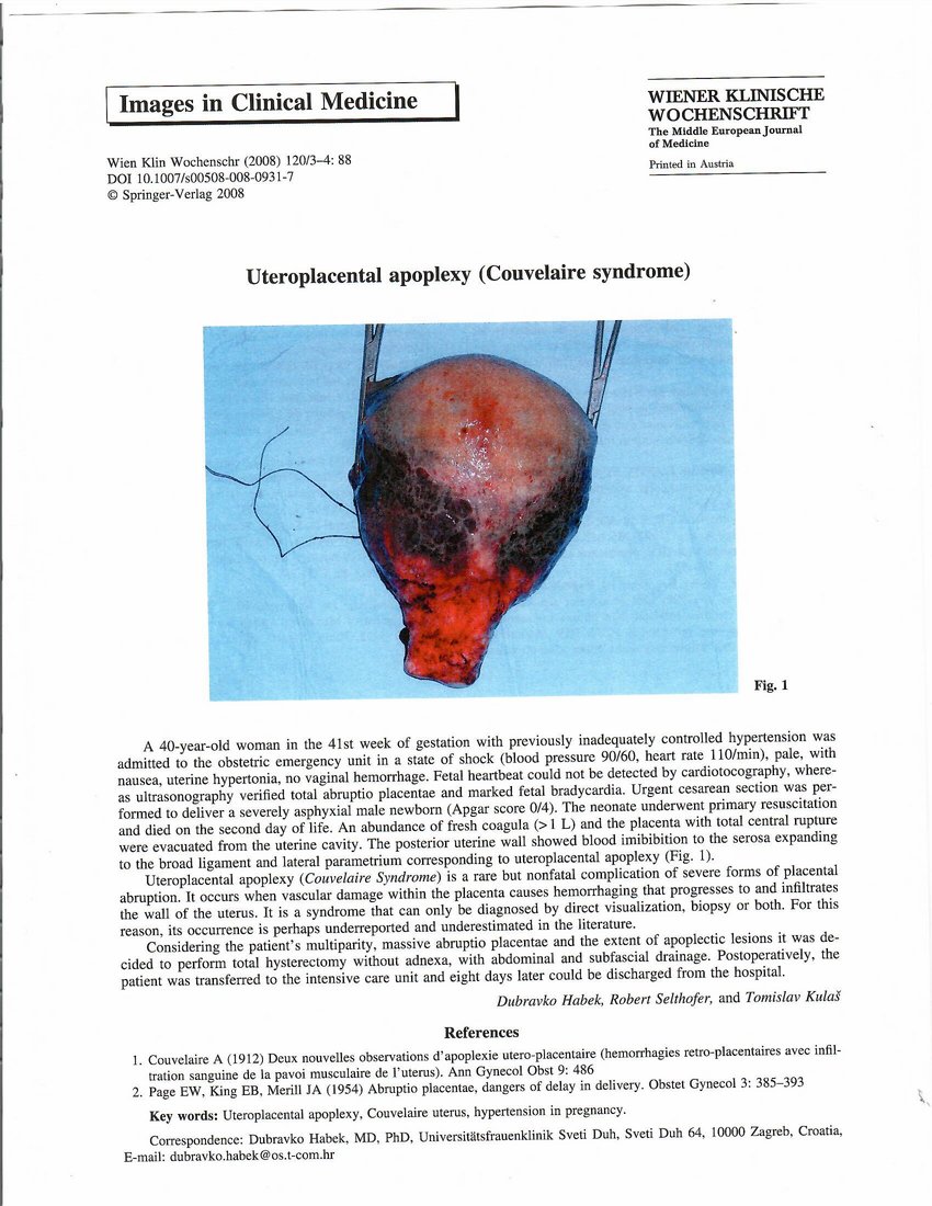
– Placental abruption is when the placenta detaches partially or completely from the wall of the uterus, causing bleeding in the mother.
– It can interfere with the baby’s supply of oxygen and nutrients from the mother’s bloodstream through the lining of the uterus.
– Prompt medical treatment is necessary to prevent dire consequences for both the mother and baby, including death.
– Worldwide, placental abruption occurs in about one pregnancy in every 100.
– About 50% of cases are mild and can be managed with ongoing monitoring. 25% are moderate, and 25% threaten the life of both the baby and the mother.
– Symptoms of moderate to severe placental abruption include bleeding, continuous abdominal and lower back pain, tender and hard uterus, very frequent uterine contractions, and fetal distress.
– In some cases, bleeding may be scanty or non-existent due to a retroplacental clot.
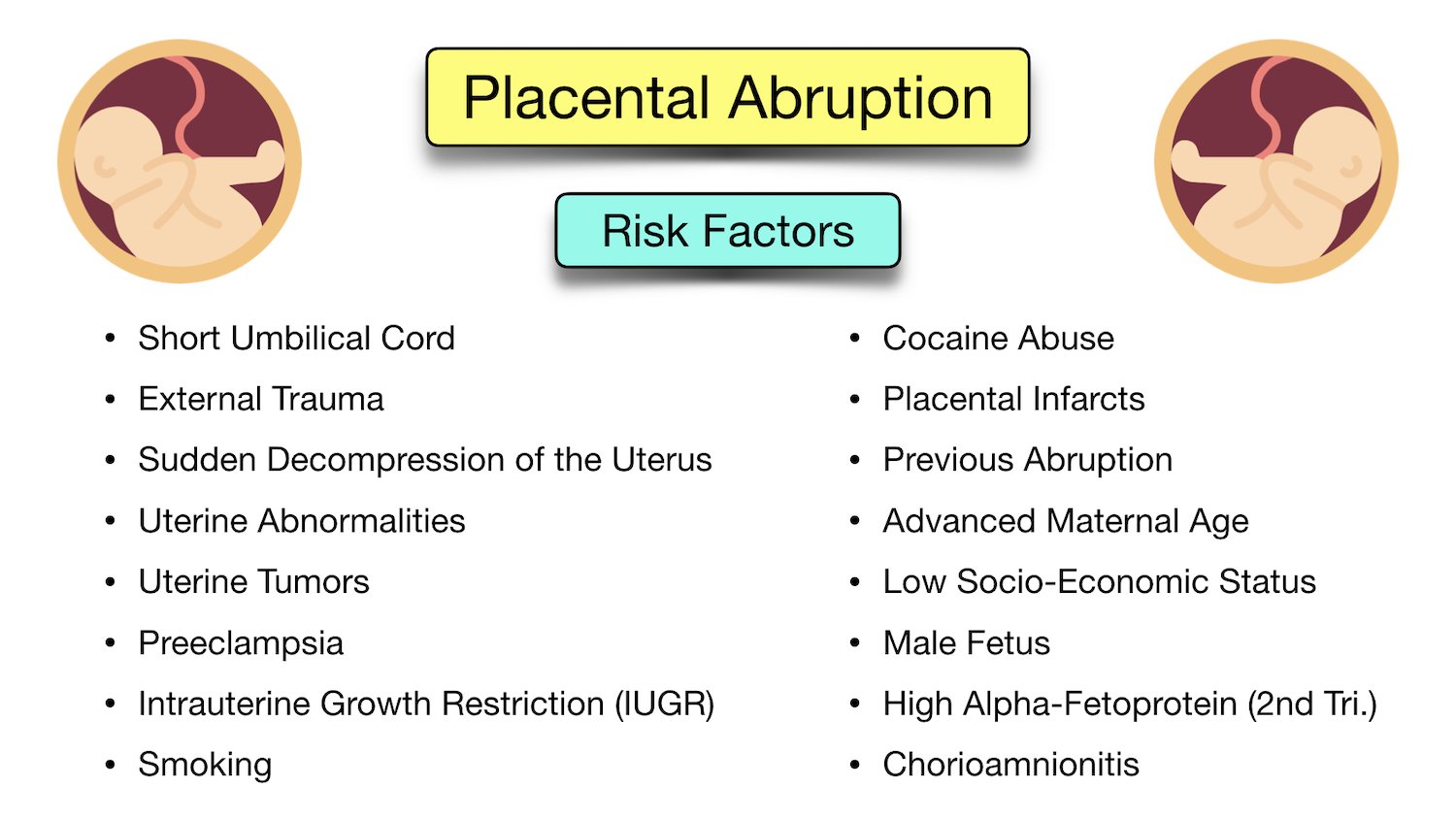
– The exact cause of placental abruption is unknown in most cases, but it is thought that abnormal blood supply in the uterus or placenta may play a role.
– Known causes of placental abruption include abdominal trauma and uterine decompression.
– Certain factors, such as advanced maternal age, prior pregnancies, and carrying multiple fetuses, increase the risk of placental abruption.
– High blood pressure increases the risk of abnormal bleeding between the placenta and the uterine wall.
– Excessive amniotic fluid increases the risk of bleeding between the placenta and the uterine wall.
– Substance use during pregnancy, such as smoking, alcohol use, and taking drugs like methamphetamine or cocaine, increase the risk of placental abruption.
– Any blood condition that affects the blood’s ability to clot can increase the risk of placental abruption.
– Procedures such as amniocentesis and amnioreduction involve a needle inserted through the mother’s abdomen into the uterus and can rarely cause bleeding.
– External cephalic version, a procedure to turn a breech baby to a head-down position, can occasionally dislodge the placenta.
– Complications in severe cases of placental abruption can include decreased oxygen to the baby, stillbirth, and maternal blood loss.
– Diagnosis of placental abruption can be done through medical history, physical examination, blood tests, ultrasound, and fetal heartbeat monitoring.
– Treatment depends on the severity of the condition and may include rest, induction of labor, vaginal birth or caesarean section, and immediate delivery.
– Severe cases may require supportive care, blood transfusion, or emergency hysterectomy.
– It is impossible to prevent placental abruption, but the risk can be reduced by avoiding substances such as cigarettes, alcohol, and street drugs, and controlling high blood pressure.

Continue Reading