Understanding and Managing Ovulatory Dysfunctional Uterine Bleeding: A Comprehensive Guide

– Aberrations in uterine bleeding are now referred to as abnormal uterine bleeding (AUB) and dysfunctional uterine bleeding (DUB)
– In 2011, the Fédération International de Gynécologie et d’Obstétrique (FIGO) defined causes of AUB using the acronym PALM-COEIN to standardize etiologies
– AUB is defined as a change in volume, regularity, or timing that has been present for 6 months or longer
– AUB affects 14-25% of women of reproductive age and is a major cause of loss of work and productivity
– Symptoms of AUB can include irregular or excessive bleeding, with excessive volume defined as an amount greater than 80 mL
– Symptoms of endometrial polyps include intermenstrual bleeding and may be asymptomatic in one-third of cases
– Symptoms of adenomyosis include heavy menses, dysmenorrhea, and midline dyspareunia
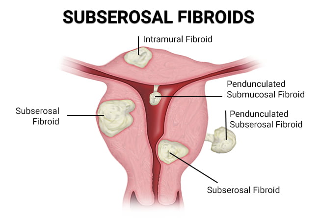
– Symptoms of leiomyomata (fibroids) depend on their size and location, with submucosal fibroids resulting in heavy unpredictable bleeding and intramural fibroids resulting in heavy but predictable bleeding
– Endometrial cancer is the most common cause of AUB-M, with postmenopausal bleeding being a common symptom
– In patients with coagulopathies, AUB typically presents in adolescence with the most common patterns being heavy menses and heavy irregular menses

The article discusses ovulatory dysfunctional uterine bleeding (AUB), which is a common type of abnormal uterine bleeding. There are different subtypes of AUB, including ovulatory dysfunction (AUB-O), primary endometrial dysfunction (AUB-E), iatrogenic causes (AUB-I), and etiologies not otherwise classified (AUB-N). A complete history and physical examination should be conducted to establish a differential diagnosis and targeted laboratory and imaging studies may be necessary. Laboratory tests may include hormone levels such as human chorionic gonadotropin (hCG), thyroid-stimulating hormone (TSH), prolactin, follicle-stimulating hormone (FSH), estradiol (E2), and luteinizing hormone (LH). Women with signs of androgen excess should have tests for testosterone, dehydroepiandrostenedione sulfate (DHEAS), and 17-hydroxyprogesterone (17OHP). Screening for certain conditions, such as Cushing disease, may also be necessary. Prothrombin time (PT), activated partial thromboplastin time (aPTT), fibrinogen, von Willebrand panel, platelet function, and coagulation factor levels should be tested for patients with AUB-C, especially if they have heavy bleeding and a history of easy bruising or bleeding from dental extraction, surgery, or the nose. Women should also have an up-to-date Pap test and human papillomavirus (HPV) testing. Imaging studies may be necessary for women with an abnormal uterus or specific bleeding patterns. This article provides information on uterine evaluation and treatment options for ovulatory dysfunctional uterine bleeding (AUB-O). The optimal imaging study depends on the specific disease process. Hysteroscopy is the best diagnostic tool for detecting polyps, while a saline infusion sonogram (SIS) is the best screening test for fibroids. Adenomyosis is best evaluated with a magnetic resonance imaging (MRI) scan, but a standard transvaginal ultrasound (TVUS) can also be used. Endometrial testing is recommended for women aged 45 and older to exclude endometrial cancer. In premenopausal patients, there are no specific ultrasound findings or endometrial thickness measurement to exclude cancer. In postmenopausal women not taking hormones, an endometrial thickness of 4 mm or less has a 94.8% sensitivity for excluding cancer. Endometrial evaluation should also be considered in women under 45 with certain conditions such as obesity, diabetes, polycystic ovary syndrome (PCOS), failed previous therapy, and prolonged estrogen exposure. Women with Lynch syndrome should undergo annual screening with endometrial biopsy and TVUS starting at age 25. Women on tamoxifen with bleeding and those with estrogen-producing ovarian tumors also require endometrial evaluation. Treatment depends on the cause of the ovulatory dysfunctional bleeding and can include hysteroscopy for polyp removal, constant progestin exposure, elimination of systemic estrogen, or surgical intervention for symptomatic adenomyosis. This article discusses the treatment options for different types of abnormal uterine bleeding. For ovulatory dysfunctional uterine bleeding, continuous progestin therapy can provide pain relief. If progestin therapy does not work, estrogen deprivation can be induced with a gonadotropin-releasing hormone agonist or an oral GnRH antagonist. Vaginal symptoms may benefit from vaginal moisturizers or low-dose vaginal estrogen. Hysterectomy is recommended for those who do not desire future pregnancy, while uterine artery embolization is an option for those who do not want a hysterectomy. Endometrial ablation is not effective for treating adenomyosis. The treatment of fibroids depends on size, location, and symptoms. Hysteroscopic myomectomy is preferred for those who desire childbearing, while hysterectomy is an option for those with complete families. Estrogen deprivation can be used to induce amenorrhea until surgery in anemic patients. Intramural fibroids may benefit from suppressing endometrial growth with progestin therapy, and surgical intervention options depend on the desire for childbearing. The management of endometrial cancer is not discussed in this article. Coagulation disorders causing abnormal bleeding can be treated according to the underlying cause, with options including tranexamic acid and desmopressin. In severe cases, von Willebrand factor concentrates or recombinant activated factor VII may be considered. Medical induction of amenorrhea is generally the preferred option for treating coagulation disorders. Summary: This article discusses the treatment options for ovulatory dysfunctional uterine bleeding. The most common bleeding disorder associated with ovulatory dysfunction is chronic anovulation due to polycystic ovary syndrome (PCOS). PCOS results in prolonged unopposed estrogen with continuous endometrial proliferation and unpredictable structural breakdown. Treatment for this condition requires chronic therapy, usually with continuous or sequential progestin exposure. Hormonal therapies such as combined oral contraceptives (COCs), progestin-secreting intrauterine devices (IUS), implants, and various progestin medications can be used. Acute treatment for heavy bleeding depends on the severity of the bleeding and the patient’s stability, and may involve the use of intravenous conjugated equine estrogens, monophasic COCs, or tranexamic acid. Common side effects of hormonal therapy include nausea, vomiting, and breast tenderness. There is a significant risk of venous thrombotic events with estrogen treatment, so non-estrogen options should be considered for women with thrombophilia or other risk factors. Tranexamic acid has an FDA warning against concomitant use with estrogen products, but studies are lacking on this interaction. The article discusses different types of ovulatory dysfunctional uterine bleeding (AUB) and their respective treatment options. AUB-E is primarily a diagnosis of exclusion and is characterized by heavy menses in ovulatory women. Treatment options for AUB-E include progestin-secreting intrauterine systems (IUS), combined oral contraceptives (COC), tranexamic acid, and nonsteroidal anti-inflammatory drugs (NSAIDs). Some authors also include chronic endometritis in this category. Endometritis can be diagnosed by the presence of plasma cells in an endometrial biopsy during the follicular phase, and empirical treatment with antibiotics such as doxycycline or a combination of a quinolone and metronidazole can be effective. For women who have completed their families, endometrial ablation or hysterectomy may be considered. AUB-I refers to iatrogenic causes of AUB, such as medications that affect the integrity of the endometrium like continuous progestins and induction of chronic endometritis with intrauterine devices. Treating AUB-I may involve short courses of vaginal estrogen or addressing inflammation related to intrauterine device use. AUB-N refers to non-specific causes of AUB, with one possibility being delayed postpartum hemorrhage due to subinvolution of the placental site. This condition can be treated with intravenous conjugated equine estrogen therapy, while dilation and curettage should be avoided.

Continue Reading

Understanding Subserous Myoma: Causes, Symptoms, and Treatment Options

– Subserosal fibroids are benign tumors that grow on the exterior of the uterus.
– The cause of subserosal fibroids is unknown, but genetics and hormones may play a role.
– African American women have a higher risk of developing fibroids.
– Women who have never had children or started puberty early (before age 12) also have a higher risk of fibroids.
– Subserosal fibroids can cause complications during pregnancy, such as lower birth weight and the need for a cesarean delivery.
– Symptoms of subserosal fibroids include a feeling of heaviness or fullness, frequent urination, constipation, and bloating.
– Subserosal uterine fibroids are diagnosed through a pelvic exam and additional tests.
– Subserosal fibroids should be treated to alleviate symptoms and avoid complications.
– Uterine Fibroid Embolization (UFE) is a non-surgical procedure that shrinks fibroids by cutting off their blood supply.
– Other treatment options include hysterectomy and myomectomy.
– Subserosal fibroids are a type of uterine fibroid that are benign and not cancerous.
– They can cause discomfort and impact nearby organs such as the bladder and bowels.
– Symptoms can include abdominal cramping, pain in the lower back and legs, and pain during sex.
– They can also lead to constipation and frequent urination.
– If subserosal fibroids are pedunculated (growing on a stalk) and the stalk becomes twisted, they can cause severe pain by cutting off the blood supply.
– Subserosal fibroids may have less impact on fertility compared to other types of fibroids, but if they grow larger during pregnancy, they can limit the space for the baby to grow and cause difficulties during childbirth.
– Fibroids are almost always non-cancerous and fibroid cancer is extremely rare.
– Subserosal fibroids can cause pain, infertility, and complications during pregnancy.
– Around 25 to 30 percent of reproductive-age women experience fibroid symptoms between the ages of 35 and 50, with most of these being subserosal fibroids.
– Symptoms of subserosal fibroids can include pain, abnormal bleeding, and abdominal discomfort.
– The causes of subserosal fibroids are not known, but hereditary factors and hormonal influence may increase the risk.
– Subserous myoma, or subserosal fibroids, are growths that appear on the uterine wall.
– Excess weight is often associated with the development of fibroids.
– Fibroids usually develop between the ages of 30 and 50.
– Subserosal fibroids can affect fertility by blocking the cervix or fallopian tubes.
– They can also cause pain and contractions, potentially leading to pre-term delivery, poor development of the fetus, miscarriage, and require a caesarian delivery.
– Treatment for subserosal fibroids depends on their condition, size, and location.
– Treatment options include close observation, subserosal fibroid removal, medications, uterine fibroid embolization, hormone treatment, fibroid surgery, and leiomyoma ablation.

Continue Reading

Infantile Uterus: Understanding the Causes, Symptoms, and Treatments

– Uterine hypoplasia is a condition that affects women’s reproductive health, where the uterus is abnormally small or underdeveloped.
– This condition can cause fertility issues, menstrual irregularities, and difficulties during pregnancy.
– Causes of uterine hypoplasia can include genetic abnormalities, exposure to toxins, hormonal imbalances, infections or inflammations of the uterus, and past surgeries or other conditions.
– Symptoms of uterine hypoplasia may include abnormal menstrual cycles, infertility, pelvic pain, and abnormalities detected through imaging tests.
– The severity of symptoms can vary depending on the degree of uterine hypoplasia.
– There are three degrees of uterine hypoplasia: first degree, second degree, and third degree.
– Treatment options for hypoplastic uterus include hormonal medications, intrauterine insemination, in vitro fertilization, surrogacy, uterine reconstruction surgery, and psychological counseling.
– Diagnostic tests that may be performed include ultrasound imaging, hysterosalpingography, and magnetic resonance imaging.
– It is important to consult with a healthcare provider for personalized recommendations based on individual needs and medical history.
– A small uterus refers to a uterus that is smaller than the average size.
– The causes of a small uterus can include genetic factors, congenital disorders, exposure to certain chemicals or radiation, and health issues such as Asherman’s Syndrome or Turner Syndrome.
– Treatment options for a small uterus can vary and commonly involve hormone therapy.
– Women with a small uterus who experience amenorrhea may receive hormonal therapy and further investigation into underlying conditions.
– Pregnancy without a uterus is possible through gestational surrogacy.
– A small uterus can present challenges during pregnancy, but with regular prenatal care and personalized care plans, successful pregnancies can still occur.
– With the right care and fertility treatments, dreams of parenthood can be realized.

Continue Reading

Congenital Absence of Uterus: Understanding the Medical Condition

– Uterine agenesis is a congenital disorder characterized by the absence of a uterus
– It may be a symptom of broader conditions like MRKH syndrome, MURCS association, or AIS
– Other reproductive organs may also be affected, such as the ovaries or fallopian tubes
– The exact cause of uterine agenesis is unknown
– Symptoms can include the absence of menstrual periods and abdominal pain
– Diagnosis is typically made during puberty when a girl fails to start menstruating
– Diagnostic tests may include blood tests, ultrasound, and MRI
– Treatment options depend on the individual and may involve creating a vagina if it is also absent
– Psychological support and counseling may be beneficial for individuals with uterine agenesis
– Regular medical follow-ups are necessary to monitor any associated conditions or complications.

Continue Reading

Demystifying the Longitudinal Vaginal Septum: Causes, Symptoms, Treatment

– A vertical or complete vaginal septum is a condition where there is a vertical wall of tissue dividing a girl’s vagina into two cavities
– Also known as “double vagina” or longitudinal vaginal septum (LVS)
– The condition is present at birth and occurs when the two parts that should join together during development don’t properly fuse
– The cause of this abnormal fetal development is unknown
– Girls with this condition may not have any symptoms and may not be aware of it until puberty
– Difficulty using tampons or discomfort during intercourse may occur during puberty
– Symptoms may include pain when inserting or removing a tampon, menstrual blood leakage even when using a tampon, and pain during intercourse
– Diagnosis involves a thorough medical history, physical exam, and additional testing such as imaging
– Treatment strategies vary
– Additional testing such as ultrasound or MRI may be used for diagnosis
– Treatment strategies vary for longitudinal vaginal septum

Continue Reading

Unlocking the Mysteries of Uterus Subseptus: A Comprehensive Guide

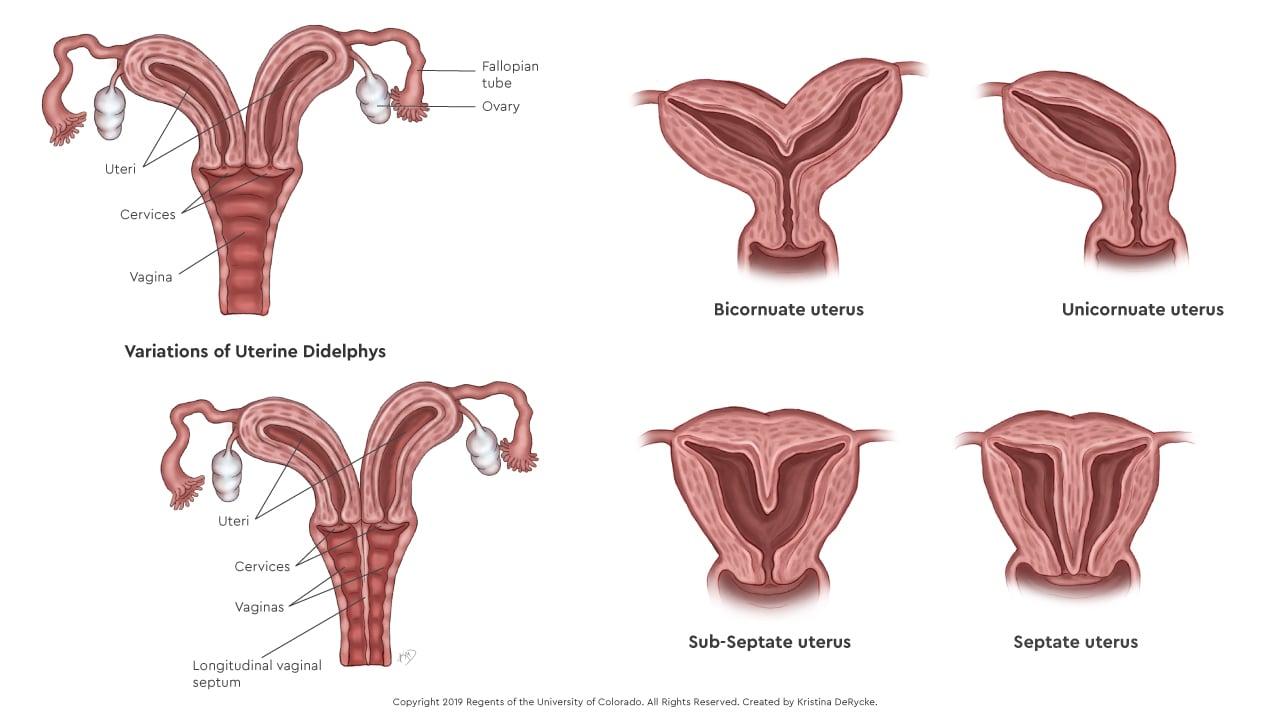
– A subseptate uterus is a mild form of congenital uterine anomaly.
– It is often considered a normal variant.
– The condition involves the presence of a partial septum within the uterus.
– The septum does not extend to the cervix.
– The angle of the central point of the septum is acute (<90°). - The external uterine contour is uniformly convex or has an indentation <10 mm. - The prevalence of a septate uterus is approximately 55% among uterine anomalies. - It is classified as a class V Müllerian duct anomaly. - A septate uterus is associated with subfertility, preterm labor, and reproductive failure in approximately 67% of cases. - 15% of women with recurrent pregnancy loss have a septate uterus. - Concurrent renal anomalies may be associated with a septate uterus. - A septate uterus is considered a type of uterine duplication anomaly that results from the partial or complete failure of resorption of the uterovaginal septum after fusion of the paramesonephric ducts. - There are different subtypes of a septate uterus, including a partial septum (subseptate uterus) and a complete septum that extends to either the internal or external cervical os. - Hysterosalpingogram alone has an accuracy of only 55% in differentiating a septate uterus from a bicornuate uterus. - Ultrasound can show that the echogenic endometrial stripe is separated at the fundus by the septum, which is isoechoic to the myometrium. The external uterine contour should be convex, flat, or mildly concave. - MRI is considered the preferred imaging modality for diagnosing a septate uterus. On MR images, the septate uterus appears normal in size, and each endometrial cavity appears smaller than a normal cavity. The septum may be composed of fibrous tissue, myometrial tissue, or both. - The treatment for a septate uterus involves shaving off the septum during hysteroscopy (metroplasty) to form a single uterine cavity without perforating the uterus. Resection of the septum in the uterus has been shown to improve outcomes, with a reported decrease in the spontaneous abortion rate from 88% to 6% after hysteroscopic metroplasty. Differential diagnosis considerations for a septate uterus include a bicornuate uterus, and it is important to differentiate between the two due to different clinical and interventional approaches. Ultrasound or MRI may also be used for diagnosis.

Continue Reading

Myoma of the uterus: Understanding causes, symptoms, treatment

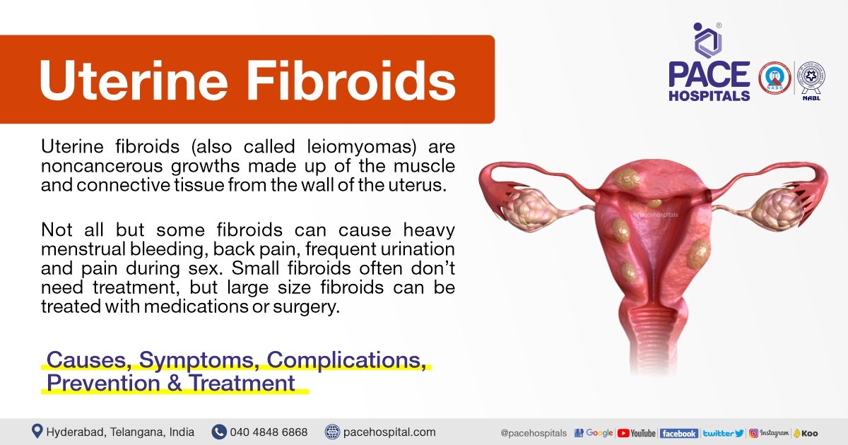
– Uterine fibroids are non-cancerous tumors that grow in the uterus, also known as myomas.
– Myomas are smooth, non-cancerous tumors made partly of muscle tissue that may develop in or around the uterus.
– Myomas in the larger, upper part of the uterus are called fibroids or leiomyomas.
– Most myomas can be seen or felt during a pelvic examination and those causing symptoms can be removed surgically or through less invasive procedures.
– The most common symptom of myomas is vaginal bleeding, which may be irregular or heavy.
– Other symptoms may include heavy bleeding, anemia, fatigue, weakness, painful intercourse, pain, bleeding, or discharge from the vagina if myomas become infected, pressure or lump in the abdomen, difficulties urinating, and urinary tract infections.
– Uterine fibroids, or myomas, affect 20 percent of women in their childbearing years.
– Uterine fibroids can cause abnormal bleeding, pelvic masses, pelvic pain, infertility, and pregnancy complications.
– There are five types of uterine fibroids: intramural, subserosal, submucosal, pedunculated, and intracavitary fibroids.
– Between 70 and 80 percent of women develop a fibroid tumor by the age of 50.
– Estrogen seems to activate the growth of uterine fibroids, and they usually shrink after menopause.
– Hormone therapy after menopause may cause fibroid symptoms to continue.
– African American women are more susceptible to developing fibroids.
– Fibroids tend to grow faster in white women younger than 35 years compared to those older than 45 years.
– Delaying pregnancy until age 30 or older increases the risk of developing fibroids.
– Early menstruation increases the risk of developing fibroids.
– Alcohol and caffeine intake may increase the risk of developing fibroids.
– Specific genetic alterations are linked to fibroid growth.
– Obesity and high blood pressure may play a role in fibroid development and growth.
– A diet rich in red meat may increase the chance of developing fibroids.
– Treatment options for uterine fibroids vary depending on the severity of symptoms and the patient’s desire to have children.
– Hormonal contraception, intrauterine devices, antifibrinolytic drugs, and nonsteroidal agents are options for managing heavy bleeding.
– Endometrial ablation can be performed if the patient does not want to have children.
– Gonadotropin-releasing hormone agonists can shrink fibroids, but they may grow back.
– Myomectomy is a procedure that removes fibroids while preserving the uterus, but fibroids may grow back.
– MRI-guided ultrasound surgery can shrink fibroids and reduce heavy bleeding.
– Uterine fibroid embolization is a minimally invasive procedure that blocks the blood supply to fibroids, causing them to shrink and die.
– Myomas can cause symptoms such as abdominal pain and heavy menstrual bleeding, but some people may remain symptom-free.
– The cause of myomas is unclear, but risk factors include a family history of myoma, obesity, and age.
– More than half of all people with a uterus will experience a myoma by age 50.
– Myomas can vary in size, from as small as a pea to as large as a melon.
– They can be located inside the uterus (intramural myomas), on the outside of the uterine wall (subserosal myomas), have a stalk or stem attaching them to the uterus (pedunculated myomas), or be found just under the lining of the uterus (submucosal myomas).
– Large myomas are considered to be 10 centimeters (cm) or more in diameter.
– Emergency room visits for myoma symptoms, such as pelvic pain and heavy bleeding, have increased from 2006 to 2017, according to a recent study.
– Myomas of the uterus are noncancerous tumors that can cause a variety of symptoms.
– Symptoms may include heavy or prolonged periods, bleeding between periods, pelvic pain, abdominal pressure, a feeling of fullness in the lower abdomen, constipation or diarrhea, frequent urination or trouble urinating, pain during sex, lower back pain, reproductive issues, fatigue, and weakness.
– Myomas are a top cause of hysterectomy surgeries, which come with their own risks and complications.
– The exact cause of myomas is unknown, but they are likely associated with hormone activity.
– High levels of estrogen and progesterone may stimulate their growth, and myomas tend to shrink when hormone levels decrease after menopause.
– Risk factors for developing myomas include a family history of myomas, obesity, high blood pressure, age, and dietary factors such as a diet high in red meat or vitamin D deficiency.
– Myomas are more common among Black individuals with a uterus, and factors such as low vitamin D levels, obesity, stress, genetics, and unequal access to healthcare have been proposed as potential risk factors.
– Medical tests used to diagnose myomas include pelvic examination, ultrasound or transvaginal ultrasound, and magnetic resonance imaging (MRI).
– Treatment for myomas depends on factors such as the severity of symptoms, the size and location of the myomas, the desire for future pregnancy, age, and proximity to menopause.
– Medications can be used to treat myomas, including over-the-counter pain medications, iron supplements for depleted iron levels, and birth control methods to control heavy menstrual bleeding.
– GnRH agonists (hormone-stimulating medications) can be used to temporarily shrink myomas, especially if surgery is planned.
– Surgical options for myomas include laparoscopic myomectomy and uterine fibroid embolization (UFE).
– No specific figures or statistics are mentioned in the article.
– One treatment option is a radiology procedure that uses injections to block blood flow to the myomas, causing them to shrink and sometimes die.
– Another option is MRI-guided ultrasound surgery, which uses ultrasound waves to shrink myomas.
– In more severe cases, other surgical options may be considered.
– Hysterectomy is a surgery to completely remove the uterus, eliminating the fibroids but also making pregnancy impossible in the future.
– Abdominal myomectomy is a surgical procedure that removes the fibroids without removing the uterus, with the possibility of future pregnancy but a risk of the fibroids returning.
– While there are no home remedies that directly treat fibroids, certain complementary therapies like acupuncture, yoga, massage, traditional Chinese medicine, and heating pads may help manage symptoms.
– Lifestyle changes such as dietary changes, exercise, stress management, and weight loss can also be beneficial.
– Myomas can cause complications related to fertility, pregnancy, and childbirth.
– These complications can include fertility issues, pregnancy complications such as miscarriage or early labor, and the need for a cesarean delivery.
– It’s important for those with myomas who want to become pregnant to discuss the condition with their healthcare provider to assess potential risks.
– It is recommended to communicate symptoms affecting one’s life to healthcare providers to determine the most suitable treatments.

Continue Reading

Tubal Abortion: Understanding the Risks, Procedures, and Recovery

List of pertinent topics to the keyword ‘tubal abortion’:

1. Ectopic pregnancy and its treatment
2. Debate surrounding treatment for ectopic pregnancy
3. Misconceptions about ectopic pregnancy management
4. Distinction between ectopic pregnancy treatment and abortion
5. Timing for trying to conceive after treatment for ectopic pregnancy
6. Methotrexate injections and their effects on fertility
7. Importance of folic acid supplementation after ectopic pregnancy treatment
8. Lack of information on tubal abortion in the article

Continue Reading