The Hidden Trauma of Genital Tract Injuries: A Comprehensive Guide

– Genital trauma refers to trauma to the genitalia, including the genital tract.
– Limited scientific data and evidence exist on genital injuries resulting from sexual assault.
– Studies on genital injuries have primarily focused on collecting evidence for legal purposes rather than medical purposes.
– Methods of studying and documenting genital injuries have improved through the use of tissue staining dyes and colposcopy.
– Vaginal trauma can occur during consensual and non-consensual intercourse, making it difficult to determine the circumstances based solely on a physical examination.
– Women are three times more likely to have vaginal injuries and intercourse-related injuries from a forced assault compared to consensual sexual experiences.
– Vaginal lacerations during intercourse may require surgery, while victims of forced assault may need additional services such as police intervention and trauma counseling.
– There is limited research on minor injuries in women of different age groups that do not require surgery or treatment.
– Factors that can predispose women to vaginal injury during consensual sex include first sexual experience, pregnancy, vigorous penetration, vaginal atrophy and spasm, previous operation or radiation therapy, disproportionate genitalia, penile ornamentation, and congenital anomalies.
– The missionary position with legs tilted all the way back during vaginal intercourse can lead to deep penetration and rotation of the uterus, potentially causing vaginal rupture.
– Vaginal tearing can occur in rape victims due to lack of vaginal lengthening and lubrication.
– Vulvar trauma is more common in prepubertal children and can occur from normal activities or sexual assault.
– Vaginal trauma can occur from the insertion of sharp objects, causing penetrating trauma.
– Severe vaginal injuries may require immediate medical attention if bleeding does not stop.
– Episiotomies can cause vaginal trauma.
– Penile trauma can occur in various forms, such as abrasions from zipper injuries or fractures from sexual activity.
– Penile amputation is a rare but emergency urological condition, often resulting from self-mutilation, accidents, or other causes.
– Micro-surgical repair is the most effective treatment method for penile trauma.
– Testicular trauma can occur from blunt, penetrating, or degloving injuries, particularly during contact sports.
– Wearing athletic cups can provide protection against testicular trauma.
– Testicular trauma can cause severe pain, bruising, swelling, and potential infertility.

Continue Reading

SertoliLeydig Cell Tumour: A Comprehensive Understanding of Diagnosis, Treatment, and Prognosis

List for the keyword ‘sertoli-leydig cell tumour’:

– The Sertoli-Leydig cell tumour (SLCT) originates in the ovaries of females.
– It is a rare form of cancer that primarily affects one ovary.
– SLCT cells release a male sex hormone, which can cause symptoms such as a deep voice, enlarged clitoris, facial hair, loss in breast size, and cessation of menstrual periods.
– Pain in the lower belly may be experienced due to the tumor pressing on nearby structures.
– SLCT typically begins in one ovary of a woman.
– The cancer cells release a male sex hormone, leading to symptoms such as a deep voice, enlarged clitoris, facial hair, loss in breast size, and stopped menstrual periods.
– Pain in the lower belly (pelvic area) can also occur due to the tumor pressing on nearby structures.

Continue Reading

Hypoplasia of uterus: Understanding its causes, symptoms, and treatment

– Uterine hypoplasia, also known as hypoplastic uterus, is a condition where a girl is born with an abnormally small uterus.
– It is a congenital disorder that occurs when the uterus fails to fully develop in the fetus.
– The cause of this abnormal development is unknown.
– Uterine hypoplasia may be associated with Mayer-Rokitansky-Küster-Hauser (MRKH) syndrome, where the girl’s uterus and vagina are absent or underdeveloped.
– Symptoms include primary amenorrhea (failure to start having periods at puberty), abdominal pain, and a small or nonexistent vaginal opening.
– Diagnosis is often made during puberty when a girl fails to start her periods, and it involves a thorough medical history, physical examination, blood tests to check for MRKH syndrome, and imaging tests such as ultrasound and MRI.
– Treatment and care for uterine hypoplasia depend on the individual and their symptoms.

Continue Reading

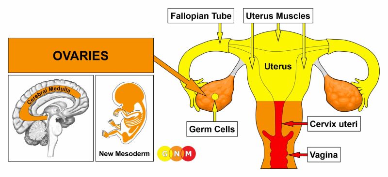
Primordial Uterus: Unveiling the Origins of Life

– Development of paramesonephric ducts in the female reproductive system
– Role of anti-Müllerian hormone (AMH) in the regression of paramesonephric ducts in males
– Persistence of paramesonephric ducts in males with mutations in AMH or AMH receptor genes
– Persistent Mullerian duct syndrome and its manifestations
– Abnormalities and complications associated with paramesonephric duct anomalies
– Difficulty in diagnosing paramesonephric duct anomalies
– Surgical advances improving the sexual function, fertility, and obstetric outcomes for women with these anomalies
– Assisted reproductive technology for women with paramesonephric duct anomalies
– Johannes Peter Müller and his discovery of paramesonephric ducts

Continue Reading

Exploring the Origins, Symptoms, and Treatment of Endometrioid Adenoma

List:

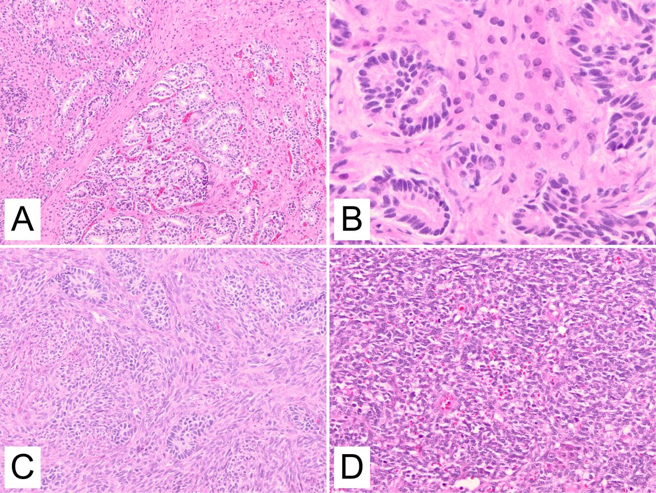
– Endometrioid adenoma
– Adenoma-malignum-like
– Rare variant
– Endometrial endometrioid adenocarcinoma
– 58 reported cases
– Microscopic examination
– Deep invasion of glandular cells
– Myometrium
– pT2 stage
– Cervical stromal involvement
– History of endometrial adenocarcinoma
– Pelvic mass
– Malignant peripheral nerve sheath tumor
– Banal glands invading the myometrium
– Prognosis of well-differentiated adenocarcinomas
– “Adenoma malignum-like” pattern of invasion
– Recurrence-free survival
– Small sample size
– Adenocarcinoma with similar pattern of invasion
– Endometrioid type minimal deviation adenocarcinoma
– Diagnostic significance
– Benign-looking endometrial glands in the myometrium
– Consider as a differential diagnosis

Continue Reading

The Mysterious Claustrum Virginale: Unveiling Its Secrets

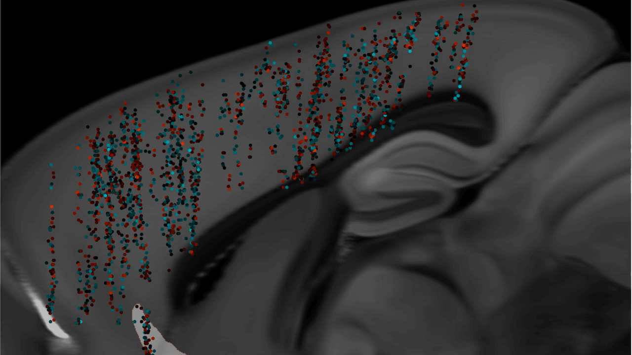
The claustrum is a thin sheet of neurons and supporting glial cells in the brain that connects to the cerebral cortex and subcortical regions. It is located between the insular cortex and putamen. The claustrum is considered to be the most densely connected structure in the brain and is hypothesized to integrate various sensory inputs into one experience. It may also play a role in salience processing and attention. The claustrum is made up of different cell types, with the principal cell type being the Golgi type I neuron. It is believed to synchronize activity in different parts of the brain and support consciousness. The claustrum may also differentiate between relevant and irrelevant information. Its precise boundaries are still debated.

The claustrum is a brain structure that has extensive connections to both cortical and subcortical regions. It is highly connected to thalamic nuclei, the basal ganglia, and various regions of the cortex. The claustrum is the most highly connected structure per regional volume in the brain and may serve as a hub to coordinate activity of cerebral circuits. It is involved in processing sensory information and the physical and emotional state of an animal. Inputs to the claustrum are organized by modality, including prefrontal, visual, auditory, and somatomotor processing areas. The claustrum possesses a distinct topological organization for each sensory modality and has dense connectivity with frontal cortices. Local connectivity within the claustrum is dominated by feed-forward disynaptic inhibition. Excitatory claustrum neurons form synapses across the anteroposterior axis and are biased toward neurons that do not share projection targets. Overall, these findings suggest that the claustrum is capable of performing local transformations of diverse input information from across the brain.

The claustrum is a part of the brain composed of various cell types. Excitatory cells in the claustrum project to different brain regions. Inhibitory neurons make up 10%-15% of the claustrum and express parvalbumin, somatostatin, or vasoactive intestinal peptide. The claustrum can be identified by its prominent plexus of parvalbumin-positive fibers. Different methods, such as electrophysiological, morphological, genetic, and connectomic approaches, have been used to study the claustrum in mice. The claustrum has widespread connectivity with cortical components associated with consciousness and sustained attention. It acts as a “conductor” in coordinating the function of all connections. The claustrum has reciprocal connections to the prefrontal cortex, visual, auditory, sensory, and motor regions. It is proposed that the claustrum functions in the gating of selective attention, selectively controlling input from different modalities. The claustrum may also implement resistance to certain inputs to prevent distraction. The claustrum integrates various sensory and motor modalities from different parts of the cortex to facilitate consciousness.

Functional imaging studies show dampened activity in the claustrum when anesthetized versus awake in rats. The claustrum has strong functional connections with the contralateral hemisphere’s claustrum, the mediodorsal thalamus, the medial prefrontal cortex, and surrounding and distant cortical areas.

The claustrum is a region in the brain that plays a role in integrating different modalities, such as sensory and motor functions. It has been shown to have connections with various parts of the cortex and is involved in processes such as spatial navigation and slow-wave sleep. The claustrum also has the ability to select between task relevant and irrelevant information for directed attention. It receives input from visual and auditory centers and can be activated by unexpected stimuli. Electrical stimulation of the claustrum can cause inhibition, leading to a blank stare and unresponsiveness. Salvinorin A, a hallucinogenic compound, can bind to Kappa Opioid Receptors in the claustrum, inducing a loss of awareness and synesthesia. High frequency stimulation of the claustrum in cats and humans has been shown to induce a decrease in awareness and consciousness.

MRI studies have also shown increased signal intensity within the claustrum. The claustrum has been associated with status epilepticus, where epileptic seizures occur without recovery of consciousness in between events. Increased signal intensity in the claustrum is associated with Focal dyscognitive seizures, which impair awareness or consciousness without convulsions. Studies have shown that the claustrum is active during REM sleep and may play a role in spatial memory consolidation. Damage to the claustrum is associated with duration of loss of consciousness in traumatic brain injuries. Stimulation of the extreme capsule, near the claustrum, can disrupt consciousness in a case study. Decreased grey matter volume in the left claustrum is associated with greater delusions in schizophrenia. Lesions to both claustrums would be needed to cause total loss of function. Electrical stimulation of the claustrum did not disrupt consciousness in a study of five patients. Damage to the claustrum may mimic other diseases or mental disorders.

Summary:

The claustrum, a region in the brain, has been found to be involved in various neurological conditions. In autism, a decrease in grey matter volume in the claustrum and insula is associated with an increase in positive symptoms. For epilepsy, increased claustral signal intensity has been observed in MRI scans of diagnosed individuals. Electrical stimulation of the claustrum has been found to disrupt consciousness, while lesions in the claustrum can lead to a loss of consciousness. However, a recent study found no disruption of consciousness with electrical stimulation. Artificial activation of the claustrum can silence brain activity across the cortex. Lesions in the claustrum have been identified as the likely origin of parkinsonism across different conditions. In mice, suppression of the claustrum attenuates anxiety and stress and increases chronic stress-resistance. The claustrum has extensive connections throughout the cortex and with sensory and motor regions in animals. The article discusses the anatomy and function of the claustrum in cats, rodents, and monkeys. In cats, high-frequency stimulation of the claustrum can alter motor activity and induce changes in awareness. The claustrum in cats responds to sensory stimuli and is connected to the motor, somatosensory, visual, and auditory cortex. Sensory input in cats is segregated based on modalities, with a preference for peripheral sensory information. In rodents, the claustrum has distinct patterning of connectivity with different cortical areas and plays specialized roles in cortical processing. In mice, inhibitory interneurons strongly modulate the activity of the claustrum and synchronize activity of claustrocortical projections to influence brain rhythms and coordinated activity of different cortical regions. In monkeys, the claustrum has widespread connections. The claustrum has connections with various cortical regions including the frontal lobe, visual cortex, temporal cortex, parieto-occipital cortex, and somatosensory areas. It also projects towards subcortical areas such as the amygdala, caudate nucleus, and hippocampus. The claustrum may have bi-directional connections with motor structures in the cortex. 70% of movement neurons in the dorsocaudal claustrum are non-selective and can fire for various types of movements, while the remaining neurons are more specific and only fire for one type of movement.

Continue Reading
HypothalamicPituitaryOvarian Axis: Navigating Female Reproductive Health

– Ovulatory disorders resulting from hypothalamic-pituitary-ovarian (HPO) axis dysfunction account for 25% of infertility diagnoses.
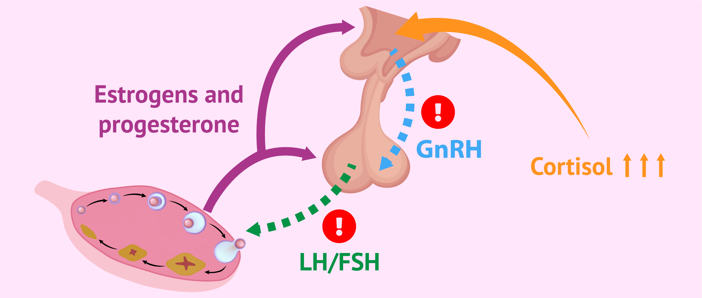
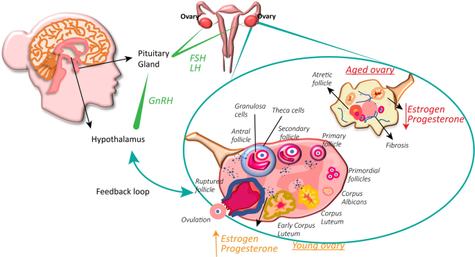
– The HPO Axis is a synchronized network of communications between the hypothalamus, the pituitary gland, and the ovaries that regulates reproductive processes.
– Ovulatory disorders manifest as abnormal, irregular, or absent ovulation and are a leading cause of infertility.
– The WHO has classified ovulatory disorders resulting from HPO dysfunction into three groups: Hypothalamic Pituitary Failure (HPF), Eugonadal Ovulatory Dysfunction, and Hypergonadotropic Ovulatory Dysfunction.

Conditions causing HPO axis dysfunction:

Group 1: Hypothalamic Pituitary Failure (HPF)
– Idiopathic hypogonadotropic hypogonadism (IHH)
– Conditions affecting the hypothalamus or pituitary gland: gene mutations, acquired panhypopituitarism, intracranial tumors, brain radiation therapy, Langerhans cell histiocytosis, De Morsier syndrome.

Group 2: Eugonadal Ovulatory Dysfunction
– Polycystic ovary syndrome (PCOS)
– Obesity
– Hyperprolactinemia
– Primary hypothyroidism

Group 3: Primary Ovarian Insufficiency or Failure (POI/POF)
– Turner Syndrome
– FMR1 gene mutation
– Autoimmune thyroiditis
– Autoimmune polyglandular syndromes
– Environmental toxins
– Cancer treatment
– Menopause

Symptoms of HPO axis dysfunction:
– Delayed puberty
– Amenorrhea
– Infertility
– Signs of hypothyroidism
– PCOS symptoms
– Insulin resistance
– Central obesity
– Hyperprolactinemia symptoms
– Symptoms of Turner Syndrome

Laboratory tests to evaluate HPO axis dysfunction:
– Dutch Cycling Mapping Test (dry urine test)
– Vibrant America’s Sex Hormones panel (blood test)
– Anti-Mullerian Hormone (AMH) test (blood test)
– Complete thyroid panels

Treatment for HPO axis dysfunction:
– Addressing the root cause
– Nutrition: fertility-based diet, calorie increase
– Herbs and supplements: Vitex, Tribulus
– Lifestyle changes: sleep, acupuncture

Note: The article briefly mentions the GnRH stimulation test as a conventional medicine test, but does not provide further details. The article also mentions genetic testing in specific cases.

Continue Reading

Exploring the Rudimentary Horn of Uterus: Anatomy, Risks, and Treatment

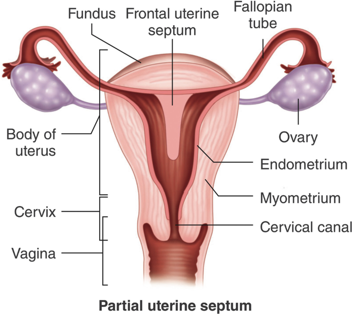
– Rudimentary horn pregnancy occurs when a fertilized egg grows in an underdeveloped part of the uterus called the rudimentary horn of a unicornuate uterus.
– Congenital uterine anomalies, including the unicornuate uterus, occur in less than 5% of all women.
– The unicornuate uterus comprises approximately 10-20% of all uterine malformations.
– Rudimentary horn pregnancy is an extremely rare type of ectopic pregnancy with an incidence of 1 in 75,000 – 150,000 pregnancies.
– Uterine anomalies result from abnormal development of embryonic structures called Mullerian ducts during fetal life.
– A unicornuate uterus results from incomplete development and failure of fusion with the opposite side of a Müllerian duct. Two-thirds of women with a unicornuate uterus may also have a rudimentary horn.
– 85% of rudimentary horn pregnancies occur in non-communicating rudimentary horns.
– Symptoms of a rudimentary horn pregnancy may include amenorrhea, vaginal bleeding (light or prolonged/intermittent), pain in the lower abdomen/pelvis/lower back, and gastrointestinal symptoms (nausea/vomiting).
– Diagnosis of a rudimentary horn pregnancy is difficult and may not be detected during regular pelvic exams.
– Transvaginal ultrasound scan (TVS) is the preferred tool for diagnosing ectopic pregnancies.
– In equivocal cases, three-dimensional ultrasound or MRI can help confirm the diagnosis.
– If left untreated, a rudimentary horn pregnancy can cause life-threatening bleeding.
– Treatment options include medical treatment with drugs, laparoscopic surgery, or abdominal surgery.
– The risk of recurrence of a pregnancy in the rudimentary horn is extremely rare with medical treatment.
– Excision of the rudimentary horn and fallopian tube is recommended to prevent future complications.
– Follow-up appointments should be scheduled, and the chances of a healthy future pregnancy can be discussed.
– The timing for attempting another pregnancy and any special precautions may be advised.
– A rudimentary horn pregnancy may not always cause symptoms and can be detected during a routine pregnancy scan.
– Diagnosis of a rudimentary horn pregnancy can be difficult and may require further medical examination.
– Symptoms of a rudimentary horn pregnancy include severe abdominal or pelvic pain, fainting, and shock.
– Prompt treatment is necessary to prevent life-threatening complications, and options include medical treatment, laparoscopic surgery, or abdominal surgery.
– Recurrence of a pregnancy in a rudimentary horn is extremely rare but possible, and routine excision of the rudimentary horn and fallopian tube may be recommended.
– Important questions to ask include the timing of follow-up appointments, chances of having a healthy future pregnancy, when to try for pregnancy again, and any special precautions to take if becoming pregnant again.

Continue Reading

Unraveling the Mysteries: Cortex of Ovary Explored

List of keywords related to ‘cortex of ovary’:

– Ovarian cortex
– Outer part of the ovary
– Ovarian follicles
– Connective tissue
– Ovarian cortex tissue transplant
– Infertility
– Primary female reproductive organs
– Pelvic cavity
– Germinal epithelium
– Tunica albuginea
– Inner medulla
– Oogenesis
– Female sex cells
– Oogonia
– Primary oocytes
– Prophase
– Follicle-stimulating hormone
– Secondary oocyte
– First polar body
– Second polar body
– Ovum
– Metaphase
– Fertilization
– Meiosis II
– Formation of an ovum
– Polar bodies
– Development of ovarian follicles
– Follicular cells
– Primordial follicles
– Antrum
– Granulosa cells
– Estrogen
– Vesicular (graafian) follicle
– Rupture of follicle
– Corpus luteum
– Progesterone
– Placenta
– Hormone secretion
– Corpus albicans.

Continue Reading