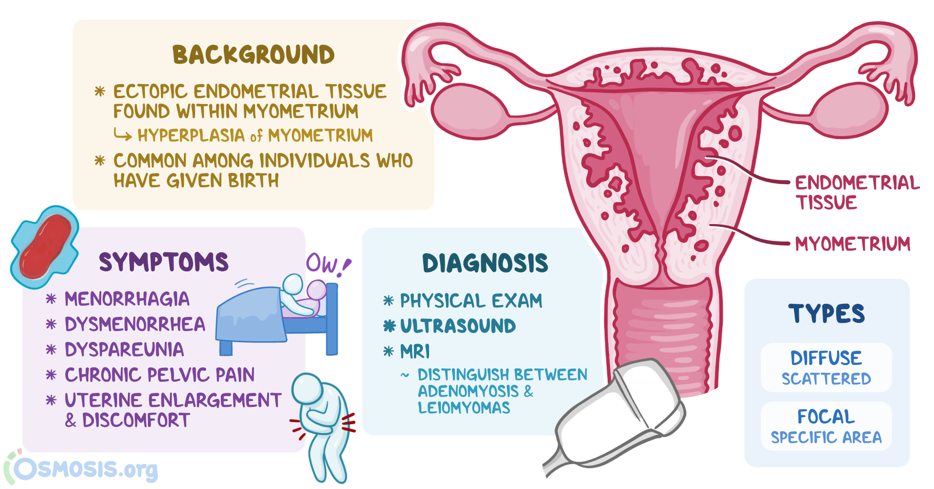
Hypoplasia of uterus: Understanding its causes, symptoms, and treatment

– Uterine hypoplasia, also known as hypoplastic uterus, is a condition where a girl is born with an abnormally small uterus.
– It is a congenital disorder that occurs when the uterus fails to fully develop in the fetus.
– The cause of this abnormal development is unknown.
– Uterine hypoplasia may be associated with Mayer-Rokitansky-Küster-Hauser (MRKH) syndrome, where the girl’s uterus and vagina are absent or underdeveloped.
– Symptoms include primary amenorrhea (failure to start having periods at puberty), abdominal pain, and a small or nonexistent vaginal opening.
– Diagnosis is often made during puberty when a girl fails to start her periods, and it involves a thorough medical history, physical examination, blood tests to check for MRKH syndrome, and imaging tests such as ultrasound and MRI.
– Treatment and care for uterine hypoplasia depend on the individual and their symptoms.

Continue Reading

Exploring the Intricate Cavity of the Uterus: Unveiling Mysteries

List of pertinent information about the cavity of the uterus (no duplications):

– The uterine cavity is the inside portion of the uterus.
– It is triangular in shape and formed by the internal surface of the body of the uterus.
– The base of the cavity is located between the openings of the fallopian tubes.
– The apex is the internal opening of the uterus that connects to the cervix.
– The part of the uterine cavity that enters the openings of the fallopian tubes is a narrow, flattened area.
– Abnormalities of the womb or congenital uterine abnormalities refer to women who have a womb that is different in shape or size from the norm.
– These abnormalities can be discovered during an ultrasound scan or if a woman experiences miscarriage, bleeding, or difficulties conceiving.
– Women with womb abnormalities may have an increased risk of miscarriage, preterm birth, or fertility problems depending on the shape of the womb.
– Women with bicornuate wombs have a slightly higher risk of miscarriage and preterm birth.
– Women with a unicornate womb have half the size of a normal womb and an increased risk of ectopic pregnancy, late miscarriage, or preterm birth.
– Women with a didelphic womb, which is split in two, may have a small increased risk of preterm birth.
– Women with a septate/subseptate womb may have an increased risk of miscarriage, preterm birth, or fertility problems.
– Septate wombs may cause difficulties with conception.
– Septate wombs have an increased risk of early miscarriage and preterm birth.
– Babies in later pregnancy with septate wombs may not be in a head-down position, leading to a higher likelihood of needing a C-section.
– Many women with congenital uterine abnormalities, like a septate uterus, are not aware of their condition.
– Surgery to resect the septum before pregnancy is common for women with a septate uterus, but it was not recommended by the consultant in this case.
– Despite having a complete septate uterus, the woman in the case was able to carry her baby to term but had an elective C-section because the baby was breech.
– An arcuate womb has a dip at the top but resembles a normal womb.
– Having an arcuate womb does not increase the risk of preterm birth or early miscarriage.
– An arcuate womb may increase the risk of late miscarriage.
– Babies in later pregnancy with an arcuate womb may not be in a head-down position, increasing the likelihood of needing a C-section.

Continue Reading

Hypoplasia of the uterus: Causes, symptoms, and treatments

– Uterine hypoplasia is a condition where a girl is born with a small uterus
– It is a congenital disorder present at birth
– The cause of abnormal fetal development leading to uterine hypoplasia is unknown
– Uterine hypoplasia may be a symptom of Mayer-Rokitansky-Küster-Hauser (MRKH), which involves underdeveloped or absent uterus and vagina
– Symptoms may include failure to start periods, abdominal pain, and a small or no vaginal opening
– Diagnosis is often not made until puberty when a girl fails to start having periods and visits a doctor
– Diagnosis involves a medical history, physical exam, pelvic exam, blood tests, ultrasound, and MRI
– Treatment depends on the individual and her symptoms

Continue Reading

Myoma of Uterus: Understanding Symptoms, Treatment, and Prevention

– A myoma, also known as a uterine fibroid or leiomyoma, is a common noncancerous tumor that grows in or around the uterus.
– Myomas can vary in size and may cause symptoms such as abdominal pain and heavy menstrual bleeding.
– Risk factors for myomas include a family history of myoma, obesity, and age.
– More than half of all people with a uterus will experience a myoma by age 50.
– Myomas can be categorized by location, size, and symptoms they may cause.
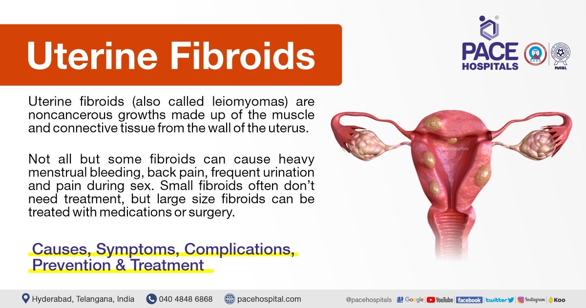
– Types of myomas include intramural myomas (located within the wall of the uterus), subserosal myomas (grow on the outside of the uterine wall), pedunculated myomas (develop a stalk or stem attaching them to the uterus), and submucosal myomas (found just under the lining of the uterus).
– A large myoma is considered to be 10 centimeters or more in diameter.
– Emergency room visits for myoma symptoms have increased from 2006 to 2017, including pelvic pain and heavy bleeding.
– Myomas are noncancerous growths that can cause various symptoms depending on their size and location.
– Symptoms of myomas include heavy and painful periods, bleeding between periods, pelvic pain, abdominal pressure, a feeling of fullness in the lower abdomen, constipation, diarrhea, frequent urination, pain during sex, lower back pain, trouble getting pregnant, fatigue, and weakness.
– Myomas are not life-threatening but can cause complications such as heavy blood loss or organ obstruction.
– Myomas are a top cause of hysterectomy surgeries.
– If a myoma bursts, immediate medical care is necessary.
– The exact cause of myomas is unknown but is likely associated with hormone activity, particularly high levels of estrogen and progesterone.
– Risk factors for myomas include a family history of the condition, obesity, high blood pressure, age, and certain dietary factors.
– Myomas are more common among Black people with a uterus.
– Diagnosis of myomas involves a series of steps, including medical history, physical examination, imaging tests such as ultrasound or MRI, and sometimes a biopsy.
– Treatment options for myomas include medication, noninvasive procedures, surgery, or a combination of therapies.
– Medications that may be used include over-the-counter pain medications, iron supplements, and birth control methods.
– Surgical options include laparoscopic myomectomy and uterine fibroid embolization (UFE).
– Lifestyle changes including dietary changes, exercise, stress management, and weight loss may improve symptoms and overall health.
– Complications of untreated myomas include fertility issues, pregnancy complications, and the need for cesarean delivery.
– It is important to discuss myomas with a healthcare provider before pregnancy for potential complications.
– Uterine fibroids, also known as myomas, are non-cancerous tumors that grow in the uterus.
– The most common symptom of myomas is vaginal bleeding.
– Other symptoms of myomas include heavy bleeding, anemia, fatigue, painful intercourse, pain, bleeding, or discharge from the vagina if myomas become infected, a feeling of pressure or lump in the abdomen, difficulties urinating, dribble at the end of urination, or urine retention if a myoma blocks the flow of urine.
– Myomas affect 20 percent of women in their childbearing years.
– The signs and symptoms of uterine fibroids include abnormal bleeding, pelvic masses, pelvic pain, infertility, and pregnancy complications.
– There are five types of uterine fibroids: intramural fibroids, subserosal fibroids, submucosal fibroids, pedunculated fibroids, and intracavitary fibroids.
– Between 70 and 80 percent of women develop a fibroid tumor by the time they reach age 50.
– Estrogen seems to activate the growth of uterine fibroids, and they shrink after menopause, but hormone therapy after menopause may cause their symptoms to continue.
– Factors associated with the development of uterine fibroids include race, age, early menstruation, caffeine and alcohol intake, genetics, obesity, high blood pressure, and diet.
– Nearly one-third of women with uterine fibroids seek treatment because of the severity of their symptoms.
– Treatment options for uterine fibroids include hormonal contraception, intrauterine devices, antifibrinolytic nonsteroidal agents, endometrial ablation, medications called gonadotropin-releasing hormone agonists, myomectomy, MRI-guided ultrasound surgery, and uterine fibroid embolization.
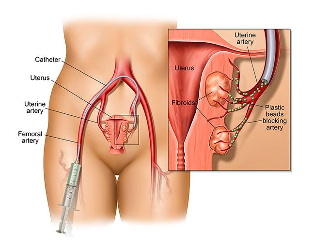
– Uterine fibroid embolization is a minimally invasive option that blocks the blood supply to fibroids, causing them to shrink and die.
– Myoma, leiomyoma, and uterine fibroids all refer to the same thing, a non-cancerous tumor of the uterus.
– The management of uterine fibroids, also known as myomas, requires further research to improve treatment outcomes.

Continue Reading

Congenital Absence of Uterus: Understanding the Medical Condition

– Uterine agenesis is a congenital disorder characterized by the absence of a uterus
– It may be a symptom of broader conditions like MRKH syndrome, MURCS association, or AIS
– Other reproductive organs may also be affected, such as the ovaries or fallopian tubes
– The exact cause of uterine agenesis is unknown
– Symptoms can include the absence of menstrual periods and abdominal pain
– Diagnosis is typically made during puberty when a girl fails to start menstruating
– Diagnostic tests may include blood tests, ultrasound, and MRI
– Treatment options depend on the individual and may involve creating a vagina if it is also absent
– Psychological support and counseling may be beneficial for individuals with uterine agenesis
– Regular medical follow-ups are necessary to monitor any associated conditions or complications.

Continue Reading

Myoma of the uterus: Understanding causes, symptoms, treatment

– Uterine fibroids are non-cancerous tumors that grow in the uterus, also known as myomas.
– Myomas are smooth, non-cancerous tumors made partly of muscle tissue that may develop in or around the uterus.
– Myomas in the larger, upper part of the uterus are called fibroids or leiomyomas.
– Most myomas can be seen or felt during a pelvic examination and those causing symptoms can be removed surgically or through less invasive procedures.
– The most common symptom of myomas is vaginal bleeding, which may be irregular or heavy.
– Other symptoms may include heavy bleeding, anemia, fatigue, weakness, painful intercourse, pain, bleeding, or discharge from the vagina if myomas become infected, pressure or lump in the abdomen, difficulties urinating, and urinary tract infections.
– Uterine fibroids, or myomas, affect 20 percent of women in their childbearing years.
– Uterine fibroids can cause abnormal bleeding, pelvic masses, pelvic pain, infertility, and pregnancy complications.
– There are five types of uterine fibroids: intramural, subserosal, submucosal, pedunculated, and intracavitary fibroids.
– Between 70 and 80 percent of women develop a fibroid tumor by the age of 50.
– Estrogen seems to activate the growth of uterine fibroids, and they usually shrink after menopause.
– Hormone therapy after menopause may cause fibroid symptoms to continue.
– African American women are more susceptible to developing fibroids.
– Fibroids tend to grow faster in white women younger than 35 years compared to those older than 45 years.
– Delaying pregnancy until age 30 or older increases the risk of developing fibroids.
– Early menstruation increases the risk of developing fibroids.
– Alcohol and caffeine intake may increase the risk of developing fibroids.
– Specific genetic alterations are linked to fibroid growth.
– Obesity and high blood pressure may play a role in fibroid development and growth.
– A diet rich in red meat may increase the chance of developing fibroids.
– Treatment options for uterine fibroids vary depending on the severity of symptoms and the patient’s desire to have children.
– Hormonal contraception, intrauterine devices, antifibrinolytic drugs, and nonsteroidal agents are options for managing heavy bleeding.
– Endometrial ablation can be performed if the patient does not want to have children.
– Gonadotropin-releasing hormone agonists can shrink fibroids, but they may grow back.
– Myomectomy is a procedure that removes fibroids while preserving the uterus, but fibroids may grow back.
– MRI-guided ultrasound surgery can shrink fibroids and reduce heavy bleeding.
– Uterine fibroid embolization is a minimally invasive procedure that blocks the blood supply to fibroids, causing them to shrink and die.
– Myomas can cause symptoms such as abdominal pain and heavy menstrual bleeding, but some people may remain symptom-free.
– The cause of myomas is unclear, but risk factors include a family history of myoma, obesity, and age.
– More than half of all people with a uterus will experience a myoma by age 50.
– Myomas can vary in size, from as small as a pea to as large as a melon.
– They can be located inside the uterus (intramural myomas), on the outside of the uterine wall (subserosal myomas), have a stalk or stem attaching them to the uterus (pedunculated myomas), or be found just under the lining of the uterus (submucosal myomas).
– Large myomas are considered to be 10 centimeters (cm) or more in diameter.
– Emergency room visits for myoma symptoms, such as pelvic pain and heavy bleeding, have increased from 2006 to 2017, according to a recent study.
– Myomas of the uterus are noncancerous tumors that can cause a variety of symptoms.
– Symptoms may include heavy or prolonged periods, bleeding between periods, pelvic pain, abdominal pressure, a feeling of fullness in the lower abdomen, constipation or diarrhea, frequent urination or trouble urinating, pain during sex, lower back pain, reproductive issues, fatigue, and weakness.
– Myomas are a top cause of hysterectomy surgeries, which come with their own risks and complications.
– The exact cause of myomas is unknown, but they are likely associated with hormone activity.
– High levels of estrogen and progesterone may stimulate their growth, and myomas tend to shrink when hormone levels decrease after menopause.
– Risk factors for developing myomas include a family history of myomas, obesity, high blood pressure, age, and dietary factors such as a diet high in red meat or vitamin D deficiency.
– Myomas are more common among Black individuals with a uterus, and factors such as low vitamin D levels, obesity, stress, genetics, and unequal access to healthcare have been proposed as potential risk factors.
– Medical tests used to diagnose myomas include pelvic examination, ultrasound or transvaginal ultrasound, and magnetic resonance imaging (MRI).
– Treatment for myomas depends on factors such as the severity of symptoms, the size and location of the myomas, the desire for future pregnancy, age, and proximity to menopause.
– Medications can be used to treat myomas, including over-the-counter pain medications, iron supplements for depleted iron levels, and birth control methods to control heavy menstrual bleeding.
– GnRH agonists (hormone-stimulating medications) can be used to temporarily shrink myomas, especially if surgery is planned.
– Surgical options for myomas include laparoscopic myomectomy and uterine fibroid embolization (UFE).
– No specific figures or statistics are mentioned in the article.
– One treatment option is a radiology procedure that uses injections to block blood flow to the myomas, causing them to shrink and sometimes die.
– Another option is MRI-guided ultrasound surgery, which uses ultrasound waves to shrink myomas.
– In more severe cases, other surgical options may be considered.
– Hysterectomy is a surgery to completely remove the uterus, eliminating the fibroids but also making pregnancy impossible in the future.
– Abdominal myomectomy is a surgical procedure that removes the fibroids without removing the uterus, with the possibility of future pregnancy but a risk of the fibroids returning.
– While there are no home remedies that directly treat fibroids, certain complementary therapies like acupuncture, yoga, massage, traditional Chinese medicine, and heating pads may help manage symptoms.
– Lifestyle changes such as dietary changes, exercise, stress management, and weight loss can also be beneficial.
– Myomas can cause complications related to fertility, pregnancy, and childbirth.
– These complications can include fertility issues, pregnancy complications such as miscarriage or early labor, and the need for a cesarean delivery.
– It’s important for those with myomas who want to become pregnant to discuss the condition with their healthcare provider to assess potential risks.
– It is recommended to communicate symptoms affecting one’s life to healthcare providers to determine the most suitable treatments.

Continue Reading
The Hidden Dangers of Red Degeneration: Untold Symptoms, Risks

– Red degeneration, also known as carneous degeneration
– Type of degeneration that can occur in uterine leiomyomas
– Most common form of degeneration during pregnancy
– Symptoms of red degeneration include abdominal pain, fever, and leukocytosis
– Red appearance of the leiomyoma
– Caused by venous thrombosis or rupture of intratumoral arteries
– Unusual signal intensity patterns on MRI, such as high signal intensity on T1-weighted images and variable signal intensity on T2-weighted images
– Contrast enhancement can vary

Continue Reading